

fig5
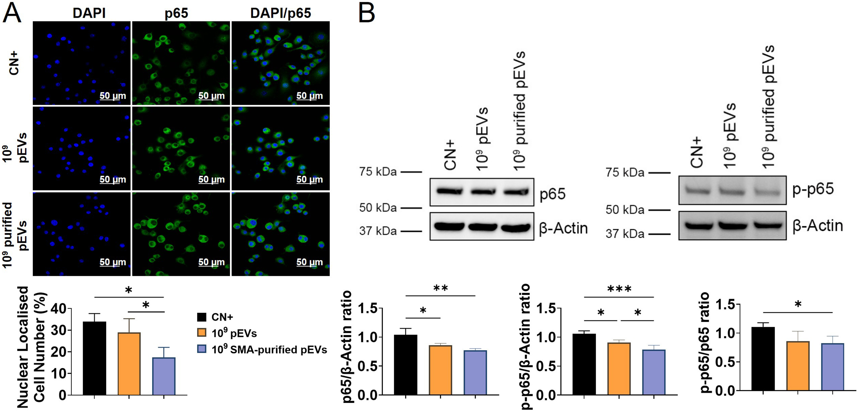
Figure 5. Effects of pEVs and purified pEVs on NF-κB-associated signalling in RAW cells. RAW cells were primed with E. coli LPS (1 µg/mL) for 2 h, washed three times with PBS, and subsequently stimulated with 100 ng/mL LPS, either alone or in combination with pEVs or purified pEVs (1 × 10⁹ particles/mL) for 24 h. (A) Representative immunofluorescence images showing p65 nuclear localisation and quantitation of the proportion of RAW cells with nuclear p65 localisation; (B) Representative Western blots of total p65, p-p65 and β-Actin, with corresponding densitometric analysis. The corresponding original, uncropped Western blot images are presented in Supplementary Figure 4. Data are expressed as the mean ± SD of three independent experiments, each using paired pEV and purified pEV preparations from a different plasma donor (n = 3). Statistical significance is indicated as *P < 0.05; **P < 0.01; ***P < 0.001. DAPI: 4’,6-diamidino-2-phenylindole; LPS: lipopolysaccharide; NF-κB: nuclear factor-κB; p65: nuclear factor-κB p65; p-p65: phosphorylated p65; pEVs: plasma-derived extracellular vesicles; PBS: phosphate-buffered saline; SD: standard deviation.