
fig6
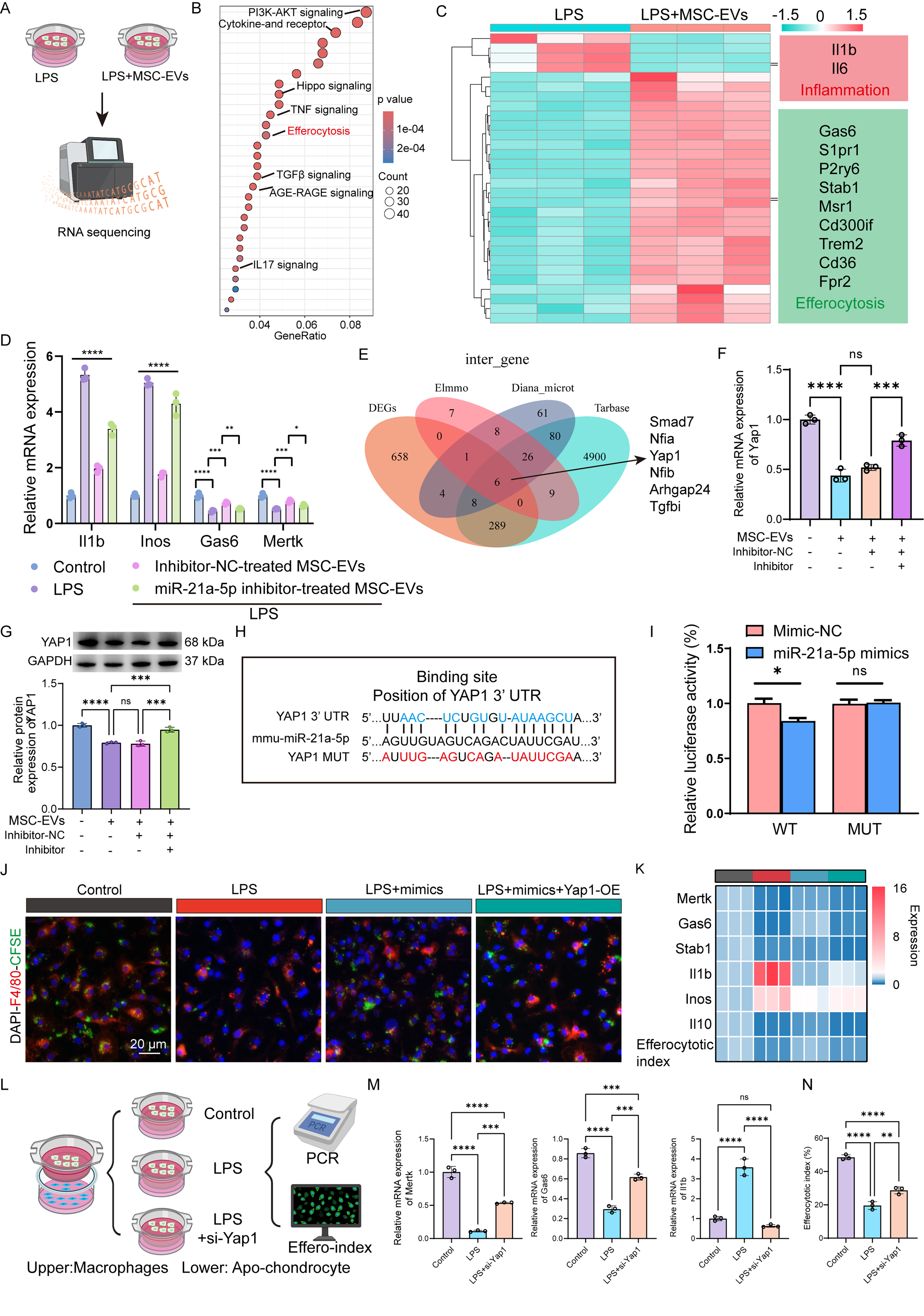
Figure 6. MSC-EVs restored the efferocytosis of macrophages and anti-inflammatory phenotype via miR-21a-5p/YAP1 axis. (A) Scheme of bulk RNA sequencing in macrophages treated with LPS or MSC-EVs; (B) KEGG pathway enrichment analysis of DEGs identified in macrophages following LPS or EV treatment; (C) Clustered heatmap illustrating altered expression patterns of genes associated with inflammation and efferocytosis in macrophages exposed to LPS or EVs; (D) Expression of inflammation- and efferocytosis-associated genes was assessed by RT-qPCR in LPS-stimulated macrophages treated with EVs obtained from BMSCs transfected with inhibitor-NC or miR-21a-5p inhibitor (n = 3 independent biological experiments); (E) Bioinformatic prediction of candidate downstream targets of miR-21a-5p; (F) Yap1 mRNA levels were determined by RT-qPCR in macrophages receiving EVs derived from inhibitor-NC- or miR-21a-5p inhibitor-transfected BMSCs (n = 3 independent biological experiments); (G) Western blot evaluation of YAP1 protein expression in LPS-stimulated macrophages treated with EVs from BMSCs transfected with inhibitor-NC or miR-21a-5p inhibitor (n = 3 independent biological experiments); (H) Predicted complementary sequence between miR-21a-5p and its putative binding site in the Yap1 3’-UTR; (I) Dual-luciferase reporter assay evaluating the interaction between miR-21a-5p and the wild-type Yap1 3’-UTR in HEK293T cells. Co-transfection with miR-21a-5p reduced the luciferase activity of the wild-type reporter construct; (J) Representative immunofluorescence images showing F4/80-positive macrophages (red) and engulfed CFSE-labeled apoptotic chondrocytes (green) following 2 h of co-incubation under the indicated treatment conditions. Scale bar: 20 µm; (K) Heatmap summarizing inflammation- and efferocytosis-related gene-expression profiles together with the efferocytosis index across the indicated macrophage treatment groups (n = 3 independent biological experiments); (L) Schematic illustration of the macrophage-apoptotic chondrocyte co-incubation model; (M and N) RT-qPCR analysis of inflammation- and efferocytosis-related genes and efferocytotic index in macrophages with varying treatments (n = 3 independent biological experiments). NC indicated inhibitor-NC or mimic-NC. One-way ANOVA followed by Tukey’s multiple-comparison test was used for comparison in multiple groups (D), (F), (G), (M), and (N). Student’s t-test was used in (I). *P < 0.05, **P < 0.01, ***P < 0.001, ****P < 0.0001. (A) and (L) were created with BioRender.com. MSC-EVs: Mesenchymal stem cell-derived extracellular vesicles; LPS: lipopolysaccharide; KEGG: Kyoto Encyclopedia of Genes and Genomes; DEGs: differentially expressed genes; EVs: extracellular vesicles; RT-qPCR: quantitative real-time polymerase chain reaction; BMSCs: bone marrow mesenchymal stem cells; NC: negative control; 3’-UTR: 3’-untranslated region; CFSE: carboxyfluorescein succinimidyl ester; ANOVA: analysis of variance.