

fig1
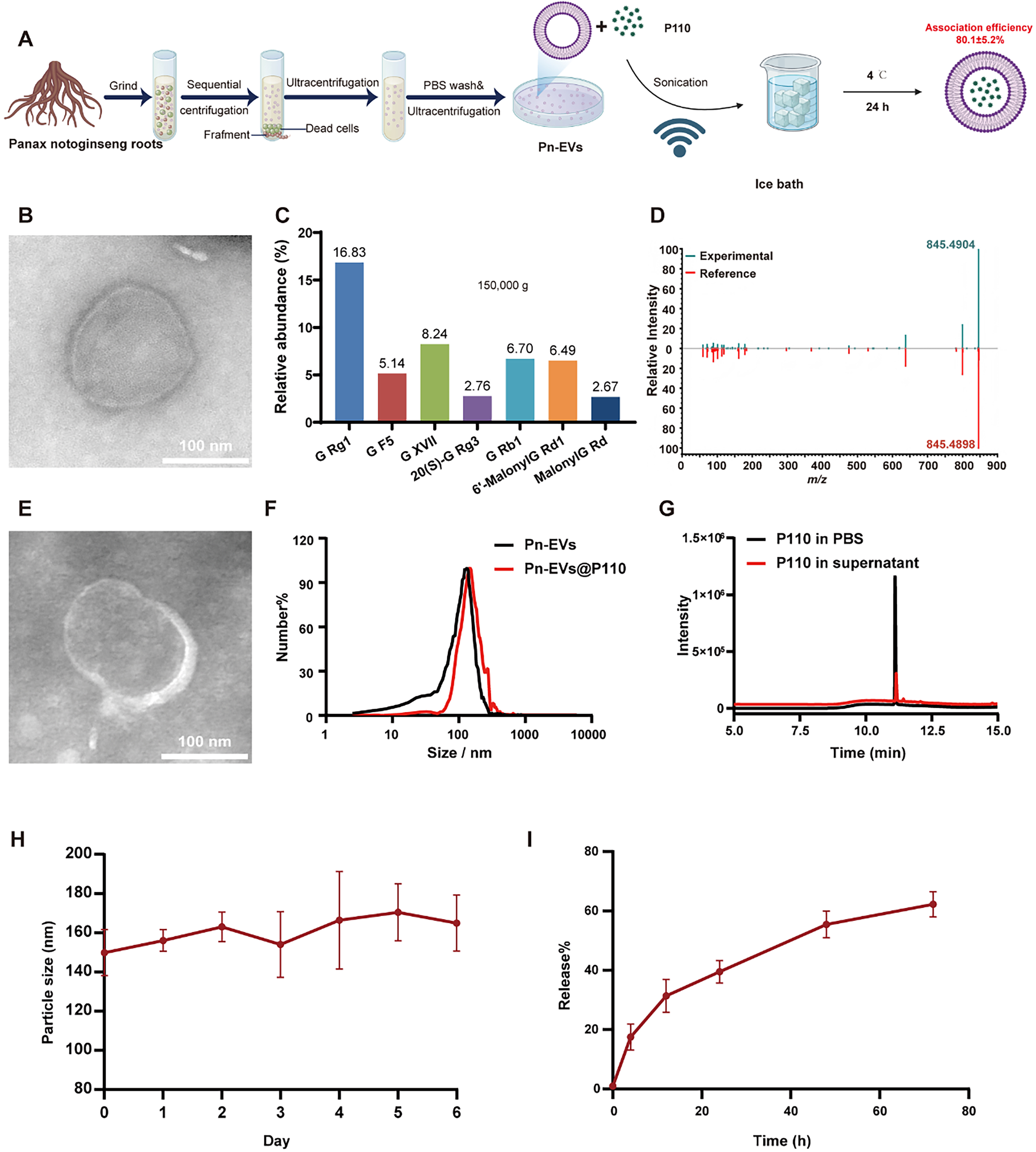
Figure 1. Preparation and characterization of Pn-EVs@P110. (A) Schematic illustration of Pn-EV isolation and Pn-EVs@P110 preparation; (B) Representative TEM image of Pn-EVs; (C) Relative abundance of representative ginsenosides detected in Pn-EVs; (D) Representative LC-MS/MS identification of ginsenoside Rg1; (E) Representative TEM image of Pn-EVs@P110; (F) Particle-size distributions of Pn-EVs and Pn-EVs@P110 measured by NTA; (G) HPLC chromatograms of P110 in PBS and P110 in the supernatant after the association procedure; (H) Particle-size stability of Pn-EVs@P110 during storage at 4 °C for 6 days; (I) In vitro release of P110 from Pn-EVs@P110 over 72 h. Data are presented as mean ± SD. The schematic in Figure 1A was adapted and modified from our previously published work[15]. Pn-EVs: Panax notoginseng-derived extracellular vesicle-like nanovesicles; TEM: transmission electron microscopy; LC-MS/MS: liquid chromatography-tandem mass spectrometry; NTA: nanoparticle tracking analysis; HPLC: high-performance liquid chromatography; PBS: phosphate-buffered saline; SD: standard deviation.




