

Animal-derived extracellular vesicles and vesicle-like nanoparticles: linking natural bioactivity to precision therapeutics
 0

Graphical Abstract
Abstract
Animal-derived extracellular vesicles (AEVs) and animal-derived vesicle-like nanoparticles (AVLNs) are emerging natural nanocarriers with diverse bioactive cargoes, potential biocompatibility, and capacity for cross-species molecular communication. This narrative review distinguishes naturally released AEVs from AVLNs obtained through tissue processing without sufficient evidence of natural extracellular release. It covers particles from glandular secretions, tissues, and cultured cells, focusing on isolation, characterization, molecular composition, biodistribution, biological functions, therapeutic mechanisms, and translational potential. Source-dependent proteins, lipids, and nucleic acids can modulate disease-relevant signaling and recipient-cell responses, contributing to immunomodulatory, anti-inflammatory, antitumor, regenerative, antioxidant, antibacterial, and antiviral activities. However, evidence derives predominantly from in vitro studies and preclinical animal models, with substantial heterogeneity in preparation, characterization, dose reporting, and outcome assessment; most reported therapeutic effects have not been clinically established. Cargo signatures may inform vesicle identity, biological activity, and therapeutic response, but their diagnostic and predictive value remains exploratory and requires clinical validation. Major translational challenges include inconsistent terminology, limited methodological standardization, cargo heterogeneity, uncertain biodistribution, and biosafety concerns, particularly immunogenicity and long-term safety. Addressing these limitations and validating relationships among vesicle sources, functional cargoes, molecular targets, and disease indications will be essential to translate intrinsic bioactivity into reproducible platforms for precision therapeutics.
Keywords


 Download PDF
INTRODUCTION
Exosomes are a subtype of extracellular vesicles (EVs) of endosomal origin that are released following the fusion of multivesicular bodies (MVBs) with the plasma membrane. Because particle size and morphology alone cannot establish an endosomal origin, the term “exosome” should be reserved for vesicles whose biogenesis is supported by appropriate experimental evidence[1,2]. Following the initial discovery of these vesicles in 1983 by Harding et al. and Pan et al. in rat and sheep reticulocytes, respectively[3,4], Johnstone et al. subsequently used the term “exosomes” for vesicles released during reticulocyte maturation in 1987[5]. Since then, accumulating evidence shows that exosomes participate in diverse physiological and pathological processes, including antigen presentation, immune response regulation, and cytokine transport[6,7].
Naturally derived EVs and vesicle-like nanoparticles can originate from a wide range of animal and plant sources[8]. Although research has traditionally focused on EVs from human stem cells and tumors, increasing attention has shifted toward non-human vesicular systems. Plant-derived vesicle-like nanoparticles have been extensively investigated, whereas animal-derived EVs and vesicle-like nanoparticles remain comparatively understudied because of source heterogeneity and challenges associated with their preparation and characterization. Nevertheless, emerging preclinical studies have reported biological activities in diverse animal-derived systems, including deer antler stem cell-derived EVs and oyster mantle-derived vesicle-like nanoparticles[9,10]. For example, the yield of vesicle-like particles isolated from oyster mantle tissue can reach as high as 3.14 × 10⁹ particles per gram of mantle tissue[10]. Moreover, the availability of plant-derived vesicle-like nanoparticles may be affected by seasonal growth patterns and variation in the supply of plant material[11,12].
Animal-derived vesicular particles have attracted increasing attention because of their diverse biological sources, bioactive cargoes, and potential applications in biomedicine and nanotechnology. However, the field remains constrained by inconsistent terminology, nonstandardized isolation and characterization methods, source-dependent heterogeneity, and an incomplete understanding of vesicle biogenesis. In accordance with the recommendations of the Minimal Information for Studies of Extracellular Vesicles (MISEV2023), naturally released vesicles isolated from animal cells or biological fluids are generally referred to in this review as animal-derived extracellular vesicles (AEVs). In contrast, nanoparticles obtained after tissue disruption, sonication, enzymatic digestion, or related processing procedures without sufficient evidence of natural extracellular release are referred to as animal-derived vesicle-like nanoparticles (AVLNs)[2]. When these heterogeneous particles are discussed collectively, the descriptive term “animal-derived vesicular particles” is used without implying a shared biogenetic origin. Based on their sources and modes of preparation, the animal-derived vesicular particles discussed in this review are classified into three major groups: those obtained from animal glandular secretions or biological fluids, those isolated from animal tissues or tissue-derived materials, and those secreted by cultured animal cells. The first group includes milk-derived EVs obtained from bovine, porcine, goat, yak, and dromedary camel milk[13], as well as vesicular particles obtained from the mucilage of Pinctada martensii[14], skin secretions of Bombina maxima[15], and honey[16]. Tissue-derived vesicle-like nanoparticles include those prepared from oyster mantle tissue[10], egg white[17], regenerating sea anemones[18], and Periplaneta americana[19]. Cell-derived EVs include those secreted by deer antler stem cells[9] and salamander skeletal muscle-derived myogenic progenitor cells[20].
We summarize current knowledge of their biological sources, isolation and characterization strategies, molecular composition, engineering strategies, administration routes, biodistribution, and source-dependent bioactivities. Particular attention is given to their anti-inflammatory, antitumor, regenerative, antibacterial, and antiviral effects, as well as the relationships among their functional cargoes, disease-relevant molecular targets, and potential therapeutic indications. We also critically discuss limitations in the current evidence, biosafety concerns, and key challenges in standardization and clinical translation, aiming to clarify their potential as naturally derived platforms for precision therapeutics. An earlier version of this manuscript was posted as a preprint[21].
OVERVIEW OF ANIMAL-DERIVED EXTRACELLULAR VESICLES AND VESICLE-LIKE NANOPARTICLES
Biogenesis and origins of animal-derived vesicular particles
The MVB pathway is a key mechanism of exosome biogenesis in animal cells. Exosome formation is closely associated with the continuous remodeling of biological membranes. The plasma membrane first undergoes invagination through endocytosis to form early endosomes, followed by the accumulation of bioactive substances within early sorting endosomes (ESEs). Subsequently, under the control of the endosomal sorting complex required for transport and associated proteins, ESEs mature into late sorting endosomes (LSEs). After a second round of invagination, LSEs develop into MVBs[8]. Based on their functional fate, MVBs can be classified into two categories: secretory MVBs and degradative MVBs. Secretory MVBs are transported to the vicinity of the plasma membrane and fuse with it in a calcium-dependent manner, releasing vesicles of relatively uniform size into the extracellular space; these vesicles are defined as exosomes[22]. In contrast, degradative MVBs fuse with lysosomes following the endocytosis of macromolecules or vesicular structures, leading to degradation of their contents, or they may fuse directly with lysosomes for degradation. Studies have indicated that cholesterol levels are a major determinant of MVB fate. Cholesterol-rich MVBs are targeted and sorted to the plasma membrane as exocytic vesicles, whereas cholesterol-poor MVBs are directed to lysosomes for degradation[23,24]. The mechanisms underlying exosome biogenesis and their hierarchical regulation are highly diverse. Current evidence increasingly supports a role for Ras-related in brain (Rab) family proteins as molecular switches that regulate intracellular vesicle formation and trafficking in eukaryotic cells[25]. However, other components, including tetraspanins and lipid rafts, have also been reported to contribute to exosome formation[26,27]. Thus, the precise mechanisms remain under debate. The major endosomal pathways involved in EV biogenesis and secretion are summarized schematically in Figure 1.
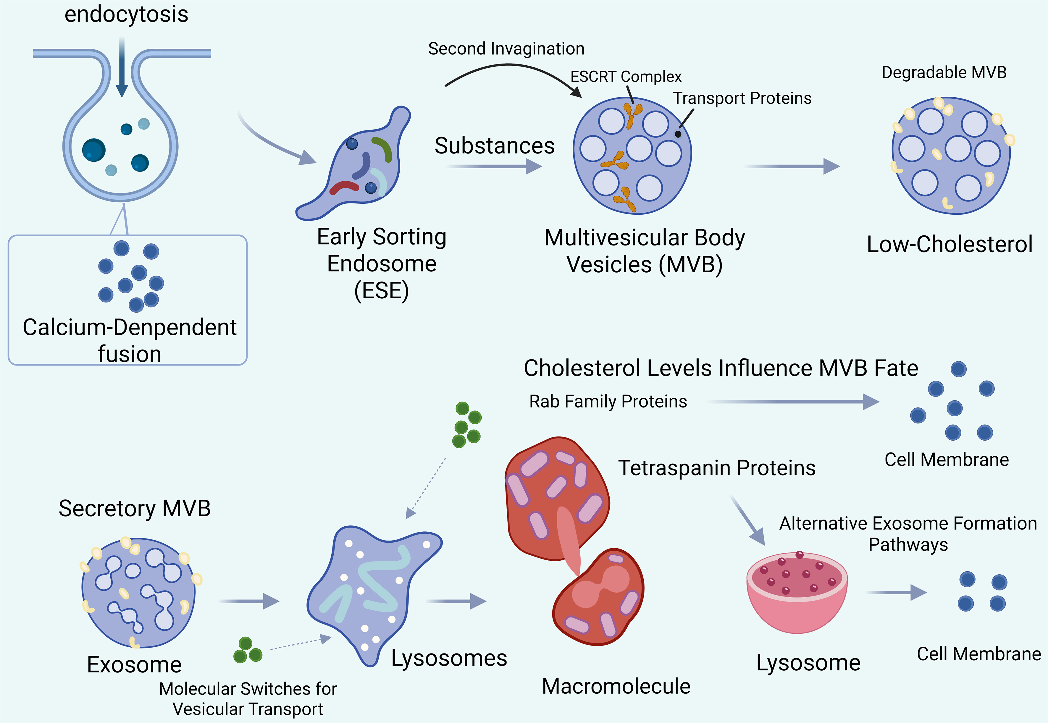
Figure 1. Biogenesis and secretion pathways of AEVs. The schematic illustrates the endocytic formation of ESEs, their maturation into MVBs, and the subsequent release of exosomes upon fusion of secretory MVBs with the plasma membrane. Alternatively, MVBs may fuse with lysosomes, resulting in degradation of their contents. This pathway applies to naturally secreted EVs with a demonstrated endosomal origin and should not be presumed to apply to vesicle-like nanoparticles obtained through tissue disruption, sonication, enzymatic digestion, or other processing procedures. Created in BioRender. LIU, Y. (2026) (https://BioRender.com/ju0anww). AEVs: Animal-derived extracellular vesicles; ESE: early sorting endosome; ESCRT: endosomal sorting complex required for transport; EVs: extracellular vesicles; MVB: multivesicular body; Rab: Ras-related in brain.
Isolation and preparation of animal-derived vesicular particles
Animal-derived vesicular particles can be obtained from heterogeneous sources, including biological fluids, glandular secretions, intact tissues, tissue-derived materials, and cultured cells. Depending on the source, preparation may involve direct isolation of EVs or tissue processing followed by enrichment of vesicle-like nanoparticles. Source-specific pretreatment is generally required before the isolation or enrichment of animal-derived vesicular particles to remove impurities and improve sample purity. For naturally secreted EVs, pretreatment may include the removal of cells, cellular debris, and soluble contaminants before EV isolation. In contrast, preparation of tissue-derived vesicle-like nanoparticles may involve tissue fragmentation, sonication, or enzymatic digestion. Because these procedures can disrupt cellular membranes and generate heterogeneous vesicular structures, the resulting particles should not be classified as exosomes without evidence of natural extracellular release and an endosomal-MVB origin[2]. The following sections summarize pretreatment, separation, and purification methods used for different categories of animal-derived vesicular particles, together with their respective advantages and limitations.
Preprocessing
Prior to the preparation of vesicle-like nanoparticles from animal tissues, tissue surfaces are generally cleaned to remove superficial contaminants and then washed with phosphate-buffered saline (PBS). Depending on the source and preparation protocol, the tissue may then undergo fragmentation, enzymatic digestion, or sonication before particle enrichment. For example, to prepare oyster mantle-derived vesicle-like nanoparticles, fresh mantle tissue is manually dissected, washed with PBS, cut into small pieces on ice, and digested with papain at 37 °C before further separation and purification of the resulting vesicular particles[10]. In contrast, EVs secreted by cultured animal cells are collected from conditioned culture media after the target cells are isolated and characterized. For example, antlerogenic stem cells are isolated from newly formed antler tissue by collagenase III digestion and flow cytometric sorting, whereas salamander myogenic precursor A1 cells are characterized using the 22/18 blastemal marker antibody[20]. During the preparation of regenerating sea anemone-derived vesicle-like nanoparticles, animals are thoroughly washed with ice-cold sterile PBS and then sonicated using a Q125 sonicator at 20 kHz, with 10 s pulses separated by 20 s intervals for a total duration of 30 min[18]. Because tissue fragmentation, enzymatic digestion, and sonication may disrupt cellular membranes and generate heterogeneous vesicular structures, particles obtained through these procedures are referred to throughout this review as vesicle-like nanoparticles rather than exosomes, unless their natural extracellular release and endosomal-MVB origin have been demonstrated[2]. Accordingly, this review distinguishes naturally secreted EVs from tissue-derived vesicle-like nanoparticles based on their biological source and preparation procedure.
Isolation and purification
Pretreatment removes superficial and relatively large impurities. However, additional contaminants may remain in vesicular particle preparations during isolation and processing, including cellular debris, soluble proteins, host-cell DNA, and co-isolated non-vesicular particles. Such impurities may compromise the purity and functional properties of the preparation, necessitating further purification. Commonly employed purification methods include ultracentrifugation, density-gradient centrifugation, ultrafiltration, commercial kit-based methods, polymer-based precipitation, and immunoaffinity purification. Because animal-derived vesicular particles are physicochemically heterogeneous, combinations of complementary separation methods are often required to balance recovery and purity[2].
Ultracentrifugation separates components according to their sedimentation rates in a homogeneous suspension. This method involves a series of centrifugation cycles performed at different centrifugal forces and durations to isolate or enrich EVs on the basis of differences in their density and size relative to other components. Generally, preliminary low-speed centrifugation is performed before ultracentrifugation to remove intact cells and cellular debris. Typically, centrifugation at 10,000 × g separates apoptotic bodies and larger vesicles; approximately 35,000 × g is used to isolate medium-sized vesicles; and 100,000 × g is suitable for smaller EVs[13]. The method yields EV-enriched preparations without the use of separation reagents. However, it has several limitations, including relatively low purity, high equipment cost, time-consuming procedures, and the potential to disrupt vesicle structure, which may affect downstream analyses[13,28].
Density-gradient centrifugation combines ultracentrifugation with density-gradient separation principles. The main approaches use sucrose discontinuous gradients or iodixanol continuous gradients. Vesicular particles with different buoyant densities sediment into distinct density fractions, allowing their separation. For example, oyster mantle-derived vesicle-like nanoparticles have been purified by differential ultracentrifugation combined with sucrose density-gradient centrifugation[10]. This approach can improve purity by separating vesicular particles from contaminants with different buoyant densities[29]. However, its limitations are similar to, and may be more pronounced than, those of ultracentrifugation[13]. The procedure involves complex and time-consuming preparation steps, depends heavily on operator expertise and specialized instrumentation, and provides relatively low EV recovery efficiency.
Polymer-based precipitation typically uses polyethylene glycol (PEG) to reduce the solubility of EVs and other nanoscale particles, thereby facilitating their collection by centrifugation[8,30]. PEG-based precipitation provides a simple and scalable approach for EV enrichment and can achieve favorable recovery for downstream molecular analyses[30]. However, co-precipitated proteins and residual polymers may reduce preparation purity and interfere with subsequent analyses.
Immunoaffinity chromatography (IAC) separates and purifies EVs through specific antibody-antigen binding. Targets typically include abundant EV-associated surface proteins, such as tetraspanin-family members, or source-specific surface molecules that enable selective recognition of EV subpopulations from particular cellular origins[31]. Compared with ultracentrifugation, IAC provides greater specificity for selected EV subpopulations and facilitates their qualitative and quantitative analysis. However, EVs isolated by IAC generally require relatively stringent handling and storage conditions. In addition, the limited binding capacity and high cost of the method limit its application to large-scale isolation[32].
Size-exclusion chromatography (SEC) exploits the inability of macromolecules to enter the pores of the gel matrix. These molecules elute early through the interstitial spaces between porous gel particles, whereas smaller molecules are retained within the pores and elute later. SEC is characterized by its rapidity, simplicity, and low cost[33]. EVs isolated using this method generally retain intact structures and exhibit relatively uniform size distributions[34], with minimal adverse effects on their biological properties; however, co-isolation of similarly sized particles may reduce purity[8]. For example, EVs from Bombina maxima skin secretions have been separated and purified by differential centrifugation combined with SEC[15]. For isolating bovine milk-derived EVs, SEC is one of the most commonly used methods[33]. Nevertheless, due to the presence of a large amount of proteins in whey, and the fact that some proteins fall within the same size range as EVs, the use of SEC alone is insufficient for effective separation[35].
Membrane filtration separates particles using filters of defined pore size and molecular weight cut-offs (MWCO)[36]. Particles larger than the MWCO are retained, whereas particles smaller than the MWCO, including small EVs, are collected. Ultrafiltration is an effective concentration step and is often used in combination with other EV isolation techniques[37]. However, shear forces generated during filtration may damage EVs and may also cause aggregation, entrapment of vesicles within membrane pores, or adsorption of EVs onto the membrane surface, resulting in sample loss.
Emerging isolation and fractionation technologies
Tangential flow filtration (TFF) is an increasingly used, scalable approach for concentrating EVs from large-volume samples. Unlike dead-end filtration, TFF directs the sample flow parallel to the membrane surface, reducing membrane blockage and enabling continuous processing. Compared with ultracentrifugation, TFF can provide higher recovery, shorter processing times, and improved batch-to-batch consistency, while exposing vesicles to relatively mild processing conditions. However, TFF concentrates particles according to membrane cut-off and does not by itself separate EVs from similarly sized proteins, lipoproteins, or other extracellular particles. It is therefore often combined with SEC or another purification method when high purity is required[38].
Asymmetric flow field-flow fractionation (AF4) is a label-free technique that separates particles within a thin channel by diffusion behavior and hydrodynamic size under the combined effects of longitudinal flow and a perpendicular cross-flow field. AF4 can resolve heterogeneous extracellular particle populations and has been used to separate small-EV fractions from smaller non-membranous nanoparticles. When coupled with multi-angle light scattering, dynamic light scattering, or ultraviolet detection, AF4 also enables detailed analysis of particle size and biophysical heterogeneity. However, AF4 requires specialized instrumentation and extensive method optimization, has limited sample capacity, and is currently better suited to analytical fractionation than for routine large-scale production[39].
Microfluidic technologies integrate size-, affinity-, acoustic-, or electrokinetic-based separation within miniaturized devices. These platforms require small sample volumes and can integrate EV capture, detection, and molecular analysis in a single workflow, offering advantages for rapid analysis and potential point-of-care applications. Affinity-based microfluidic devices may capture EV subpopulations using antibodies against surface proteins, whereas label-free devices separate particles according to physical properties. However, microfluidic platforms may suffer from limited throughput, device-to-device variability, channel blockage, and dependence on specialized fabrication. Affinity-based designs may also introduce selection bias by enriching only marker-positive subpopulations[40].
Immunomagnetic isolation uses antibody-conjugated magnetic beads to capture EV subpopulations expressing selected surface proteins, such as cluster of differentiation 9 (CD9), cluster of differentiation 63 (CD63), and cluster of differentiation 81 (CD81), or source-specific antigens. Magnetic separation is rapid, technically straightforward, and compatible with downstream flow cytometry, immunoblotting, and molecular analysis. Its specificity helps isolate defined EV subpopulations from complex samples. However, the method is limited by antibody availability, bead-binding capacity, marker heterogeneity, and the possibility that bead attachment interferes with subsequent functional studies. Because canonical mammalian EV markers may not be conserved in non-mammalian species, immunomagnetic isolation of animal-derived EVs requires source-appropriate and experimentally validated surface targets[41].
These emerging approaches offer complementary advantages in scalability, analytical resolution, processing speed, and subpopulation specificity. However, no single method simultaneously optimizes recovery, purity, throughput, and biological representativeness. Method selection should therefore be guided by sample type, required scale, downstream application, and the degree of purity or subpopulation specificity needed, in accordance with the MISEV2023 principle of fit-for-purpose EV separation.
In addition to conventional isolation methods, several commercial kits are available, such as the exoEasy Maxi Kit (QIAGEN), MagCaptureTM Exosome Isolation Kit PS (Wako), and the MinuteTM High-Efficiency Exosome Precipitation Kit (Invent)[8]. The ExoVista Exosome Isolation Kit (PerciaVista Biotechnology Co., Iran) has also been used to prepare vesicle-like nanoparticles from regenerating sea anemones. Kit-based methods are rapid, provide relatively high yields, and preserve vesicle integrity[18]. However, these kits are relatively expensive, and their performance may vary considerably across products [Table 1].
Methods for the isolation and purification of animal-derived vesicular particles
| Animal source | Initial treatment | Isolation method | Ref. |
| Apis mellifera honey | Dilution, centrifugation | UC | [67] |
| Aulactinia stella | ICW washing, PBS suspension | US + DC + Precip | [18] |
| Bee products | Centrifugation | UC | [16] |
| Bombina maxima | NR | DC + SEC | [15] |
| Bovine colostrum | Filtration, centrifugation | UC | [93] |
| Bovine milk | Centrifugation, filtration | UC | [77] |
| Bovine milk | Centrifugation, filtration | Precip | [99] |
| Bovine milk | Centrifugation, enzymatic treatment, filtration | UC | [94] |
| Chicken eggs | Dilution, centrifugation | DC + GUC | [63] |
| Cobra venom | Centrifugation | UC | [97] |
| Cooked pork | Boiling, filtration, centrifugation | UC | [102] |
| Deer antler MSCs | NR | UC | [9] |
| Guinea pig blood | Centrifugation, incubation | UC | [117] |
| Human breastmilk | Centrifugation, fractionation | NR | [75] |
| Oyster mantle | Washing, tissue digestion, centrifugation | NR | [10] |
| Periplaneta americana | Soaking, centrifugation | UC | [19] |
| Reticulocytes | Centrifugation | UC | [5] |
| Yak milk | NR | UC | [95] |
Characterization and quality assessment
In accordance with the MISEV2023 recommendations, the identity and quality of EV preparations should be assessed using complementary physical and biochemical methods, as no single technique is sufficient to establish EV identity, purity, or biogenetic origin[2]. Characterization should include particle morphology, size distribution and concentration, EV-associated proteins, and possible non-vesicular contaminants. Pre-analytical conditions, sample source, isolation and purification procedures, storage conditions, and instrument settings should also be reported in sufficient detail to support reproducibility.
Particle morphology should be evaluated using transmission electron microscopy (TEM), cryogenic electron microscopy, or another appropriate high-resolution imaging method to demonstrate the presence of membrane-delimited vesicular structures. However, the cup-shaped morphology frequently observed by conventional TEM is an artifact of dehydration and fixation and should not be regarded as definitive evidence of EV identity. Particle size distribution and concentration can be assessed using nanoparticle tracking analysis (NTA), tunable resistive pulse sensing, or nano-flow cytometry. Representative physicochemical characteristics of animal-derived vesicular particles, including particle size and zeta potential, are summarized below [Table 2]. Because conventional NTA detects light-scattering particles rather than EVs specifically, NTA data should be interpreted alongside imaging and biochemical analyses and should not be used alone to confirm the presence or purity of EVs.
Physical characteristics of animal-derived vesicular particles
| Animal source | Particle size (nm) | Zeta potential (mV) | Buffer | Ref. |
| Apis mellifera honey | ~120-190 | NR | PBS | [67] |
| Aulactinia stella | ~99.38 | NR | PBS | [18] |
| Bee products | ~120-190 | NR | PBS | [16] |
| Bombina maxima | ~30-450 | NR | PBS | [15] |
| Bovine colostrum | ~30-150 | -15.5 ± 0.7 | PBS | [93,118] |
| Bovine milk | ~100-150 | -9.6 ± 1.0 | PBS | [77,119] |
| Bovine milk | ~80-190 | -9.8 | PBS | [99,120] |
| Bovine milk | ~30-200 | -10.9 ± 0.8 | PBS | [94,121] |
| Chicken eggs | ~90-130 | NR | PBS | [63] |
| Cobra venom | ~100-150 | NR | PBS | [97] |
| Cooked pork | ~20-200 | NR | PBS | [102] |
| Deer antler MSCs | ~100 | -35 | NR | [9,66] |
| Guinea pig blood | ~50 | NR | PBS | [117] |
| Human breastmilk | NR | -7.7 ± 1.0 | NR | [75,122] |
| Oyster mantle | ~30-300 | NR | PBS | [10] |
| Periplaneta americana | ~105 | -9.6 ± 1.8 | PBS | [19] |
| Reticulocytes | ~40-100 | NR | PBS | [5] |
| Yak milk | ~50-200 | NR | NR | [95] |
Protein-marker validation should include detection of source-appropriate positive EV markers and assessment of negative or contaminant markers. For mammalian cell-derived EVs, commonly evaluated positive markers include transmembrane or lipid-associated proteins such as CD9, CD63, and CD81, together with cytosolic EV-associated proteins such as tumor susceptibility gene 101 (TSG101), apoptosis-linked gene 2-interacting protein X (ALIX), or syntenin-1. Negative or contaminant markers may include intracellular proteins associated with the endoplasmic reticulum or Golgi apparatus, such as calnexin and Golgi matrix protein 130 (GM130), as well as source-dependent co-isolates such as albumin, apolipoproteins, or other abundant soluble proteins. Marker selection should be adapted to the biological source, because canonical mammalian EV markers may be neither conserved nor experimentally validated in non-mammalian species. Purity assessment should therefore combine particle concentration, total protein or lipid measurements, marker analysis, and evaluation of source-specific contaminants, rather than relying exclusively on a single particle-to-protein ratio. For tissue-derived vesicle-like nanoparticles, these characterization methods describe the physical and biochemical properties of the preparation but do not by themselves demonstrate natural extracellular release or an endosomal origin.
Composition of animal-derived vesicular particles
Animal-derived vesicular particles carry diverse bioactive cargoes, including proteins, nucleic acids, lipids, and carbohydrates, which can mediate intercellular communication and regulate recipient-cell functions[1,42,43]. Their molecular composition is strongly source-dependent and differs from that of plant-derived vesicle-like nanoparticles, which are enriched in plant-specific lipids and secondary metabolites[6,44]. Multi-omics analyses have identified source-specific functional cargoes that may underlie distinct biological activities and therapeutic effects[45]. Different types of animal-derived vesicular particles exert their functions through distinct classes of biomolecules.
Lipids
Lipids not only form the structural framework of EVs but also participate in key biological processes, including vesicle biogenesis, stability maintenance, intercellular communication, and targeted delivery[46]. EVs are enclosed by a phospholipid bilayer membrane, and their lipid composition influences their functional properties and cellular uptake[47]. Animal-derived EVs are enriched in various lipid species, including sphingolipids, cholesterol, phosphatidylserine, and ceramides. These features contribute to enhanced membrane fluidity and optimized lipid raft structures, which may facilitate fusion with animal cell membranes. Their lipid composition is consistent with roles in mammalian intercellular communication and immune regulation. In contrast, plant-derived vesicle-like nanoparticles contain glycolipids, phytosterols, and phosphatidic acid, reflecting the membrane characteristics of plant cells and their involvement in physiological processes such as stress responses and pathogen defense[48,49]. Analytical techniques such as thin-layer chromatography, gas-liquid chromatography, and mass spectrometry have been widely applied to identify and characterize lipids in EVs and vesicle-like nanoparticles[50].
Lipidomic analysis of oyster mantle-derived vesicle-like nanoparticles (OM-VLNs) indicates that the major lipid classes in these particles include phosphatidylcholine (PC, ~25%), phosphatidylethanolamine (PE, ~23%), phosphatidylserine (PS, ~15%), and lysophosphatidylcholine (LPC, ~14%). The relatively high PC content is thought to contribute to the enhanced membrane fluidity and structural stability of the vesicle membrane[10].
Proteins
Animal-derived EVs and vesicle-like nanoparticles contain diverse functional proteins involved in intercellular communication, immune regulation, and target recognition. In recent years, vesicle-associated proteins have become a central focus for elucidating functional mechanisms and exploring clinical applications. Proteomic approaches, including mass spectrometry and Western blotting, have identified numerous membrane-associated and luminal proteins in EV preparations, including tetraspanins CD9, CD63, and CD81, heat shock proteins, and proteins involved in membrane trafficking. According to curated proteomic datasets in the ExoCarta database, 9,769 proteins have been reported in vesicles classified as exosomes across different cell types and organisms[51]. Vesicle-associated proteins exhibit diverse biological functions, including targeting and adhesion, anti-apoptotic activity, membrane fusion, signal transduction, metabolism, and structural dynamics[47,52].
Liquid chromatography-tandem mass spectrometry (LC-MS/MS) has been used to identify and quantify the protein composition of OM-VLNs. A total of 3,468 proteins were identified in OM-VLNs. Gene Ontology (GO) analysis showed enrichment of these proteins in categories such as binding, catalytic activity, organismal systems, cellular processes, and metabolic processes[53]. Kyoto Encyclopedia of Genes and Genomes (KEGG) pathway analysis further indicated that the 20 most abundant proteins involved in osteogenic differentiation-related signaling pathways have been implicated in the regulation of osteogenesis[10]. Proteomic analysis of EVs derived from deer antler stem cells showed that, among the 1,432 identified proteins, a distinct proteomic profile was observed in EVs derived from antler blastema progenitor cell-derived EVs (EVsABPC) compared with EVs derived from embryonic mouse neural stem cells (EVsNSC). Notably, 79 upregulated proteins in EVsABPC were associated with processes such as wound healing (PPIA, ANXA5, and TIMP1), synaptic vesicle maturation, and immune responses[54].
Although protein composition appears broadly similar across species, proteomic data from EVs isolated from murine and human dendritic cells (DCs) indicate that approximately 80% of proteins are conserved between the two species[47,55]. However, EVs secreted by different cell types have distinct protein profiles[56], and the protein composition of EVs from a given cell type also differs from that of their parent cells. In studies of EVs derived from Bombina maxima skin secretions, comparison with the total protein composition of the secretions identified 62 proteins specifically associated with the EV fraction[15]. Among these, stress-related heat shock proteins constitute the majority and contribute to ensuring proper protein synthesis and degradation, as well as maintaining normal cellular function under stress conditions such as heat, oxidative stress, nitrosative stress, and bacterial infection[57]. These findings suggest that EVs may play a critical role in maintaining the homeostasis of protein synthesis and degradation during stress responses. Such source- and cell-dependent variations may provide candidate cargo signatures for vesicle characterization, quality control, and biological potency assessment. However, their value as diagnostic, prognostic, or predictive biomarkers requires independent clinical validation. Tetraspanins such as CD63, CD81, and CD9 are commonly used as positive markers for characterizing mammalian cell-derived EVs, although no single marker can establish an exosomal origin. EVs also contain numerous glycoproteins; these are proteins modified by glycosylation rather than a separate molecular class. EV-associated glycoproteins play important roles in membrane stability, cellular recognition, receptor binding, and intercellular communication. Emerging evidence suggests that glycosylation patterns may influence EV uptake, biodistribution, and biological activity[58]. In contrast, although the protein composition and functions of plant-derived EVs have been partially characterized, specific markers remain poorly defined[59].
Nucleic acids
EVs contain a variety of RNA species, including mRNA, tRNA, microRNA (miRNA), and long non-coding RNA, which contribute to intercellular communication and regulate gene expression. In contrast, the association of DNA with EVs remains controversial. Although several studies have reported DNA associated with EV preparations, it remains unclear whether this DNA is encapsulated within the vesicle lumen, attached to the vesicle surface, or derived from co-isolated non-vesicular material. Methodological differences in EV isolation and characterization have contributed to these discrepancies[42,47]. Furthermore, DNA-associated EVs have been reported in both tumor-derived and non-tumor-derived cells, including immune cells, stem cells, and normal tissues, although tumor-derived vesicles remain the most extensively studied owing to their potential applications in liquid biopsy and cancer diagnostics. As potential mediators of intercellular communication, animal-derived vesicular particles exhibit complex and source-dependent nucleic acid profiles. Among these, miRNAs appear to play a central role in the therapeutic effects of most EV-based treatments[60,61]. In porcine milk-derived EVs, miRNAs such as miR-219, miR-338, and miR-4334 have been reported to attenuate lipopolysaccharide (LPS)-induced apoptosis and inflammatory responses in intestinal epithelial cells by modulating the Toll-like receptor 4 (TLR4)/nuclear factor-κB (NF-κB) and p53 pathways[62]. A large number of miRNAs, including miR-22 and miR-200a, have been identified in chicken egg yolk-derived vesicle preparations, which exhibit protective effects against oxidative stress-induced apoptosis[63,64]. Furthermore, miR-22 and miR-200a have been reported to suppress inflammatory activation and intestinal epithelial cell apoptosis, thereby inhibiting the onset and progression of inflammatory bowel disease (IBD)[65]. In studies of egg white-derived vesicle-like nanoparticles, treatment with proteinase K and RNase revealed that the RNase-treated group exhibited significantly reduced cell viability, along with increased expression levels of interleukin-8 (IL-8), interleukin-1 beta (IL-1β), interleukin-6 (IL-6), tumor necrosis factor alpha (TNF-α), TP53, NF-κB, and FAS. These findings indicate that, compared with proteins, miR-22 exerts a more pronounced therapeutic effect in LPS-induced intestinal epithelial injury[17]. In studies of wound healing promoted by antler mesenchymal stem cell-derived EVs (AMSC-EVs), favorable outcomes have been associated with specific vesicle-associated miRNAs[9]. For instance, miR-21-5p enhances cell proliferation and migration by targeting SPRY2 and promotes angiogenesis and granulation tissue formation by inducing macrophage polarization toward the alternatively activated macrophage (M2) phenotype. Collectively, these findings suggest that the therapeutic relevance of vesicle-associated miRNAs depends on the correspondence among vesicle source, cargo abundance, recipient-cell uptake, molecular target engagement, and disease context. Therefore, cargo detection alone is insufficient to establish a precision-therapeutic indication without functional validation of the relevant cargo-target relationship.
Functional cargoes, molecular targets, and precision-therapeutic implications
The precision-therapeutic potential of animal-derived vesicular particles depends on the relationships among their biological sources, functional cargo, molecular targets, and disease contexts. For example, porcine milk-derived EV miRNAs, including miR-219, miR-338, and miR-4334, have been reported to attenuate intestinal epithelial apoptosis and inflammatory responses through modulation of the TLR4/NF-κB and p53 pathways, suggesting potential relevance to inflammatory intestinal disorders[62]. Egg white-derived vesicle-like nanoparticles contain miR-22, which has been associated with regulation of the ataxia-telangiectasia mutated (ATM)/p53/NF-κB axis in models of intestinal epithelial inflammation[17]. Antler mesenchymal stem cell-derived EVs carry miR-21-5p, which promotes cell proliferation, migration, angiogenesis, and wound repair through SPRY2- and signal transducer and activator of transcription 3 (STAT3)-associated signaling[9,66]. Oyster mantle-derived vesicle-like nanoparticles contain osteogenesis-associated proteins and lipids and have been reported to promote osteoblast differentiation while suppressing osteoclast activity through the phosphoinositide 3-kinase (PI3K)/Akt/β-catenin and NF-κB pathways, respectively, supporting their potential application in osteoporosis and bone-defect repair[10]. In addition, antimicrobial proteins and peptides carried by honey- and royal jelly-derived vesicular particles, including major royal jelly protein 1 (MRJP1), defensin-1, and jellein-3, have been associated with bacterial membrane disruption, metabolic interference, and antibiofilm activity[16,67]. These examples indicate that therapeutic indications should not be assigned solely according to vesicle origin; instead, they should be linked to experimentally verified cargo-target-function relationships.
Evidence supporting animal-derived vesicular particles as diagnostic or predictive biomarkers is more limited than that for their therapeutic use. Source-dependent proteins, lipids, and miRNAs may provide candidate signatures for vesicle identity, batch consistency, biological potency, or therapeutic response. For example, osteogenesis-associated proteins in oyster mantle-derived vesicle-like nanoparticles[10], regeneration-associated proteins in antler cell-derived EVs[9,66], and inflammation-regulating miRNAs in milk-derived EVs[62] may help define functionally distinct vesicle preparations. However, these molecules have been identified mainly in exploratory proteomic, lipidomic, or transcriptomic analyses, and their diagnostic sensitivity, specificity, reproducibility, and association with clinical outcomes remain unestablished. Therefore, they should currently be regarded as exploratory cargo signatures or candidate potency markers rather than validated disease biomarkers. Future studies should integrate standardized multi-omics profiling, independent validation cohorts, quantitative potency assays, and disease-specific outcome measures to determine whether these cargoes can support predictive patient stratification or treatment selection.
Engineering strategies for animal-derived vesicular particles
Beyond their intrinsic biological activities, engineering approaches in the broader EV field offer opportunities to enhance the therapeutic performance of animal-derived vesicular particles. These strategies include surface modification, membrane engineering, exogenous or endogenous cargo loading, genetic engineering of vesicle-producing cells, and targeted-delivery design[68]. Surface and membrane engineering can alter vesicle-cell interactions, tissue tropism, circulation behavior, and therapeutic specificity by introducing functional moieties such as targeting peptides, antibodies, polymers, lipids, or other ligands onto vesicle membranes[69]. These modifications can be achieved through covalent or non-covalent conjugation, lipid insertion, membrane fusion, or hybridization with synthetic nanomaterials[70]. However, because membrane composition and surface proteins vary substantially among animal sources, engineering strategies established for conventional mammalian cell-derived EVs cannot be assumed to be directly applicable to all AEVs or AVLNs and require source-specific validation.
Cargo engineering provides an additional strategy for expanding vesicle functions beyond their endogenous molecular repertoire. Therapeutic nucleic acids, proteins, peptides, and small-molecule drugs can be incorporated into EVs through endogenous or exogenous loading strategies[71]. Endogenous loading generally involves manipulation of vesicle-producing cells to enrich selected therapeutic cargoes during vesicle biogenesis, whereas exogenous loading introduces cargoes into isolated vesicles through approaches such as passive incubation, electroporation, sonication, extrusion, or membrane permeabilization[72]. The choice of loading method depends on the physicochemical properties of both the cargo and the vesicle platform. Importantly, loading efficiency should be evaluated alongside potential changes in vesicle size, membrane integrity, aggregation, cargo stability, and biological potency.
Genetic engineering of vesicle-producing cells represents a complementary strategy for controlling vesicle cargo and surface properties[69]. Genetic manipulation of donor cells can promote incorporation of selected RNAs or proteins into secreted EVs, whereas fusion of targeting ligands to EV-associated membrane proteins can direct their display on the vesicle surface[73]. Such producer-cell engineering is principally applicable to EVs generated from cultured animal cells and is less directly applicable to AVLNs obtained through tissue disruption or other processing procedures. Researchers have already explored targeted engineering in animal-derived EV systems. In the spinal cord injury model discussed above, antler reserve mesenchyme progenitor cell-derived EVs were engineered with an activatable cell-penetrating peptide (ACPP) to generate ACPP@EVsABPC with enhanced lesion-site targeting and regenerative activity[54]. This example illustrates how engineering may combine the intrinsic bioactivity of animal-derived EVs with improved tissue-specific delivery.
Targeted-delivery engineering integrates surface modification, membrane engineering, cargo optimization, and producer-cell manipulation to increase vesicle accumulation in disease-relevant tissues while reducing nonspecific exposure[70]. Targeting ligands displayed on the vesicle surfaces may promote interactions with receptors on selected recipient cells, whereas hybrid vesicle-nanoparticle systems may combine the biological properties of vesicular membranes with the controllable physicochemical characteristics of synthetic nanocarriers[69]. Nevertheless, engineering introduces further translational challenges, including variable modification efficiency, structural damage during processing, altered biodistribution or immunogenicity, incomplete control of cargo loading, increased manufacturing complexity, and difficulty in defining reproducible critical quality attributes[71]. These concerns may be particularly important for animal-derived vesicular particles because their membrane composition, endogenous cargoes, and surface markers are highly source-dependent. Future studies should therefore determine whether engineering strategies established for conventional EV platforms can be reproducibly adapted to individual AEV and AVLN systems without compromising their intrinsic biological functions, safety, or manufacturing consistency.
Comparative membrane composition and cross-species communication
Human stem cell-derived EVs, vertebrate-derived vesicular particles, invertebrate-derived vesicular particles, and plant-derived vesicle-like nanoparticles exhibit distinct membrane components, lipid profiles, and mechanisms of interaction with mammalian recipient cells. Human stem cell-derived EVs commonly express well-characterized mammalian EV-associated proteins and may retain donor-cell surface molecules involved in cellular recognition, whereas animal-derived particles display greater source-dependent diversity. Selected vertebrate EV populations express mammalian-compatible proteins such as CD47, but this feature is not universal and should not be generalized to all animal-derived EVs or tissue-derived vesicle-like nanoparticles. Invertebrate-derived particles may mediate cross-species effects through conserved lipids, proteins, miRNAs, or glycosylated surface structures, although their identity markers and immune-recognition mechanisms remain incompletely characterized. Plant-derived vesicle-like nanoparticles contain distinctive glycolipids, phosphatidic acid, phytosterols, small RNAs, and secondary metabolites, which may support gastrointestinal stability and interactions with mammalian cells or the gut microbiota. However, direct head-to-head evidence is insufficient to conclude that any category has generally superior targeting, immune evasion, biocompatibility, or therapeutic efficacy. These platforms should therefore be regarded as complementary, and their suitability evaluated according to source, preparation method, molecular cargo, administration route, target tissue, and intended therapeutic application [Table 3].
Balanced comparison of animal-derived vesicular particles and plant-derived vesicle-like nanoparticles
| Comparative feature | Animal-derived vesicular particles | Plant-derived vesicle-like nanoparticles | Ref. |
| Typical sources | Mammalian milk and colostrum, animal tissues, cultured animal cells, marine organisms, insects, and animal secretions | Fruits, vegetables, medicinal plants, edible plants, and other plant tissues | [8,13,16,19] |
| Biological origin and terminology | Includes naturally released AEVs and processing-derived AVLNs; biological origin and terminology depend on source and preparation method | Generally described as plant-derived vesicle-like nanoparticles because their biogenesis and equivalence to mammalian EV subclasses are not always fully established | [2,8] |
| Membrane and lipid features | Animal-derived EVs contain phospholipids such as phosphatidylcholine, phosphatidylethanolamine, phosphatidylserine, sphingolipids, cholesterol, and ceramides; lipid profiles are source-dependent | Characterized by plant-associated lipids such as glycolipids, phosphatidic acid, and phytosterols; composition varies by plant species and tissue | [10,48,49] |
| Major bioactive cargoes | Proteins, lipids, miRNAs and other nucleic acids, carbohydrates, and source-specific bioactive molecules | Small RNAs, proteins, lipids, and plant-specific secondary metabolites | [42,44] |
| Interaction with mammalian cells | Some animal-derived particles may interact with mammalian recipient cells through conserved membrane components, proteins, lipids, and nucleic acids; cellular uptake is highly source-dependent | Can interact with mammalian intestinal cells and the gut microbiota through cross-kingdom molecular communication, although the mechanisms remain incompletely understood | [28,44,90] |
| Targeting and tissue tropism | Selected preparations exhibit source- and route-dependent tissue accumulation; for example, oyster mantle-derived vesicle-like nanoparticles have shown preferential bone accumulation. However, generalized intrinsic targeting superiority has not been established | Tissue and cellular uptake has been reported for selected plant-derived preparations, particularly after oral administration, but generalized targeting superiority has likewise not been established | [10,84-85,90] |
| Immune interaction and immune evasion | Selected mammalian EV populations may express surface proteins such as CD47, which can reduce macrophage-mediated clearance through CD47-SIRPα signaling; this feature is not universal among animal-derived particles | Plant-derived particles do not rely on mammalian CD47 signaling; plant-specific lipids and secondary metabolites may modulate host immune responses, but their effects are source- and composition-dependent | [48,101] |
| Cross-species/Cross-kingdom communication | Conserved proteins, lipids, and miRNAs may mediate cross-species communication between animal-derived particles and mammalian recipient cells | Small RNAs, lipids, and secondary metabolites may participate in cross-kingdom communication with mammalian cells and gut microbiota | [28,90,102] |
| Potential advantages | Diverse endogenous bioactive cargoes; some mammalian-derived EVs may exhibit favorable compatibility with mammalian cells; broad regenerative, immunomodulatory, antibacterial, antiviral, and antitumor activities have been reported in preclinical models | Renewable and widely available biological sources; plant-associated lipid composition may support gastrointestinal stability and oral delivery; secondary metabolites may provide additional biological activities | [13,43,90] |
| Major limitations | Strong source heterogeneity; inconsistent isolation and characterization; possible xenogeneic immune responses, allergenicity, pathogen transmission, batch variation, and uncertain long-term safety | Strong species-, tissue-, cultivation-, and season-dependent variability; incomplete standardization and biogenesis characterization; plant-specific secondary metabolites may influence host responses | [2,48,90,107,108] |
| Current therapeutic evidence | Anti-inflammatory, regenerative, antitumor, antibacterial, and antiviral effects have been reported, but most evidence is derived from in vitro experiments and animal models | Anti-inflammatory, gastrointestinal, drug-delivery, and other therapeutic effects have also been reported predominantly in experimental and preclinical studies | [10,19,90,93] |
| Overall interpretation | No general superiority can currently be concluded. Suitability depends on source, cargo composition, preparation method, administration route, target tissue, and therapeutic purpose | No general superiority can currently be concluded. Animal- and plant-derived platforms should be regarded as complementary rather than intrinsically superior or inferior | [44,48] |
ADMINISTRATION ROUTES AND IN VIVO BIODISTRIBUTION OF ANIMAL-DERIVED VESICULAR PARTICLES
Animal-derived vesicular particles can be administered through various routes, and their in vivo distribution is influenced by their physicochemical properties and by physiological barriers. Oral administration, intravenous injection, and local delivery represent the primary strategies, and the chosen route markedly influences biodistribution and therapeutic potential of these vesicular particles.
Oral delivery has emerged as an important strategy due to its non-invasive nature[74]. Notably, EVs and their RNA cargo are resistant to the acidic conditions of the stomach[75,76]. This stability allows milk-derived EVs to protect labile cargo against the harsh conditions of the gastrointestinal tract, so that the vesicles can enter the systemic circulation and subsequently modulate gene expression in distal cells. Depending on the administration route, milk-derived EVs accumulate in organs such as the liver, spleen, and brain, and vesicle-associated miRNAs have been detected in multiple tissues, including the intestinal mucosa, spleen, liver, heart, and brain. For example, 6 h after oral gavage, the miR-320a signal was significantly higher in the liver, spleen, and kidney than in controls, whereas miR-34a and miR-155-5p were enriched in the brain and spleen, respectively[77]. Chicken egg yolk-derived small EVs and their RNA cargo are also reported to be absorbed after oral administration and distributed to the intestine, brain, and lung in mice[63].
Intravenous administration is one of the most extensively investigated delivery routes in preclinical EV studies. Intravenous administration enables systemic distribution of therapeutic cells or EV-based preparations through the circulatory system and their potential action at multiple targets and organs[78]. However, this route is associated with rapid organ clearance and limited targeting specificity[79]. Following tail vein injection, deer antler cell-derived EVs preferentially accumulate at sites of spinal cord injury, guided by inflammatory chemokine gradients, reaching concentrations at the target site that are 2.3-fold higher than those in plasma within 72 h[54]. When administered intravenously, mesenchymal stem cell (MSC)-derived EVs show natural homing capability and low immunogenicity[80], allowing systemic distribution and targeted enrichment at lesions in systemic lupus erythematosus (SLE). By delivering bioactive molecules such as miRNAs, these EVs regulate immune cell functions, including promoting regulatory T cell (Treg) differentiation and inhibiting B cell activation, thereby achieving systemic immunomodulation[81]. Following intravenous administration, a substantial proportion of mammalian cell-derived EVs, including those derived from bovine mammary epithelial cells, are preferentially cleared by the mononuclear phagocyte system, particularly liver sinusoidal endothelial cells and macrophages, which limits their circulation time and reduces delivery efficiency[78,82].
Local administration may enhance the local biological activity of EVs or vesicle-like nanoparticles at the target site while minimizing systemic exposure, thereby achieving multiple benefits including targeted and sustained release as well as low immunogenicity. For example, Periplaneta americana-derived vesicle-like nanoparticles (PA-VLNs), when locally injected into diabetic ulcer sites, are internalized by macrophages within 72 h and significantly reduce expression of the pro-inflammatory cytokine TNF-α[19]. Bone marrow MSC-derived EVs incorporated into an injectable gelatin methacrylate hydrogel and delivered locally to a spinal cord lesion showed enhanced EV retention and sustained release at the injury site. This hydrogel-based delivery system promoted neurological functional recovery and neurogenesis while attenuating glial scar formation[83].
The in vivo biodistribution of animal-derived vesicular particles is influenced by particle size, membrane surface proteins, and cellular uptake[1,25]. Studies have shown that honey-derived EVs can be internalized by intestinal epithelial cells and exert biological activity[16]. EVs derived from antler stem cells show prolonged retention at sites of spinal cord injury, suggesting a potential capacity for immune evasion[54]. In addition, PA-VLNs have been shown to promote tissue repair through modulation of the local inflammatory microenvironment[19]. These findings indicate that animal-derived EVs and vesicle-like nanoparticles from different sources may exhibit distinct tissue distribution patterns and biological behaviors.
Animal-derived vesicular particles show route- and source-dependent biodistribution patterns through distinct administration strategies[84]. Non-invasive routes, including oral delivery, are generally preferred when feasible because they improve patient compliance, reduce procedural burden, and facilitate long-term treatment. Oral administration primarily targets the gastrointestinal tract and contributes to the regulation of gut microbiota. Although intravenous administration remains one of the most extensively investigated delivery routes in preclinical studies owing to its ability to achieve systemic distribution, its targeting efficiency and off-target accumulation require further optimization[85]. In contrast, local delivery can enhance therapeutic efficacy by increasing vesicular particle retention and bioavailability at sites of injury or disease. Future studies should focus on carrier-engineering strategies, including biomimetic modifications and stimulus-responsive systems[86], as well as the development of in vivo dynamic tracking technologies such as live imaging[87], in order to further improve the precision and safety of vesicular particle-based therapies. The major biological sources and reported therapeutic effects of animal-derived vesicular particles are summarized schematically [Figure 2].
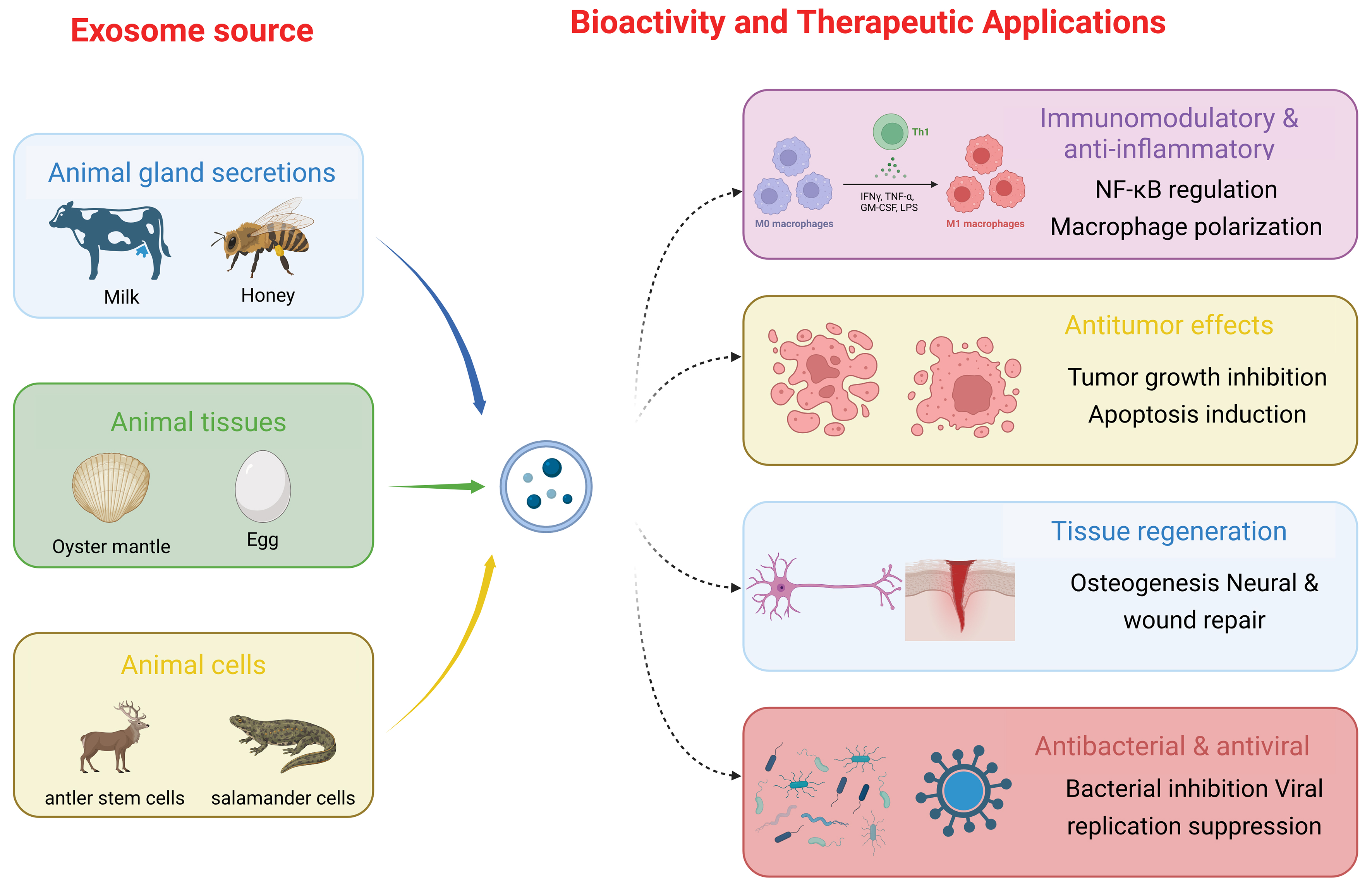
Figure 2. Major biological sources and reported therapeutic effects of animal-derived vesicular particles. The diagram summarizes the principal biological sources of animal-derived vesicular particles, including glandular secretions, tissues, and cultured animal cells, together with their reported therapeutic effects. Major activities include immunomodulatory, anti-inflammatory, antitumor, regenerative, antibacterial, and antiviral effects, which are mediated by source-dependent cargoes and molecular mechanisms. Created in BioRender. LIU, Y. (2026) (https://BioRender.com/9kqlmu3). GM-CSF: Granulocyte-macrophage colony-stimulating factor; IFNγ: interferon gamma; LPS: lipopolysaccharide; M0: unpolarized macrophages; M1: classically activated macrophages; NF-κB: nuclear factor kappa B; Th1: T helper 1 cell; TNF-α: tumor necrosis factor alpha.
THERAPEUTIC EFFECTS
Recent studies have shown that certain animals possess remarkable regenerative capacities. For example, deer antlers are among the most extensively studied models of mammalian regeneration[9], whereas adult salamanders can regenerate entire limbs and substantial portions of the jaw, retina, and heart after injury[88]. EVs from these animals are closely associated with their organisms of origin and may reflect some of their distinctive regenerative properties. For instance, EVs derived from skin secretions are enriched in heat shock proteins (HSPs), redox enzymes such as Prdx6, and antimicrobial peptides (AMPs). These vesicles are internalized by fibroblasts through endocytic pathways, suggesting a role in maintaining amphibian skin homeostasis, including water balance, respiration, immune defense, and antioxidative functions[15]. Moreover, increasing evidence indicates that animal-derived vesicles may participate in cross-species molecular communication. In particular, bovine milk-derived EVs are absorbed after oral or intravenous administration and can alleviate osteoporosis-related phenotypes[89].
Animal-derived vesicular particles can transfer bioactive cargoes to recipient cells and may participate in cross-species molecular communication[90,91]. Preclinical studies have reported immunomodulatory, anti-inflammatory, antitumor, regenerative, antibacterial, and antiviral activities across multiple disease models. However, most evidence remains limited to in vitro experiments and animal studies. The following sections summarize these reported effects, with particular emphasis on functional cargoes, molecular mechanisms, therapeutic indications, and the limitations of current evidence. Unless otherwise specified, these findings should not be interpreted as evidence of established clinical efficacy.
Anti-inflammatory effects
Inflammation is a complex host immune response to injury, infection, or foreign substances. It is characterized by extensive neutrophil accumulation, profound alterations in tissue metabolism[92], the release of pro-inflammatory cytokines such as IL-1β and TNF-α, immune cell infiltration, and the initiation of tissue repair processes. EVs are natural nanoscale mediators of intercellular communication, whereas tissue-derived vesicle-like nanoparticles may also transfer bioactive cargoes to recipient cells. The bioactive components they transport - including proteins, nucleic acids, and lipids - may modulate inflammatory responses. Animal-derived vesicular particles have shown anti-inflammatory potential in preclinical studies, although their biocompatibility and efficacy remain source-, dose-, and model-dependent. These vesicles appear to act mainly by modulating NF-κB signaling, promoting macrophage polarization toward the anti-inflammatory M2 phenotype, and downregulating pro-inflammatory cytokine expression.
Milk-derived EVs and egg white-derived vesicle-like nanoparticles have shown anti-inflammatory activity in preclinical intestinal inflammation models[93-96]. Specifically, bovine milk-derived EVs containing transforming growth factor-β1 (TGF-β1) were reported to inhibit T helper 17 (Th17) cell differentiation and promote Treg cells, thereby reducing pro-inflammatory cytokine expression such as IL-6 and TNF-α in experimental colitis models. In addition, yak milk-derived EVs reduce the levels of IL-1β and TNF-α by inhibiting the PI3K/Akt signaling, further supporting their anti-inflammatory effects. These findings suggest that milk-derived EVs may help to maintain intestinal immune homeostasis through coordinated regulation of multiple signaling pathways.
Antler stem cell-derived EVs[9] have demonstrated anti-inflammatory potential in models of osteoarthritis and spinal cord injury. After intra-articular administration, these vesicles deliver bioactive molecules that downregulate upstream regulators of pro-inflammatory cytokines tumor necrosis factor (TNF), IL-1β, and interferon gamma (IFN-γ), thereby inhibiting NF-κB signaling while promoting TGF-β1-mediated anti-inflammatory responses[9]. This mechanism, characterized by the suppression of inflammation associated with degenerative conditions, not only facilitates cartilage regeneration but also produces systemic anti-inflammatory effects by restoring the balance between Th17 and Treg cell populations. Furthermore, as reported by Yang et al., engineered antler stem cell-derived EVs inhibited NF-κB pathway activation by reducing the expression of phosphorylated IκBα (p-IκBα) and phosphorylated p65 (p-p65)[54]. This modulation promotes the phenotypic transition of macrophages from the pro-inflammatory, classically activated (M1) phenotype toward the anti-inflammatory, tissue-repair-associated M2 phenotype.
PA-VLNs have shown anti-inflammatory activity in a preclinical diabetic wound model. PA-VLN treatment reduced the expression of the pro-inflammatory cytokines TNF-α and IL-6 and of CD68 in wound tissue, indicating attenuation of persistent local inflammation[19].
Notably, the biological effects of animal-derived EVs are not universally beneficial. Cobra venom-derived EV preparations and their miRNA cargo have been implicated in cobra envenomation. In particular, a vesicle-associated miRNA, designated CV-exo-miR-2904 in the original study, induced envenomation-like symptoms and liver injury in mice, and its overexpression inhibited hepatocyte proliferation and promoted apoptosis in vitro[97]. These findings highlight the functional heterogeneity of EV cargo and underscore the importance of carefully evaluating the source and biological effects of EVs before therapeutic application.
Antitumor effects
Evidence for the antitumor activity of animal-derived vesicular particles remains limited and is derived primarily from preclinical studies of glandular secretion-derived EVs and mucilage-derived vesicle-like nanoparticles. Available data suggest source-dependent antitumor mechanisms involving apoptosis induction, modulation of tumor-associated signaling pathways, and alterations in the tumor microenvironment. In preclinical studies, mammalian milk-derived EVs, including camel milk-derived EVs, were associated with increased tumor-cell apoptosis, reduced oxidative stress, and modulation of the inflammatory tumor microenvironment. Oral and local administration reduced breast cancer xenograft growth in the animal models tested, and local injection produced greater tumor accumulation than systemic administration[98]. Vesicle-like nanoparticles from marine organisms show additional mechanistic characteristics. For example, mucilage-derived vesicle-like nanoparticles from Pinctada martensii inhibit osteosarcoma cell proliferation by targeting the ERK1/2 and Wnt signaling pathways, while simultaneously promoting the osteogenic differentiation of mesenchymal stem cells[14]. Animal-derived vesicular particles have also been investigated as carriers of miRNAs with potential regulatory activity. Milk EV-associated miR-2478 has been reported to inhibit melanogenesis in melanoma cells by suppressing the Rap1a-PI3K/Akt signaling axis, and its cross-species regulatory activity has also been validated in human cells[99,100]. In terms of therapeutic strategies, Bovine milk-derived EVs have also been reported to alter the proteomic and metabolic profiles of neuroblastoma cells and to exert antiproliferative effects[100].
Animal-derived EVs and plant-derived vesicle-like nanoparticles differ in their membrane composition, cargo profiles, and interactions with mammalian cells; however, direct comparative evidence is currently insufficient to conclude that either category has generally superior tissue penetration or immune-regulatory specificity. Although plant-derived vesicle-like nanoparticles have intrinsically stable lipid membranes and prolonged circulation properties[6], the secondary metabolites they carry may trigger host immune responses. In contrast, some mammalian EV populations may express surface proteins such as CD47, which can contribute to reduced macrophage-mediated clearance through CD47-signal regulatory protein alpha (SIRPα) signaling[101]. Nevertheless, animal-derived EV-based therapies face several challenges. The lack of standardized isolation and purification procedures results in substantial batch-to-batch heterogeneity[6]. In addition, their nanoscale size may limit penetration efficiency within solid tumors, and long-term systemic administration may induce autoimmune responses. Furthermore, although several studies have demonstrated tumor growth inhibition, clinical translation remains limited by their poorly defined in vivo biodistribution and narrow therapeutic dosage window. For example, the antitumor activity of P. martensii mucilage-derived vesicle-like nanoparticles, including inhibition of proliferation, migration, and invasion as well as induction of apoptosis, increased in a concentration-dependent manner over the range 50-200 μg/mL, whereas higher concentrations (≥ 500 μg/mL) may be cytotoxic to normal cells such as osteoblasts[14]. Future studies should further evaluate whether targeted engineering can improve tumor-specific delivery and therapeutic efficacy while maintaining the safety and biological integrity of animal-derived vesicular particles.
Growth-promoting and regenerative effects
Animal-derived vesicular particles, as natural nanoscale carriers, have demonstrated growth-promoting and regenerative potential in preclinical models involving skeletal, neural, and cutaneous tissues. These vesicles carry distinctive bioactive molecular repertoires acquired during the evolution of their source organisms and can exert cross-species regulatory effects on mammalian cells. By delivering functional cargoes such as proteins and miRNAs, these vesicular particles can modulate the biological behavior of recipient cells and reshape the tissue microenvironment, thereby providing novel therapeutic strategies for orthopedics and regenerative medicine[10].
Regulation of skeletal system repair and metabolic homeostasis
In the regulation of skeletal system repair and metabolic homeostasis, animal-derived vesicular particles exhibit dual mechanisms involving the direct stimulation of bone formation and the systemic modulation of bone metabolism. Among them, OM-VLNs have been the most extensively investigated. OM-VLNs not only directly promote osteoblast differentiation in vitro, but also preferentially accumulate in bone tissue following oral administration in vivo. By simultaneously activating the PI3K/Akt/β-catenin signaling pathway in osteoblasts and suppressing the NF-κB pathway in osteoclast precursors, OM-VLNs were associated with enhanced osteogenesis and reduced osteoclastogenesis and promoted bone regeneration in the investigated animal models of osteoporosis and bone defects[10]. In addition, milk- and colostrum-derived EVs have also demonstrated therapeutic potential in improving bone metabolism. Oral administration of these EVs alleviated glucocorticoid-induced osteoporosis, possibly through both direct inhibition of osteoclast differentiation and systemic regulation via remodeling of the gut microbiota. These findings highlight the potential importance of the “gut-bone axis” in mediating the biological functions of animal-derived vesicular particles[89].
Regeneration and functional reconstruction of the nervous system
In the field of nervous system regeneration and functional reconstruction, EVs derived from organisms with extraordinary regenerative capacity have shown regenerative potential in preclinical models. EVsABPC are enriched in unique proteins associated with nervous system development and markedly promote neural stem cell proliferation and axonal growth. Within the complex microenvironment of spinal cord injury, engineered ACPP@EVsABPC can specifically target the lesion site and synergistically enhance neural regeneration by coordinating activation of the Wnt/β-catenin and Hippo/Yes-associated protein (YAP) signaling pathways. These vesicles also promote macrophage polarization toward the anti-inflammatory M2 phenotype by suppressing NF-κB signaling, thereby relieving the inflammatory microenvironment that inhibits regeneration and improving recovery of motor function[54]. Salamander A1 cell-derived EVs deliver mitochondria-associated proteins such as translocase of outer mitochondrial membrane 70 (TOM70), enhancing oxidative phosphorylation in recipient cells and supporting rapid axonal growth[20]. Although these mechanisms depend partly on amphibian-specific metabolic characteristics, including anaerobic glycolysis, partial functional reconstruction has nonetheless been observed in murine models.
Wound healing and anti-aging effects in skin and soft tissues
In skin and soft tissue wound healing and in anti-aging applications, multiple animal-derived vesicular particles promote tissue repair through multi-target mechanisms. PA-VLNs can be internalized by endothelial cells and fibroblasts, thereby promoting their proliferation and migration and ultimately accelerating wound healing in diabetic mouse models[19]. AMSC-EVs have broad regenerative effects. Their miR-21-5p cargo has been reported to modulate STAT3-associated signaling, enhance cellular activity and angiogenesis, and influence collagen remodeling, improving wound healing and reducing scar formation in preclinical models[66]. In addition, bovine colostrum-derived EVs and regeneration-associated sea anemone-derived vesicle-like nanoparticles have demonstrated anti-photoaging effects and the ability to promote fibroblast migration, respectively, further broadening the potential applications of animal-derived vesicular particles in skin repair and regeneration.
These studies suggest that animal-derived vesicular particles may contribute to regenerative repair through a three-tiered regulatory network encompassing the “organelle-tissue-organ” axis. At the cellular level, these vesicular particles may deliver genetic information that regulates recipient cell phenotypes; at the tissue level, they remodel the immune-stromal microenvironment; and at the organ level, they integrate metabolic and biomechanical signaling pathways. The evolutionary conservation of cross-species communication also has implications for xenogeneic therapeutic applications. For example, porcine skeletal muscle-derived vesicles have been shown to mediate cross-species transfer of miRNAs, thereby inducing metabolic syndrome-like phenotypes in mice[102]. The oral stability and immunomodulatory properties of glandular secretion-derived EVs further expand their translational potential. For instance, bovine milk-derived EVs can improve gut microbiota diversity, including increasing the abundance of Lactobacillus species, enhancing the expression of tight junction proteins, and restoring intestinal epithelial barrier integrity[13,89]. Future studies should focus on elucidating the spatiotemporal dynamics of vesicular cargo composition and the mechanisms underlying vesicle-host microenvironment interactions, in order to advance precision regenerative medicine based on natural nanovesicles.
Antibacterial and antiviral effects
In addition to their anti-inflammatory, antitumor, growth-promoting, and regenerative properties, animal-derived vesicular particles - particularly milk-derived EVs and vesicular particles obtained from bee-derived products such as honey and royal jelly - have been reported to show antibacterial and antiviral activity in experimental studies, suggesting potential applications that require further validation. The following sections discuss the mechanisms underlying the antibacterial and antiviral effects of milk-derived EVs from cattle and goats and of vesicular particles derived from bee products.
Milk-derived EVs from mammals such as cattle and sheep are enriched with immunomodulatory factors, including TGF-β1 and interleukin-10 (IL-10), as well as antimicrobial peptides such as lysozyme and lactoferrin. These bioactive components can suppress pathogen colonization by regulating intestinal immune homeostasis[13]. For instance, bovine milk-derived EVs have been shown to reduce the expression of pro-inflammatory cytokines, including IL-6 and TNF-α, while promoting the secretion of anti-inflammatory factors, thereby alleviating intestinal inflammatory injury in dextran sulfate sodium (DSS)-induced murine colitis models[93,103]. Milk-derived EVs also help to maintain intestinal mucosal barrier integrity by delivering regulatory sequences such as miR-200a-3p, which suppresses expression of the C-X-C motif chemokine ligand 9 (CXCL9) induced by pathogenic bacteria such as Escherichia coli[104]. Vesicular particles derived from bee products, including honey and royal jelly, contain antimicrobial peptides such as MRJP1, defensin-1, and jellein-3, which exert bactericidal effects through disruption of bacterial membrane integrity[67]. Atomic force microscopy (AFM) revealed that Staphylococcus aureus treated with honey-derived vesicular particles displayed cell wall shrinkage and perforation, impaired cell division, and a significantly reduced elastic modulus[16]. In vitro experiments further demonstrated that the minimum inhibitory concentration (MIC) of honey-derived vesicular particles against methicillin-resistant Staphylococcus aureus (MRSA) was 0.05:1 (vesicles per colony-forming unit), and that Eucryphia cordifolia honey-derived EVs (HEc-EVs) effectively inhibited biofilm formation[16]. Royal jelly-derived vesicular particles suppress bacterial growth by interfering with bacterial metabolic pathways. Specifically, the MRJP1 protein carried by these vesicular particles can bind to bacterial ATP synthase, thereby disrupting energy metabolism. They also induce elevated levels of reactive oxygen species (ROS), causing DNA damage and cell cycle arrest[16].
Virus-specific mechanisms underlying antiviral activity
Bovine milk-derived EVs can target nonstructural protein 3 (NS3) of dengue virus (DENV), thereby inhibiting its RNA helicase activity and suppressing viral replication. Quantitative real-time polymerase chain reaction (PCR) analysis demonstrated that treatment with goat milk-derived EVs (GM-EVs) reduced DENV RNA copy numbers by 66.4%. Moreover, heat inactivation or RNase treatment abolished this antiviral effect, supporting the involvement of RNA-associated components[105]. GM-EVs also inhibited adsorption of Newcastle disease virus (NDV-K) by interfering with the interaction between viral envelope proteins and host cell receptors. However, no significant inhibition of human immunodeficiency virus type 1 (HIV-1) was observed, indicating that the antiviral activity of GM-EVs is virus-specific.
Animal-derived vesicular particles may exert antibacterial and antiviral effects through several mechanisms. Milk-derived EVs act mainly through immune regulation and metabolic intervention, whereas vesicular particles from honey and other bee products rely mainly on antimicrobial peptides for direct bactericidal activity. In contrast, virus-specific inhibitory effects are associated with mechanisms involving RNA interference and protein blockade. The reported gastrointestinal stability and preliminary biocompatibility of selected animal-derived vesicular particles support their investigation as drug-delivery vehicles, although their safety, targeting efficiency, and clinical applicability require further validation[106]. Representative animal-derived vesicular particles investigated for potential therapeutic applications are summarized below [Table 4].
Animal-derived vesicular particles investigated for potential therapeutic applications
| Animal source | Contents | Cellular accumulation | Potential application | Biological function | Ref. |
| Apis mellifera honey | Protein, RNA | Oral streptococci | Antibacterial and antibiofilm activity | Inhibits bacterial growth and biofilm formation, particularly in Streptococcus mutans response | [67] |
| Aulactinia stella | Protein, RNA | Fibroblasts/Skin cells | Anti-inflammatory | Reduce inflammatory responses | [18] |
| Bee products | Protein | MSCs | Therapeutic | Inhibit bacterial growth, suppress biofilm formation, promote cell migration | [16] |
| Bombina maxima | Protein, RNA, DNA | Fibroblasts | Wound healing | Promote fibroblast proliferation and migration | [15] |
| Bovine colostrum | Proteins, RNA | Keratinocytes, melanocytes, fibroblasts | Skin repair | Promote proliferation, reduce oxidative stress, inhibit melanin production | [123] |
| Bovine milk | Proteins, RNA | Liver, spleen, brain | Drug delivery | Deliver miRNA cargo, regulate gene expression, enable cross-species communication | [77] |
| Bovine milk | Proteins, RNA | NR | Skin whitening | Deliver miRNA cargo, inhibit melanogenesis, regulate gene expression | [99] |
| Bovine milk | Proteins, RNA | Intestine (colon) | Ulcerative colitis treatment | Regulate immune response, inhibit inflammation, restore gut microbiota, rebalance Treg/Th17 cells | [94] |
| Chicken eggs | Lipids, protein, RNA | Immune cells/macrophages | Anti-inflammatory | Regulate gut microbiota and inflammation | [63] |
| Cobra venom | Protein, miRNA | Hepatocytes/liver | Envenomation-associated pathogenic effects | Induce envenomation-like symptoms and liver injury; inhibit hepatocyte proliferation and promote apoptosis | [97] |
| Cooked pork | Proteins, RNA | Intestinal cells | Metabolic disorder model | Induce insulin resistance, alter glucose metabolism, promote lipid accumulation | [102] |
| Deer antler MSCs | Protein, lipid, RNA | Mesenchymal stem cells, joint cells | Anti-aging, osteoarthritis | Anti-senescence, pro-proliferation, anti-inflammatory, cartilage repair | [9] |
| Guinea pig blood | Protein | NR | Mechanistic study | Regulate membrane phospholipid asymmetry during maturation | [117] |
| Human breastmilk | miRNAs | Intestinal epithelial cells, immune cells | Infant development, immune protection | Regulate gene expression, modulate immune system, support development | [75] |
| Oyster mantle | Proteins, lipids, RNA | Osteoblasts, osteoclasts | Bone regeneration | Promote proliferation, inhibit differentiation, regulate bone homeostasis | [10] |
| Periplaneta americana | Protein, RNA | Fibroblasts, endothelial cells, macrophages | Wound healing | Promote cell proliferation, enhance cell migration, inhibit inflammation, regulate autophagy | [19] |
| Reticulocytes | Protein, lipids | Blood circulation | Mechanistic study | Remove membrane proteins, mediate protein sorting, regulate membrane remodeling | [5] |
| Yak milk | Proteins, RNA | Intestinal epithelial cells | Anti-inflammatory | Promote proliferation, reduce inflammation, enhance barrier function | [95] |
CRITICAL APPRAISAL OF EVIDENCE QUALITY AND REPRODUCIBILITY
The current evidence base for animal-derived vesicular particles is predominantly preclinical. A major strength of the available literature is that several studies have linked source-dependent cargoes and signaling pathways to measurable biological outcomes. In particular, the anti-inflammatory effects of milk-derived EVs in intestinal inflammation models have been reported across multiple milk sources and experimental studies[93-96]. However, most other applications, including many antitumor, regenerative, antibacterial, and antiviral effects, are supported by only one or a few source-specific studies. Reproducibility is further limited by substantial differences in animal source, tissue-processing procedure, isolation and purification method, particle-characterization criteria, storage conditions, dose units, administration routes, and outcome measures. The frequent use of differential ultracentrifugation or precipitation may also co-isolate non-vesicular components, making it difficult to attribute the observed effects exclusively to vesicular particles.
Reported biological effects are also source- and context-dependent and are not uniformly beneficial. Although milk-derived EVs have shown anti-inflammatory effects in several models of intestinal inflammation, long-term administration of cooked pork-derived vesicular particles was associated with insulin resistance, hepatic lipid accumulation, and excessive weight gain in mice[102]. Cobra venom-derived EV preparations have been implicated in envenomation-related liver injury and hepatocyte apoptosis rather than therapeutic protection[97]. Antiviral findings are similarly pathogen-specific: goat milk-derived EVs reduced DENV replication and interfered with NDV adsorption but showed no significant inhibitory activity against HIV-1[105]. Biological activity therefore cannot be generalized from particle source or morphology alone and must be evaluated in relation to specific cargoes, doses, administration routes, recipient cells, and disease contexts.
Accordingly, the available evidence should be interpreted at several levels. Reproducible findings across more than one preclinical study or model may be considered relatively consistent preclinical evidence, but they do not establish clinical efficacy. Effects reported for a single vesicle source, laboratory, or disease model should be regarded as preliminary. Mechanisms inferred mainly from cargo detection, pathway enrichment, or cellular uptake should be considered hypothesis-generating unless confirmed by cargo-depletion, gain- or loss-of-function, receptor-blocking, or pathway-rescue experiments. Future studies should prioritize standardized particle characterization and dose reporting, appropriate non-vesicular controls, blinded and adequately powered designs, independent replication, and clinically relevant long-term outcomes.
SAFETY EVALUATION AND TOXICITY ANALYSIS OF ANIMAL-DERIVED VESICULAR PARTICLES
Safety evaluation of animal-derived vesicular particles represents a critical foundation for their clinical translation. Current studies have conducted preliminary investigations into the biocompatibility and potential toxicity of different animal-derived vesicular particles in both in vitro and in vivo models. However, because vesicle sources, preparation procedures, and experimental design differ widely, safety profiles must be interpreted with reference to the specific origin of each preparation.
PA-VLNs showed no observable acute or chronic toxicity following oral administration in a rat diabetic wound model. The treatment group exhibited enhanced skin tissue regeneration and significantly reduced inflammatory cytokine levels, including TNF-α and IL-6. However, the study included neither blood biochemical analyses, such as liver and kidney function assessments, nor histopathological evaluation of major organs to support systemic safety. Similarly, OM-VLNs prepared from Crassostrea hongkongensis did not cause abnormal body weight change or hepatic and renal damage in ovariectomized mice following daily oral administration of 25-50 mg/kg for six weeks. Nevertheless, immune responses and hematological toxicity markers were not evaluated. These findings suggest that some animal-derived vesicular particles may exhibit preliminary biocompatibility in the tested preclinical models, although standardized toxicological validation remains necessary. In healthy mice, plasma biochemical analyses showed that goat milk-derived EVs did not alter liver function indicators, including alanine aminotransferase (ALT) and aspartate aminotransferase (AST), nor did they affect inflammation-related proteins such as C-reactive protein and albumin. Notably, only triglyceride levels were significantly reduced (treatment group: 144.13 ± 52.43 mg/dL vs. control group: 228.14 ± 86.85 mg/dL), suggesting a potential metabolic regulatory effect rather than toxicity. In cytotoxicity assays, bovine colostrum-derived EVs (ColM-EVs) could be isolated at yields several dozen times higher than those obtained from cell-derived EVs from the same sample volume, while exhibiting no detectable cytotoxicity even at high concentrations. Studies investigating the anti-osteosarcoma effects of mucilage-derived vesicle-like nanoparticles from Pinctada martensii demonstrated no significant toxicity toward 143B osteosarcoma cells, HepG2 hepatocellular carcinoma cells, or other tested cell lines at concentrations ≤ 200 μg/mL. In addition, no abnormalities in body weight or organ pathology were observed in animal experiments. However, long-term oral administration of pork-derived vesicular particles, equivalent to 5 g of pork per day, induced metabolic disturbances in mice, including insulin resistance, hepatic lipid accumulation, and excessive weight gain. These effects were attributed to vesicle-associated miRNAs such as miR-1 and miR-133a-3p, which may disrupt host homeostasis by modulating lipid metabolism. Deer cell-derived EVs, including EVsABPC and antler stem cell-derived EVs (AnSC-EVs), caused no reported acute adverse reactions after local injection in osteoarthritis and trauma models. However, data regarding immunogenicity and long-term toxicity remain unavailable. Although salamander A1 cell-derived EVs (A1-EVs) have demonstrated protective effects against oxidative stress-induced cardiomyocyte injury, their dose-dependent toxicity and host immune responses have not yet been evaluated[20]. Parasite-derived EVs, such as Schistosoma japonicum egg-derived EV-associated Sja-miR-71a, have shown antifibrotic potential, but their pathogenicity and potential immunomodulatory adverse effects remain unclear.
Current studies generally emphasize the potential advantages of animal-derived vesicular particles as cell-free therapeutic agents. However, comprehensive toxicological evidence is lacking, particularly on immunogenicity, xenogeneic immune responses, repeated-dose toxicity, and long-term safety. Importantly, the absence of acute toxicity after a single administration should not be interpreted as evidence of long-term immune tolerance, as repeated EV administration has been reported to induce EV-specific antibody responses that were not apparent after initial exposure[107]. Moreover, EV-induced immune responses may vary with recipient species and genetic background, which limits extrapolation of immunotoxicity findings across species[108]. Although certain milk- and bovine colostrum-derived EVs appear safe based on basic biochemical and physiological measures, the metabolic and immunological effects of their complex xenogeneic cargoes, including proteins, lipids, and nucleic acids, warrant further investigation. This concern is particularly relevant to vesicular particles derived from evolutionarily distant or potentially bioactive sources, such as Periplaneta americana and cobra venom, for which comprehensive long-term in vivo immunogenicity and allergenicity assessments remain necessary before therapeutic translation. Accordingly, source-, dose-, and administration-route-specific therapeutic windows should be established to distinguish therapeutic activity from immunological, metabolic, or organ-specific toxicity.
Biological contamination represents another important safety concern for clinical translation. Because animal-derived vesicular particles may originate from biological fluids, glandular secretions, tissues, or tissue-derived materials, their safety depends on the health status of the source animal, the conditions of collection, and the processing procedures used. Potential transmission of source-associated pathogens or adventitious agents should therefore be considered, particularly for materials obtained directly from animals rather than from well-characterized cell culture systems. Source-specific screening for relevant bacteria, fungi, mycoplasma, viruses, and other adventitious agents, together with appropriate donor-animal health assessment and raw-material traceability, should be incorporated into preclinical and manufacturing workflows. Endotoxin introduced by raw materials, reagents, or processing procedures may also confound experimentally observed inflammatory responses and pose a potential pyrogenic risk. Accordingly, validated aseptic processing, sterility testing, endotoxin testing, and contamination-control procedures should be incorporated into product quality-control and release criteria[109].
Long-term biodistribution and clearance represent additional unresolved safety issues. Current studies characterize mainly short-term tissue localization; the persistence, degradation, and clearance of animal-derived vesicular particles after prolonged or repeated administration remain poorly understood. Across animal studies, administered EVs localize predominantly to the liver, lung, kidney, and spleen, although methodological variability among biodistribution studies is substantial[85]. Particular attention should be paid to potential accumulation in clearance organs such as the liver and spleen, unintended exposure of non-target tissues, and alterations in pharmacokinetic behavior after repeated dosing. Future studies should therefore combine quantitative biodistribution and clearance analyses with long-term biochemical monitoring and histopathological assessment. Labeling approaches should also, where possible, distinguish intact vesicular particles from free label or degraded vesicular components, to avoid overestimating long-term tissue retention[110].
From a regulatory perspective, the clinical translation of animal-derived vesicular products will require source-specific and risk-based quality-control frameworks. Critical quality attributes should include source identity and traceability, particle identity and purity, biological potency, sterility, endotoxin levels, adventitious-agent testing, batch-to-batch consistency, and storage stability. Manufacturing processes intended for clinical use should be standardized under good manufacturing practice (GMP) conditions, with predefined release specifications and validated analytical methods[111]. Standardized safety-evaluation frameworks should integrate acute and repeated-dose toxicity, immunogenicity, long-term biodistribution, and source-specific biological risks. Overall, favorable short-term biocompatibility alone is insufficient to establish clinical safety, and comprehensive preclinical safety assessment, together with robust manufacturing and quality-control standards, will be essential before animal-derived vesicular particles can advance toward human therapeutic applications.
SUMMARY AND PERSPECTIVES
Animal-derived vesicular particles have shown potential as natural nanocarriers for cross-species signal transmission and have been investigated in preclinical models of skeletal regeneration, neural repair, and immune regulation. This rapid development nevertheless calls for critical appraisal across three key dimensions: molecular mechanisms, clinical translation, and theoretical paradigms.
Molecular mechanisms and evolutionary paradoxes underlying cross-species signal transmission
The cross-species bioactivity of animal-derived vesicular particles challenges conventional immunological frameworks regarding the incompatibility of xenogeneic biomolecules. For example, AMSC-EVs promote skin repair through the miR-21-5p/STAT3 signaling axis, a mechanism similar to that reported for mammalian MSC-derived EVs. This functional conservation may reflect convergent selection of critical signaling pathways under evolutionary pressure, such as the central role of the Wnt/β-catenin pathway in both antler regeneration and human bone metabolism. Furthermore, OM-VLNs have been reported to regulate osteogenic differentiation in mammalian models through activation of the PI3K/Akt signaling pathway, suggesting previously unrecognized conserved molecular docking mechanisms across phylogenetically distant species. Such mechanisms may be mediated by specific sphingomyelin-to-cholesterol ratios within the lipid bilayer or by mimicry-related structures of transmembrane proteins.
This phenomenon also raises critical concerns about how the nucleic acid components of xenogeneic vesicular particles, particularly miRNAs, evade recognition by mammalian RNA interference and innate immune surveillance. Recent studies have shown that PA-VLNs can mimic the surface characteristics of host-derived EVs through membrane glycosylation, such as α-2,6-linked sialic acid residues, thereby avoiding Toll-like receptor 7/8 (TLR7/8)-mediated immune recognition. Although this “molecular mimicry” may enhance biocompatibility, it may also increase the risk of misrecognition involving pathogen-associated molecular patterns (PAMPs). Consequently, there is an urgent need to establish predictive models for evaluating the immunogenicity of cross-species vesicular particles.
The interplay between biological origin and engineering modification
The compositional heterogeneity of animal-derived vesicular particles is both a foundation of their multifunctionality and a major obstacle to clinical standardization. For example, although bovine milk-derived EVs contain anti-inflammatory components such as miR-219 and miR-338, proteomic analyses have reported batch-to-batch variation of up to 30%. This variability is likely attributable to the combined effects of individual animal differences, breeding conditions, and isolation methods. In contrast, plant-derived vesicle-like nanoparticles can be obtained through mechanical disruption of plant tissues; however, their purity, composition, and biological properties may vary according to the source material and preparation method.
Current engineering strategies seek to balance preservation of natural properties with functional optimization. For instance, antler cell-derived EVs modified with distearoyl phosphoethanolamine-polyethylene glycol (DSPE-PEG)/ACPP achieved targeted accumulation at sites of spinal cord injury with a 2.6-fold higher targeting efficiency than unmodified EVs. However, the loss of surface CD47 during the modification process may compromise their intrinsic immune evasion capability. Such trade-offs, in which enhancement of one function may occur at the expense of another, suggest that future engineering approaches should adopt multiparametric optimization models rather than relying on linear modifications aimed at a single functional objective.
Spatiotemporal dynamics of metabolic reprogramming and microenvironmental remodeling
The therapeutic efficacy and safety of EVs are strongly influenced by their spatiotemporal biodistribution. Across preclinical studies, systemically administered EVs commonly accumulate in organs such as the liver, spleen, lungs, and kidneys, although the relative distribution varies substantially depending on vesicle source, administration route, particle properties, and experimental conditions[84,85]. In conventional mammalian EV models, membrane proteins may contribute to organ-specific distribution. For example, exosomal integrins α6β4 and α6β1 have been associated with lung tropism, whereas integrin αvβ5 has been associated with liver tropism. However, these relationships were established mainly in tumor-derived exosome models, and whether comparable integrin-dependent organotropism applies to animal-derived vesicular particles from diverse biological sources remains to be determined[84,112]. Most existing studies also rely on static biodistribution measurements, underscoring the need for quantitative, dynamic tracking approaches to better characterize the pharmacokinetics, tissue accumulation, and clearance of animal-derived vesicular particles. For example, egg-derived vesicular particles, due to the absence of CD47, show a circulatory half-life approximately one-third that of synthetic liposomes. Therefore, the development of real-time imaging technologies based on radioisotope labeling or quantum dot tracking will be essential to elucidate the pharmacokinetics and metabolic dynamics of animal-derived vesicular particles.
Bioethical and ecological challenges associated with large-scale production
Marine invertebrates provide a representative example of the potential for large-scale production of AVLNs. Approximately 3.14 × 10⁹ vesicle-like particles can be obtained from each gram of oyster mantle tissue, suggesting that large-scale production may be feasible. However, the ecological burden of intensive aquaculture, such as coastal eutrophication, has not yet been incorporated into current evaluation frameworks. More importantly, although deer antlers have ethical advantages as renewable organs, their growth is highly dependent on seasonal hormonal regulation, and industrial-scale expansion may alter vesicular composition. The establishment of heterologous expression systems based on synthetic biology, such as recombinant expression of antler-specific miRNAs in Chinese hamster ovary (CHO) cells, may offer a promising strategy for balancing production yield and product quality.
Clinical translation, manufacturing, and regulatory considerations
The clinical translation of animal-derived vesicular particles requires scalable, reproducible manufacturing under GMP conditions. Large-scale production should incorporate qualified and traceable animal sources, standardized feeding or culture conditions, controlled collection and preprocessing procedures, closed or aseptic processing systems, and validated purification methods. Source-dependent variables, including animal species, age, diet, health status, season, tissue condition, and collection time, may alter particle yield, molecular composition, and biological activity. These variables, together with differences in isolation procedures, can result in substantial batch-to-batch variation. Therefore, critical process parameters and acceptance criteria should be defined throughout manufacturing, and any process modifications should be accompanied by comparability studies confirming that product identity, purity, potency, and safety are unchanged[113,114].
Long-term storage and quality control represent additional translational challenges. Storage temperature, formulation buffer, container material, storage duration, transportation conditions, and repeated freeze-thaw cycles may affect particle concentration, membrane integrity, cargo stability, aggregation, and biological potency. Although storage at -80 °C is commonly used, optimal conditions are likely to be source- and product-specific, and alternatives such as cryoprotectants or lyophilization require systematic validation. Stability studies should therefore evaluate not only particle size and concentration but also morphology, molecular cargoes, sterility, endotoxin levels, residual host- or tissue-derived contaminants, and mechanism-relevant biological potency over the intended shelf life. Product release criteria should include validated assays for identity, quantity, purity, potency, and safety, supported by reference standards and predefined acceptance ranges[115].
Regulatory classification and commercialization pathways also remain uncertain. Depending on their composition, mechanism of action, degree of manipulation, and intended use, vesicular particle-based products may be regulated as biological medicinal products, drug-delivery systems, or combination products in different jurisdictions. Developers must therefore define the active substance and proposed mechanism of action, and provide evidence on source qualification, manufacturing consistency, biodistribution, pharmacology, toxicity, immunogenicity, tumorigenicity, and the potential transmission of zoonotic or other adventitious agents. Early consultation with regulatory authorities will be necessary because specific regulatory frameworks for animal-derived EVs and vesicle-like nanoparticles have not yet been harmonized. Commercialization will additionally depend on cost-effective production, scalable purification, validated cold-chain or stabilized formulations, intellectual-property strategies, and clinically meaningful advantages over existing biologics or synthetic nanocarriers. Accordingly, successful translation will require coordinated development of manufacturing, quality, regulatory, and commercial strategies rather than demonstration of preclinical efficacy alone[116].
Reconstruction of theoretical paradigms: from “drug delivery” to “ecological medicine”
Current studies largely regard animal-derived vesicular particles as independent functional entities, overlooking their ecological significance within their organisms of origin. For example, skin secretion-derived EVs from Bombina maxima are enriched in heat shock proteins and antimicrobial peptides, which fundamentally constitute part of the symbiotic defense system of amphibian skin. When such EVs are applied as anti-inflammatory agents, they may inadvertently disrupt the allelopathic interaction networks within the human microbiota. Future research should therefore adopt an ecological perspective and evaluate the cascading effects of these particles on interactions among humans, microorganisms, and the environment. This will require the development of dynamic systems models that integrate multi-omics analyses.
CONCLUSION
AEVs and AVLNs exhibit diverse biological activities linked to their source-dependent molecular cargoes, supporting their potential as natural therapeutic platforms. Distinguishing naturally released AEVs from processing-derived AVLNs is essential for interpreting their functions and comparing evidence across studies. Although preclinical findings suggest opportunities for immune modulation, tissue regeneration, and disease-oriented interventions, clinical efficacy and long-term safety remain unestablished. Translation toward precision therapeutics will require experimentally validated relationships among particle sources, functional cargoes, molecular targets, and disease indications. Future research should prioritize standardized isolation and characterization, reproducible manufacturing and quality control, quantitative biodistribution assessment, and comprehensive safety evaluation. These efforts will be critical to determining whether the intrinsic bioactivity of AEVs and AVLNs can support safe, reproducible, and clinically meaningful therapeutic applications.
DECLARATIONS
Acknowledgments
The Graphical Abstract was Created in BioRender. LIU, Y. (2026) (https://BioRender.com/802df7i).
Authors’ contributions
Conceptualized and designed the review: Liu Y, Wang X, Chen Z
Performed the literature search and collected relevant studies: Liu Y, Wang X, Chen Z
Organized and interpreted the literature and prepared the tables: Liu Y, Wang X
Prepared and revised the figures: Chen Z, Xu P, Yao C
Drafted the original manuscript: Liu Y, Wang X
Critically reviewed and revised the manuscript for important intellectual content: Liu Y, Wang X, Chen Z, Xu P, Yao C
Provided technical and methodological guidance: Xu P, Yao C
Conceived the review, defined its scope, and developed the manuscript framework: Fan L, Li Z
Contributed to the interpretation and critical evaluation of the literature, particularly the relationships among biological activities, therapeutic mechanisms, and translational challenges: Fan L, Li Z
Substantively revised the manuscript for important intellectual content: Fan L, Li Z
Supervised the preparation of the manuscript: Fan L, Li Z
Contributed to the critical evaluation and interpretation of the literature and substantively revised the manuscript for important intellectual content during revision: Zhang Z, Mao H, Shi Z
All authors read and approved the final manuscript.
Availability of data and materials
Not applicable.
AI and AI-assisted tools statement
During the preparation of this manuscript, the AI tool ChatGPT (version GPT-5.6 Sol, released 2026-07-09) was used solely for language editing. The tool did not influence the study design, data collection, analysis, interpretation, or the scientific content of the work. All authors take full responsibility for the accuracy, integrity, and final content of the manuscript.
Financial support and sponsorship
None.
Conflicts of interest
All authors declared that there are no conflicts of interest.
Ethical approval and consent to participate
Not applicable.
Consent for publication
Not applicable.
Copyright
© The Author(s) 2026.
REFERENCES
1. Kalluri R, LeBleu VS. The biology, function, and biomedical applications of exosomes. Science. 2020;367:eaau6977.
2. Welsh JA, Goberdhan DCI, O’Driscoll L, et al. Minimal information for studies of extracellular vesicles (MISEV2023): from basic to advanced approaches. J Extracell Vesicles. 2024;13:e12404.
3. Harding C, Heuser J, Stahl P. Receptor-mediated endocytosis of transferrin and recycling of the transferrin receptor in rat reticulocytes. J Cell Biol. 1983;97:329-39.
4. Pan BT, Johnstone RM. Fate of the transferrin receptor during maturation of sheep reticulocytes in vitro: selective externalization of the receptor. Cell. 1983;33:967-78.
5. Johnstone RM, Adam M, Hammond JR, Orr L, Turbide C. Vesicle formation during reticulocyte maturation. Association of plasma membrane activities with released vesicles (exosomes). J Biol Chem. 1987;262:9412-20.
6. Zhao B, Lin H, Jiang X, et al. Exosome-like nanoparticles derived from fruits, vegetables, and herbs: innovative strategies of therapeutic and drug delivery. Theranostics. 2024;14:4598-621.
7. Han Y, Guo X, Ji Z, et al. Colon health benefits of plant-derived exosome-like nanoparticles via modulating gut microbiota and immunity. Crit Rev Food Sci Nutr. 2025;65:7718-38.
8. Zhang Y, Bi J, Huang J, Tang Y, Du S, Li P. Exosome: a review of its classification, isolation techniques, storage, diagnostic and targeted therapy applications. Int J Nanomedicine. 2020;15:6917-34.
9. Lei J, Jiang X, Li W, et al. Exosomes from antler stem cells alleviate mesenchymal stem cell senescence and osteoarthritis. Protein Cell. 2022;13:220-6.
10. Hu Y, Hou Z, Liu Z, et al. Oyster mantle-derived exosomes alleviate osteoporosis by regulating bone homeostasis. Biomaterials. 2024;311:122648.
11. Song Y, Feng N, Yu Q, et al. Exosomes in disease therapy: plant-derived exosome-like nanoparticles current status, challenges, and future prospects. Int J Nanomedicine. 2025;20:10613-44.
12. Zhang GK, Zhou F. Exosomes-loaded with traditional Chinese medicine constituents: a novel therapeutic avenue for aplastic anemia. Chin Med. 2026;21:53.
13. Mecocci S, Trabalza-Marinucci M, Cappelli K. Extracellular vesicles from animal milk: great potentialities and critical issues. Animals. 2022;12:3231.
14. Li P, Ma L, Zhan W, et al. Exosome-like nanovesicles derived from the mucilage of Pinctada martensii exhibit antitumor activity against 143B osteosarcoma cells. ACS Appl Mater Interfaces. 2023;15:26227-40.
15. Wei XS, Liu LZ, Bian XL, et al. Protein composition of extracellular vesicles from skin secretions of the amphibian Bombina maxima. Zool Res. 2022;43:687-90.
16. Schuh CMAP, Aguayo S, Zavala G, Khoury M. Exosome-like vesicles in Apis mellifera bee pollen, honey and royal jelly contribute to their antibacterial and pro-regenerative activity. J Exp Biol. 2019;222:jeb208702.
17. Zhang F, Yue Y, Chen J, et al. Albumen exosomes alleviate LPS-induced inflammation of intestinal epithelial cells via miR-22/ATM/p53/NF-κB axis. Int J Biol Macromol. 2024;267:131241.
18. Jafari N, Afshar A, Zare A, et al. Proliferating and migrating effects of regenerating sea anemone Aulactinia stella cells-derived exosomes on human skin fibroblasts. Nat Prod Res. 2025;39:4916-23.
19. Liao Q, Su L, Pang L, et al. Natural exosome-like nanoparticles derived from ancient medicinal insect Periplaneta americana L. as a novel diabetic wound healing accelerator. J Nanobiotechnology. 2023;21:169.
20. Middleton RC, Rogers RG, De Couto G, et al. Newt cells secrete extracellular vesicles with therapeutic bioactivity in mammalian cardiomyocytes. J Extracell Vesicles. 2018;7:1456888.
21. Liu Y, Wang X, Chen Z, et al. Animal-derived vesicles: nanobridges linking the wisdom of natural evolution with precision medicine. SciErxiv 2026.
22. Huang C, Li J, Xie Z, Hu X, Huang Y. Relationship between exosomes and cancer: formation, diagnosis, and treatment. Int J Biol Sci. 2025;21:40-62.
23. Buschow SI, Nolte-’t Hoen EN, van Niel G, et al. MHC II in dendritic cells is targeted to lysosomes or T cell-induced exosomes via distinct multivesicular body pathways. Traffic. 2009;10:1528-42.
24. van Niel G, Wubbolts R, Stoorvogel W. Endosomal sorting of MHC class II determines antigen presentation by dendritic cells. Curr Opin Cell Biol. 2008;20:437-44.
25. Raposo G, Stoorvogel W. Extracellular vesicles: exosomes, microvesicles, and friends. J Cell Biol. 2013;200:373-83.
26. Rana S, Zöller M. Exosome target cell selection and the importance of exosomal tetraspanins: a hypothesis. Biochem Soc Trans. 2011;39:559-62.
27. de Gassart A, Geminard C, Fevrier B, Raposo G, Vidal M. Lipid raft-associated protein sorting in exosomes. Blood. 2003;102:4336-44.
28. Sanwlani R, Fonseka P, Chitti SV, Mathivanan S. Milk-derived extracellular vesicles in inter-organism, cross-species communication and drug delivery. Proteomes. 2020;8:11.
29. Van Deun J, Mestdagh P, Sormunen R, et al. The impact of disparate isolation methods for extracellular vesicles on downstream RNA profiling. J Extracell Vesicles. 2014;3:24858.
30. Rider MA, Hurwitz SN, Meckes DG Jr. ExtraPEG: a polyethylene glycol-based method for enrichment of extracellular vesicles. Sci Rep. 2016;6:23978.
31. Tauro BJ, Greening DW, Mathias RA, Mathivanan S, Ji H, Simpson RJ. Two distinct populations of exosomes are released from LIM1863 colon carcinoma cell-derived organoids. Mol Cell Proteomics. 2013;12:587-98.
32. Zarovni N, Corrado A, Guazzi P, et al. Integrated isolation and quantitative analysis of exosome shuttled proteins and nucleic acids using immunocapture approaches. Methods. 2015;87:46-58.
33. Böing AN, van der Pol E, Grootemaat AE, Coumans FA, Sturk A, Nieuwland R. Single-step isolation of extracellular vesicles by size-exclusion chromatography. J Extracell Vesicles. 2014;3:23430.
34. Baranyai T, Herczeg K, Onódi Z, et al. Isolation of exosomes from blood plasma: qualitative and quantitative comparison of ultracentrifugation and size exclusion chromatography methods. PLoS One. 2015;10:e0145686.
35. Liangsupree T, Multia E, Riekkola ML. Modern isolation and separation techniques for extracellular vesicles. J Chromatogr A. 2021;1636:461773.
36. Cheruvanky A, Zhou H, Pisitkun T, et al. Rapid isolation of urinary exosomal biomarkers using a nanomembrane ultrafiltration concentrator. Am J Physiol Renal Physiol. 2007;292:F1657-61.
37. Lobb RJ, Becker M, Wen SW, et al. Optimized exosome isolation protocol for cell culture supernatant and human plasma. J Extracell Vesicles. 2015;4:27031.
38. Busatto S, Vilanilam G, Ticer T, et al. Tangential flow filtration for highly efficient concentration of extracellular vesicles from large volumes of fluid. Cells. 2018;7:273.
39. Zhang H, Freitas D, Kim HS, et al. Identification of distinct nanoparticles and subsets of extracellular vesicles by asymmetric flow field-flow fractionation. Nat Cell Biol. 2018;20:332-43.
40. Kanwar SS, Dunlay CJ, Simeone DM, Nagrath S. Microfluidic device (ExoChip) for on-chip isolation, quantification and characterization of circulating exosomes. Lab Chip. 2014;14:1891-900.
41. Oksvold MP, Kullmann A, Forfang L, et al. Expression of B-cell surface antigens in subpopulations of exosomes released from B-cell lymphoma cells. Clin Ther. 2014;36:847-62.e1.
42. Valadi H, Ekström K, Bossios A, Sjöstrand M, Lee JJ, Lötvall JO. Exosome-mediated transfer of mRNAs and microRNAs is a novel mechanism of genetic exchange between cells. Nat Cell Biol. 2007;9:654-9.
43. Mateescu B, Kowal EJ, van Balkom BW, et al. Obstacles and opportunities in the functional analysis of extracellular vesicle RNA - an ISEV position paper. J Extracell Vesicles. 2017;6:1286095.
44. Xu H, Zhang J, Li M. Cargo-driven extracellular vesicles as pharmaceutical nanocarriers: a pharmaceutics-oriented comparison of animal exosomes and plant-derived exosome-like nanoparticles. Int J Pharm X. 2026;11:100530.
45. EL Andaloussi S, Mäger I, Breakefield XO, Wood MJ. Extracellular vesicles: biology and emerging therapeutic opportunities. Nat Rev Drug Discov. 2013;12:347-57.
46. Record M, Subra C, Silvente-Poirot S, Poirot M. Exosomes as intercellular signalosomes and pharmacological effectors. Biochem Pharmacol. 2011;81:1171-82.
47. Hade MD, Suire CN, Suo Z. Mesenchymal stem cell-derived exosomes: applications in regenerative medicine. Cells. 2021;10:1959.
48. Sergazy S, Adekenov S, Khabarov I, Adekenova K, Maikenova A, Aljofan M. Harnessing mammalian- and plant-derived exosomes for drug delivery: a comparative review. Int J Mol Sci. 2025;26:4857.
49. Wang Q, Ren Y, Mu J, et al. Grapefruit-derived nanovectors use an activated leukocyte trafficking pathway to deliver therapeutic agents to inflammatory tumor sites. Cancer Res. 2015;75:2520-9.
50. Skotland T, Sagini K, Sandvig K, Llorente A. An emerging focus on lipids in extracellular vesicles. Adv Drug Deliv Rev. 2020;159:308-21.
51. Keerthikumar S, Chisanga D, Ariyaratne D, et al. ExoCarta: a web-based compendium of exosomal cargo. J Mol Biol. 2016;428:688-92.
52. Colombo M, Raposo G, Théry C. Biogenesis, secretion, and intercellular interactions of exosomes and other extracellular vesicles. Annu Rev Cell Dev Biol. 2014;30:255-89.
53. Ashburner M, Ball CA, Blake JA, et al. Gene ontology: tool for the unification of biology. The Gene Ontology Consortium. Nat Genet. 2000;25:25-9.
54. Yang S, Xue B, Zhang Y, et al. Engineered extracellular vesicles from antler blastema progenitor cells: a therapeutic choice for spinal cord injury. ACS Nano. 2025;19:5995-6013.
55. Théry C, Boussac M, Véron P, et al. Proteomic analysis of dendritic cell-derived exosomes: a secreted subcellular compartment distinct from apoptotic vesicles. J Immunol. 2001;166:7309-18.
56. Simpson RJ, Lim JW, Moritz RL, Mathivanan S. Exosomes: proteomic insights and diagnostic potential. Expert Rev Proteomics. 2009;6:267-83.
57. Simoncelli F, Lucentini L, La Porta G, Belia S, Di Rosa I, Fagotti A. Small heat shock proteins in the amphibian Pelophylax bergeri: cloning and characterization of Hsp27 and Hsp30 cDNAs and their expression analysis in ex vivo skin exposed to abiotic stresses. Comp Biochem Physiol A Mol Integr Physiol. 2019;235:90-101.
58. Williams C, Royo F, Aizpurua-Olaizola O, et al. Glycosylation of extracellular vesicles: current knowledge, tools and clinical perspectives. J Extracell Vesicles. 2018;7:1442985.
59. Liu H, Dong T, Dong C, et al. Plant-derived exosome-like nanovesicles: a novel therapeutic perspective for skin diseases. J Nanobiotechnology. 2025;23:640.
60. Zhang ZG, Buller B, Chopp M. Exosomes - beyond stem cells for restorative therapy in stroke and neurological injury. Nat Rev Neurol. 2019;15:193-203.
61. Yin K, Wang S, Zhao RC. Exosomes from mesenchymal stem/stromal cells: a new therapeutic paradigm. Biomark Res. 2019;7:8.
62. Xie MY, Hou LJ, Sun JJ, et al. Porcine milk exosome MiRNAs attenuate LPS-induced apoptosis through inhibiting TLR4/NF-κB and p53 pathways in intestinal epithelial cells. J Agric Food Chem. 2019;67:9477-91.
63. Fratantonio D, Munir J, Shu J, et al. The RNA cargo in small extracellular vesicles from chicken eggs is bioactive in C57BL/6 J mice and human peripheral blood mononuclear cells ex vivo. Front Nutr. 2023;10:1162679.
64. Saadeldin IM, Tanga BM, Bang S, et al. Isolation, characterization, proteome, miRNAome, and the embryotrophic effects of chicken egg yolk nanovesicles (vitellovesicles). Sci Rep. 2023;13:4204.
65. Peng S, Shen L, Yu X, et al. miR-200a attenuated oxidative stress, inflammation, and apoptosis in dextran sulfate sodium-induced colitis through activation of Nrf2. Front Immunol. 2023;14:1196065.
66. Meng D, Li Y, Chen Z, Guo J, Yang M, Peng Y. Exosomes derived from antler mesenchymal stem cells promote wound healing by miR-21-5p/STAT3 axis. Int J Nanomedicine. 2024;19:11257-73.
67. Leiva-Sabadini C, Alvarez S, Barrera NP, Schuh CMAP, Aguayo S. Antibacterial effect of honey-derived exosomes containing antimicrobial peptides against oral Streptococci. Int J Nanomedicine. 2021;16:4891-900.
68. Jia X, Tang J, Yao C, Yang D. Recent progress of extracellular vesicle engineering. ACS Biomater Sci Eng. 2021;7:4430-8.
69. Mohammadi AH, Ghazvinian Z, Bagheri F, Harada M, Baghaei K. Modification of extracellular vesicle surfaces: an approach for targeted drug delivery. BioDrugs. 2023;37:353-74.
70. Zhao S, Di Y, Fan H, et al. Targeted delivery of extracellular vesicles: the mechanisms, techniques and therapeutic applications. Mol Biomed. 2024;5:60.
71. Ahmed W, Mushtaq A, Ali S, Khan N, Liang Y, Duan L. Engineering approaches for exosome cargo loading and targeted delivery: biological versus chemical perspectives. ACS Biomater Sci Eng. 2024;10:5960-76.
72. Zeng H, Guo S, Ren X, Wu Z, Liu S, Yao X. Current strategies for exosome cargo loading and targeting delivery. Cells. 2023;12:1416.
73. Richter M, Vader P, Fuhrmann G. Approaches to surface engineering of extracellular vesicles. Adv Drug Deliv Rev. 2021;173:416-26.
74. Munagala R, Aqil F, Jeyabalan J, Gupta RC. Bovine milk-derived exosomes for drug delivery. Cancer Lett. 2016;371:48-61.
75. Alsaweed M, Hartmann PE, Geddes DT, Kakulas F. MicroRNAs in breastmilk and the lactating breast: potential immunoprotectors and developmental regulators for the infant and the mother. Int J Environ Res Public Health. 2015;12:13981-4020.
76. Benmoussa A, Lee CH, Laffont B, et al. Commercial dairy cow milk microRNAs resist digestion under simulated gastrointestinal tract conditions. J Nutr. 2016;146:2206-15.
77. Manca S, Upadhyaya B, Mutai E, et al. Milk exosomes are bioavailable and distinct microRNA cargos have unique tissue distribution patterns. Sci Rep. 2018;8:11321.
78. Wiklander OP, Nordin JZ, O’Loughlin A, et al. Extracellular vesicle in vivo biodistribution is determined by cell source, route of administration and targeting. J Extracell Vesicles. 2015;4:26316.
79. Imai T, Takahashi Y, Nishikawa M, et al. Macrophage-dependent clearance of systemically administered B16BL6-derived exosomes from the blood circulation in mice. J Extracell Vesicles. 2015;4:26238.
80. Phinney DG, Pittenger MF. Concise review: MSC-derived exosomes for cell-free therapy. Stem Cells. 2017;35:851-8.
81. Shen Z, Huang W, Liu J, Tian J, Wang S, Rui K. Effects of mesenchymal stem cell-derived exosomes on autoimmune diseases. Front Immunol. 2021;12:749192.
82. Kooijmans SA, Aleza CG, Roffler SR, van Solinge WW, Vader P, Schiffelers RM. Display of GPI-anchored anti-EGFR nanobodies on extracellular vesicles promotes tumour cell targeting. J Extracell Vesicles. 2016;5:31053.
83. Cheng J, Chen Z, Liu C, et al. Bone mesenchymal stem cell-derived exosome-loaded injectable hydrogel for minimally invasive treatment of spinal cord injury. Nanomedicine. 2021;16:1567-79.
84. Choi H, Choi Y, Yim HY, Mirzaaghasi A, Yoo JK, Choi C. Biodistribution of exosomes and engineering strategies for targeted delivery of therapeutic exosomes. Tissue Eng Regen Med. 2021;18:499-511.
85. Kang M, Jordan V, Blenkiron C, Chamley LW. Biodistribution of extracellular vesicles following administration into animals: a systematic review. J Extracell Vesicles. 2021;10:e12085.
86. Chen Z, Xiong M, Tian J, Song D, Duan S, Zhang L. Encapsulation and assessment of therapeutic cargo in engineered exosomes: a systematic review. J Nanobiotechnology. 2024;22:18.
87. Aimaletdinov AM, Gomzikova MO. Tracking of extracellular vesicles’ biodistribution: new methods and approaches. Int J Mol Sci. 2022;23:11312.
88. Brockes JP, Kumar A. Appendage regeneration in adult vertebrates and implications for regenerative medicine. Science. 2005;310:1919-23.
89. Yun B, Maburutse BE, Kang M, et al. Short communication: dietary bovine milk-derived exosomes improve bone health in an osteoporosis-induced mouse model. J Dairy Sci. 2020;103:7752-60.
90. Lian MQ, Chng WH, Liang J, et al. Plant-derived extracellular vesicles: Recent advancements and current challenges on their use for biomedical applications. J Extracell Vesicles. 2022;11:e12283.
91. Liu JH, Chen CY, Liu ZZ, et al. Extracellular vesicles from child gut microbiota enter into bone to preserve bone mass and strength. Adv Sci. 2021;8:2004831.
92. Taylor CT, Colgan SP. Regulation of immunity and inflammation by hypoxia in immunological niches. Nat Rev Immunol. 2017;17:774-85.
93. Reif S, Elbaum-Shiff Y, Koroukhov N, Shilo I, Musseri M, Golan-Gerstl R. Cow and human milk-derived exosomes ameliorate colitis in DSS murine model. Nutrients. 2020;12:2589.
94. Tong L, Hao H, Zhang Z, et al. Milk-derived extracellular vesicles alleviate ulcerative colitis by regulating the gut immunity and reshaping the gut microbiota. Theranostics. 2021;11:8570-86.
95. Gao HN, Hu H, Wen PC, et al. Yak milk-derived exosomes alleviate lipopolysaccharide-induced intestinal inflammation by inhibiting PI3K/AKT/C3 pathway activation. J Dairy Sci. 2021;104:8411-24.
96. Gao HN, Ren FZ, Wen PC, et al. Yak milk-derived exosomal microRNAs regulate intestinal epithelial cells on proliferation in hypoxic environment. J Dairy Sci. 2021;104:1291-303.
97. Liao T, Gan M, Qiu Y, et al. miRNAs derived from cobra venom exosomes contribute to the cobra envenomation. J Nanobiotechnology. 2023;21:356.
98. Badawy AA, El-Magd MA, AlSadrah SA. Therapeutic effect of camel milk and its exosomes on MCF7 cells in vitro and in vivo. Integr Cancer Ther. 2018;17:1235-46.
99. Bae IS, Kim SH. Milk exosome-derived microRNA-2478 suppresses melanogenesis through the Akt-GSK3β pathway. Cells. 2021;10:2848.
100. Fonseka P, Kang T, Chee S, et al. Temporal quantitative proteomics analysis of neuroblastoma cells treated with bovine milk-derived extracellular vesicles highlights the anti-proliferative properties of milk-derived extracellular vesicles. Cells. 2021;10:750.
101. Ben XY, Wang YR, Zheng HH, et al. Construction of exosomes that overexpress CD47 and evaluation of their immune escape. Front Bioeng Biotechnol. 2022;10:936951.
102. Shen L, Ma J, Yang Y, et al. Cooked pork-derived exosome nanovesicles mediate metabolic disorder-microRNA could be the culprit. J Nanobiotechnology. 2023;21:83.
103. Stremmel W, Weiskirchen R, Melnik BC. Milk exosomes prevent intestinal inflammation in a genetic mouse model of ulcerative colitis: a pilot experiment. Inflamm Intest Dis. 2020;5:117-23.
104. Wu D, Kittana H, Shu J, et al. Dietary depletion of milk exosomes and their microRNA cargos elicits a depletion of miR-200a-3p and elevated intestinal inflammation and chemokine (C-X-C Motif) ligand 9 expression in Mdr1a-/- mice. Curr Dev Nutr. 2019;3:nzz122.
105. Yenuganti VR, Afroz S, Khan RA, et al. Milk exosomes elicit a potent anti-viral activity against dengue virus. J Nanobiotechnology. 2022;20:317.
106. Adriano B, Cotto NM, Chauhan N, Jaggi M, Chauhan SC, Yallapu MM. Milk exosomes: nature’s abundant nanoplatform for theranostic applications. Bioact Mater. 2021;6:2479-90.
107. Driedonks TAP, Jiang L, Gololobova O, Liao Z, Witwer KW. ELISA-based detection of immunoglobulins against extracellular vesicles in blood plasma. J Extracell Biol. 2024;3:e129.
108. Kim DO, Nguyen QT, Choi YC, et al. Strain- and species-specific immune responses to human adipose stem cell-derived extracellular vesicles: a comparative pharmacological evaluation in mice and human PBMCs. Eur J Pharm Biopharm. 2026;222:115008.
109. Nonaka CKV, Costa-Ferro ZSM, Arraes ACP, et al. Validation of an automated quality control method to test sterility of two advanced therapy medicinal products: Mesenchymal stromal cells and their extracellular vesicles. Hematol Transfus Cell Ther. 2025;47:103727.
110. Skotland T, Iversen TG, Llorente A, Sandvig K. Biodistribution, pharmacokinetics and excretion studies of intravenously injected nanoparticles and extracellular vesicles: possibilities and challenges. Adv Drug Deliv Rev. 2022;186:114326.
111. Silva AKA, Morille M, Piffoux M, et al. Development of extracellular vesicle-based medicinal products: a position paper of the group “Extracellular Vesicle translatiOn to clinicaL perspectiVEs - EVOLVE France”. Adv Drug Deliv Rev. 2021;179:114001.
112. Hoshino A, Costa-Silva B, Shen TL, et al. Tumour exosome integrins determine organotropic metastasis. Nature. 2015;527:329-35.
113. Ng CY, Kee LT, Al-Masawa ME, et al. Scalable production of extracellular vesicles and its therapeutic values: a review. Int J Mol Sci. 2022;23:7986.
114. Takakura Y, Hanayama R, Akiyoshi K, et al. Quality and safety considerations for therapeutic products based on extracellular vesicles. Pharm Res. 2024;41:1573-94.
115. Jeyaram A, Jay SM. Preservation and storage stability of extracellular vesicles for therapeutic applications. AAPS J. 2017;20:1.
116. Lener T, Gimona M, Aigner L, et al. Applying extracellular vesicles based therapeutics in clinical trials - an ISEV position paper. J Extracell Vesicles. 2015;4:30087.
117. Vidal M, Sainte-Marie J, Philippot JR, Bienvenue A. Asymmetric distribution of phospholipids in the membrane of vesicles released during in vitro maturation of guinea pig reticulocytes: evidence precluding a role for “aminophospholipid translocase”. J Cell Physiol. 1989;140:455-62.
118. Moholkar DN, Kandimalla R, Saeed M, et al. Tumor-targeted exosomal delivery of celastrol for enhanced therapeutic efficacy in NSCLC. Theranostics. 2026;16:5125-49.
119. Zhang Y, Belaid M, Luo X, et al. Probing milk extracellular vesicles for intestinal delivery of RNA therapies. J Nanobiotechnology. 2023;21:406.
120. Wu L, Wang L, Liu X, et al. Milk-derived exosomes exhibit versatile effects for improved oral drug delivery. Acta Pharm Sin B. 2022;12:2029-42.
121. Matic S, Dia VP. Bovine milk exosomes affected proliferation of macrophages under hypoxia. Curr Res Food Sci. 2022;5:2108-13.
122. Gómez-Ferrer M, Amaro-Prellezo E, Albiach-Delgado A, Ten-Domenech I, Kuligowski J, Sepúlveda P. Identification of omega-3 oxylipins in human milk-derived extracellular vesicles with pro-resolutive actions in gastrointestinal inflammation. Front Immunol. 2023;14:1293737.
Cite This Article
 Open Access
How to Cite
Liu Y, Wang X, Chen Z, Xu P, Yao C, Zhang Z, Mao H, Shi Z, Li Z, Fan L. Animal-derived extracellular vesicles and vesicle-like nanoparticles: linking natural bioactivity to precision therapeutics. Extracell Vesicles Circ Nucleic Acids. 2026;7:1927-59. https://dx.doi.org/10.20517/evcna.2026.103
Download Citation
If you have the appropriate software installed, you can download article citation data to the citation manager of your choice. Simply select your manager software from the list below and click on download.
Export Citation File
Type of Import
Tips on Downloading Citation
Citation Manager File Format
Type of Import
Direct Import: When the Direct Import option is selected (the default state), a dialogue box will give you the option to Save or Open the downloaded citation data. Choosing Open will either launch your citation manager or give you a choice of applications with which to use the metadata. The Save option saves the file locally for later use.
Indirect Import: When the Indirect Import option is selected, the metadata is displayed and may be copied and pasted as needed.
About This Article
Copyright

Data & Comments
Data
0










Comments
Comments must be written in English. Spam, offensive content, impersonation, and private information will not be permitted. If any comment is reported and identified as inappropriate content by OAE staff, the comment will be removed without notice. If you have any queries or need any help, please contact us at [email protected].