


fig3
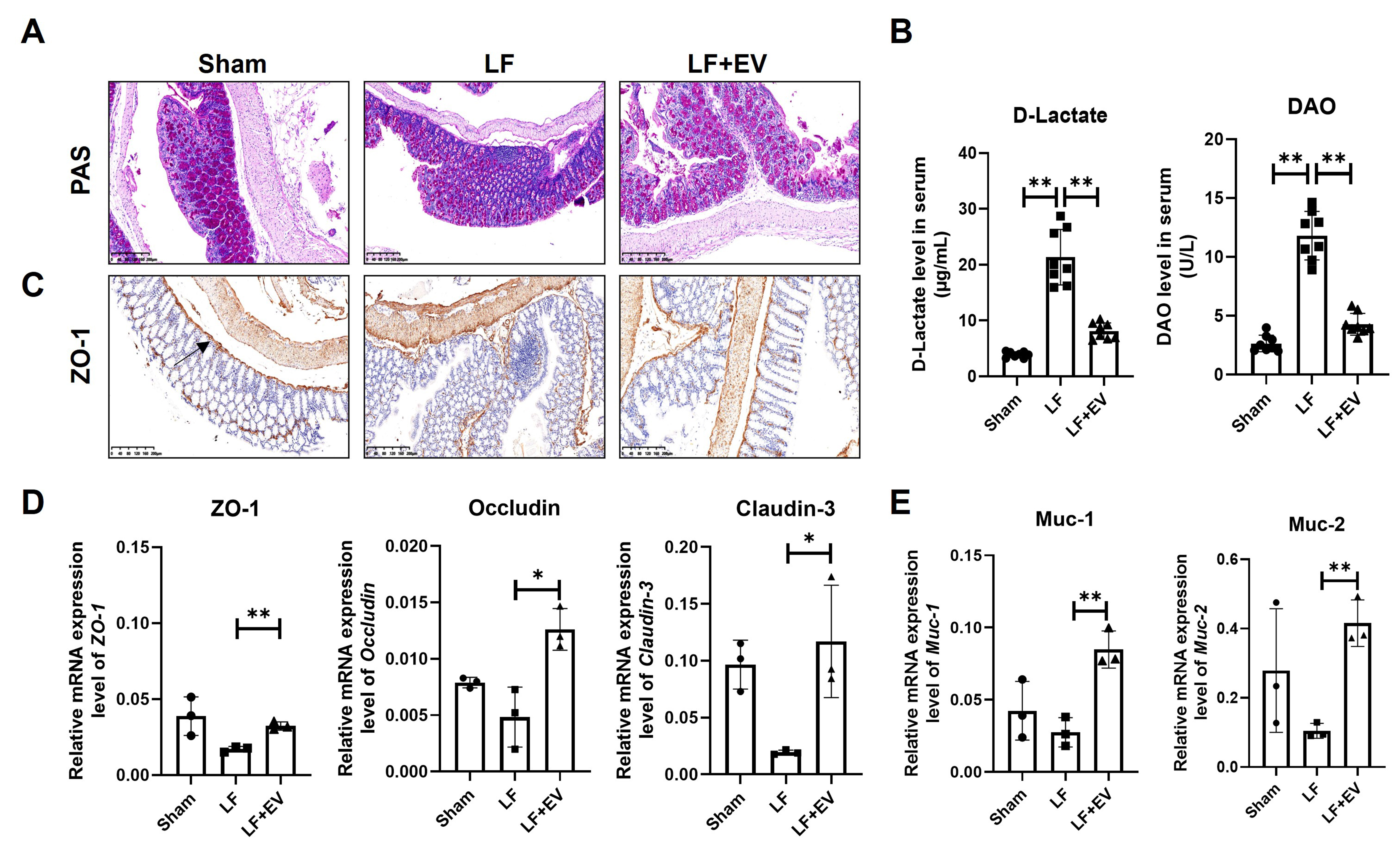
Figure 3. ADSC-EVs improve the intestinal barrier integrity by reducing intestinal permeability and reinforcing the mucus barrier in mice with LF. (A) PAS staining of colonic mucosa in Sham, LF and LF + EV group mice (scale bar: 200 µm); (B) The intestinal injury marker D-Lactate and DAO level in the serum of mice in each group (n = 8); (C) Representative images of ZO-1 expression in liver tissues by IHC staining (scale bar: 200 µm); (D) mRNA levels of tight junction associated genes ZO-1, Occludin, and Claudin-3 in colonic tissue were evaluated by q-PCR (n = 3); (E) The mRNA levels of mucosal barrier-associated genes Muc-1 and Muc-2 in colonic tissue were evaluated by q-PCR (n = 3). The expression was normalized to Gapdh. All data are shown as the mean ± SD. *P < 0.05; **P < 0.01. ADSC-EVs: Adipose-derived stem cell extracellular vesicles; LF: liver fibrosis; PAS: periodic acid Schiff; DAO: diamine oxidase; ZO-1: zonula occludens-1; IHC: immunohistochemistry; Occludin: tight junction protein Occludin; Claudin-3: tight junction protein 3; q-PCR: quantitative polymerase chain reaction; Muc-1: Mucin 1; Muc-2: Mucin 2; SD: standard deviation; LF + EV: liver fibrosis + ADSC-EV group.


