


fig2
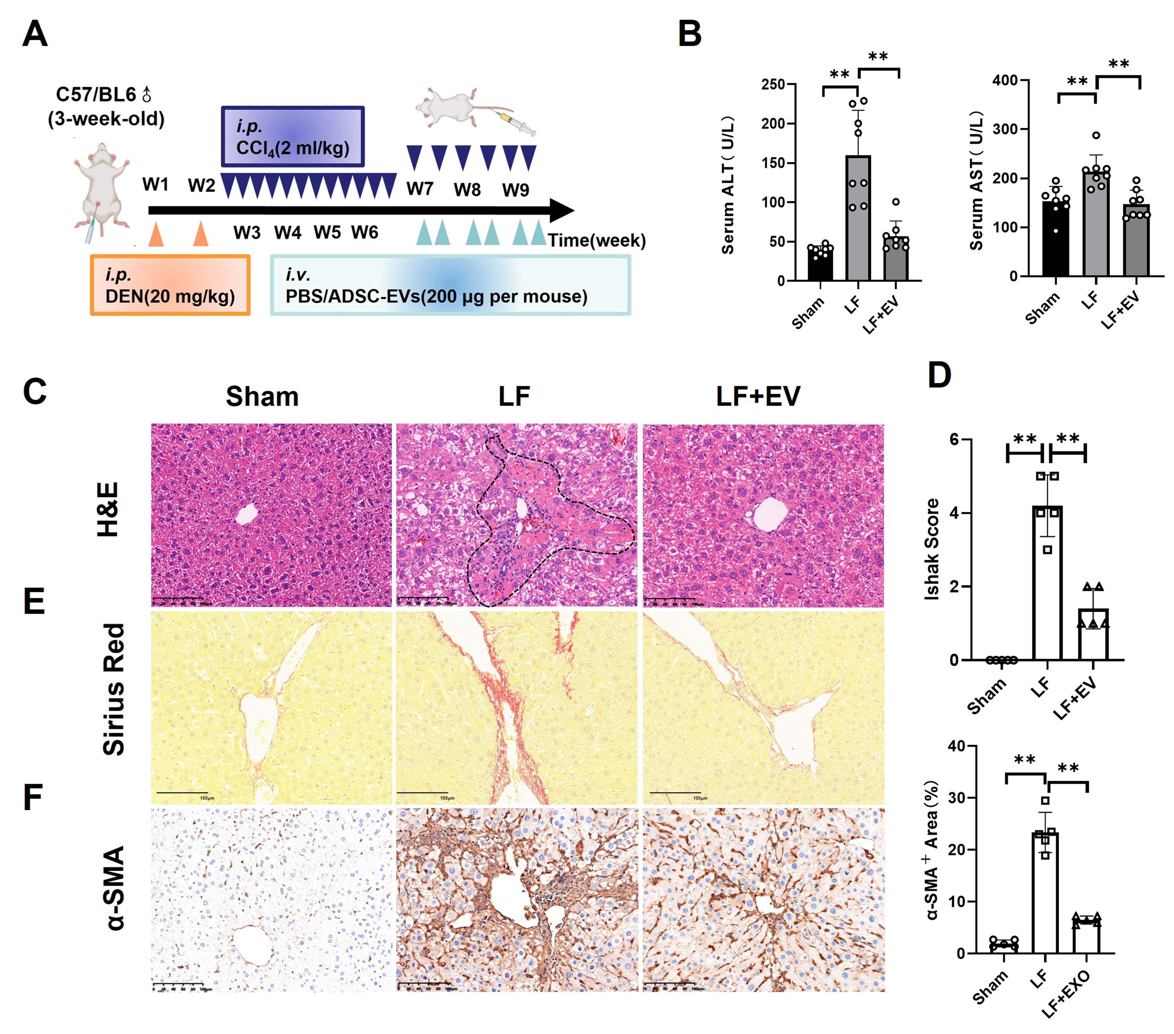
Figure 2. ADSC-EVs inhibit DEN/CCl4-induced hepatic fibrosis in mice. (A) Schematic diagram of DEN/CCl4-induced LF animal model establishment (i.p.) and ADSC-EV treatment strategy (i.v.); (B) The biochemical parameters measurement of ALT and AST in the mouse serum of each group (n = 8); (C) Representative histomorphology images of liver tissues by H&E staining (scale bar: 100 µm). The dotted line indicates the severely damaged area; (D) LF stage assessed using the Ishak score system (n = 5); (E) Representative images of liver tissues by Sirius Red staining (scale bar: 100 µm); (F) Expression of the activated HSCs marker α-SMA in liver tissues by IHC staining (scale bar: 100 µm). All data are shown as the mean ± SD. **P < 0.01. ADSC-EVs: Adipose-derived stem cell extracellular vesicles; DEN: diethylnitrosamine; LF: liver fibrosis; i.p.: intraperitoneal injection; i.v.: intravenous injection; ALT: alanine aminotransferase; AST: aspartate aminotransferase; H&E: hematoxylin and eosin; HSCs: hepatic stellate cells; α-SMA: alpha smooth muscle actin; IHC, immunohistochemistry; SD: standard deviation; PBS: phosphate-buffered saline; LF + EV: liver fibrosis + ADSC-EV group.


