


fig1
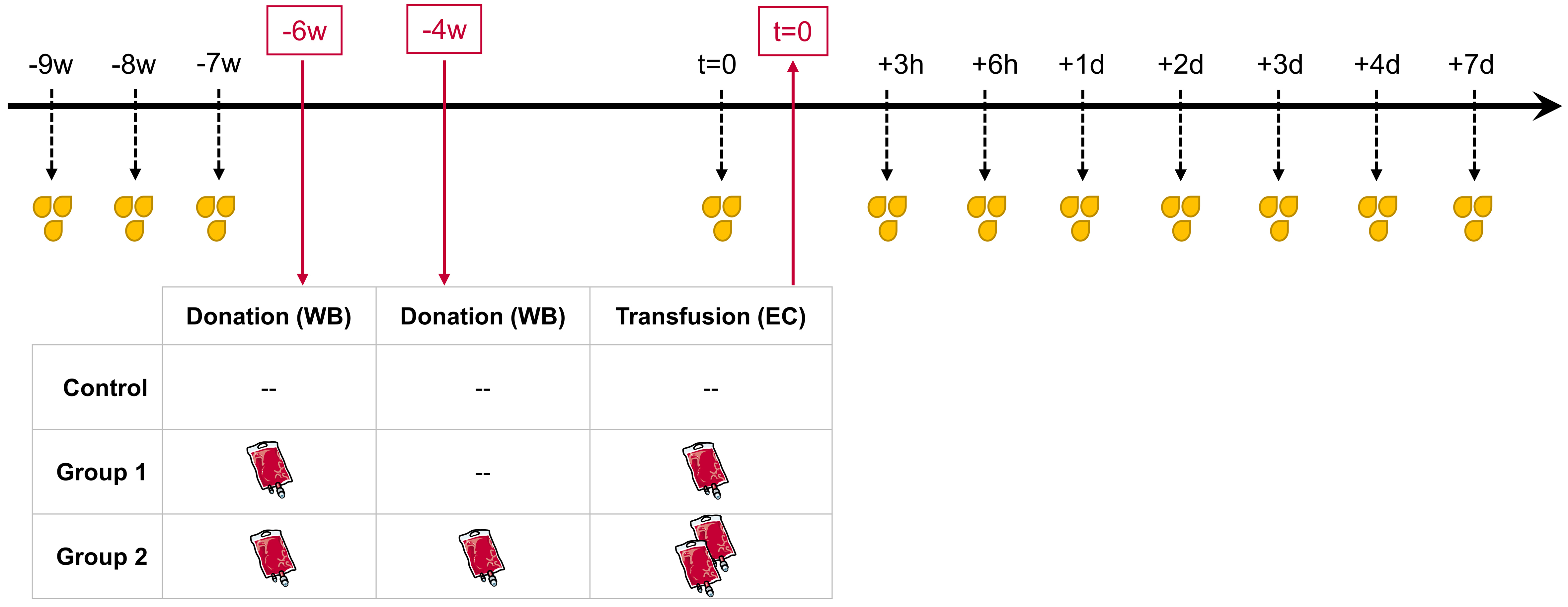
Figure 1. Longitudinal study design. All male subjects (n = 30) were randomly divided into three equal groups. WB bags (~ 500 mL, each) were donated six weeks prior to re-transfusion (-6 w) in Groups 1 and 2, and a second whole blood bag was donated in Group 2 four weeks prior to re-transfusion (-4 w). Depending on the grouping, either one (Group 1) or two (Group 2) processed and stored EC were re-transfused (t = 0). The Control group was analogously sampled for urine but without any blood donation or re-transfusion. Urine samples were collected before blood donation (-9 w, -8 w, -7 w) to create baseline measurements as well as shortly before re-transfusion (t = 0), and after re-transfusion (+3 h, +6 h, +1 d, +2 d, +3 d, +4 d, and +7 d) to identify transfusion-dependent variations.


