


fig4
From: Assessment of the reproducibility of bacterial membrane vesicle isolation and characterization
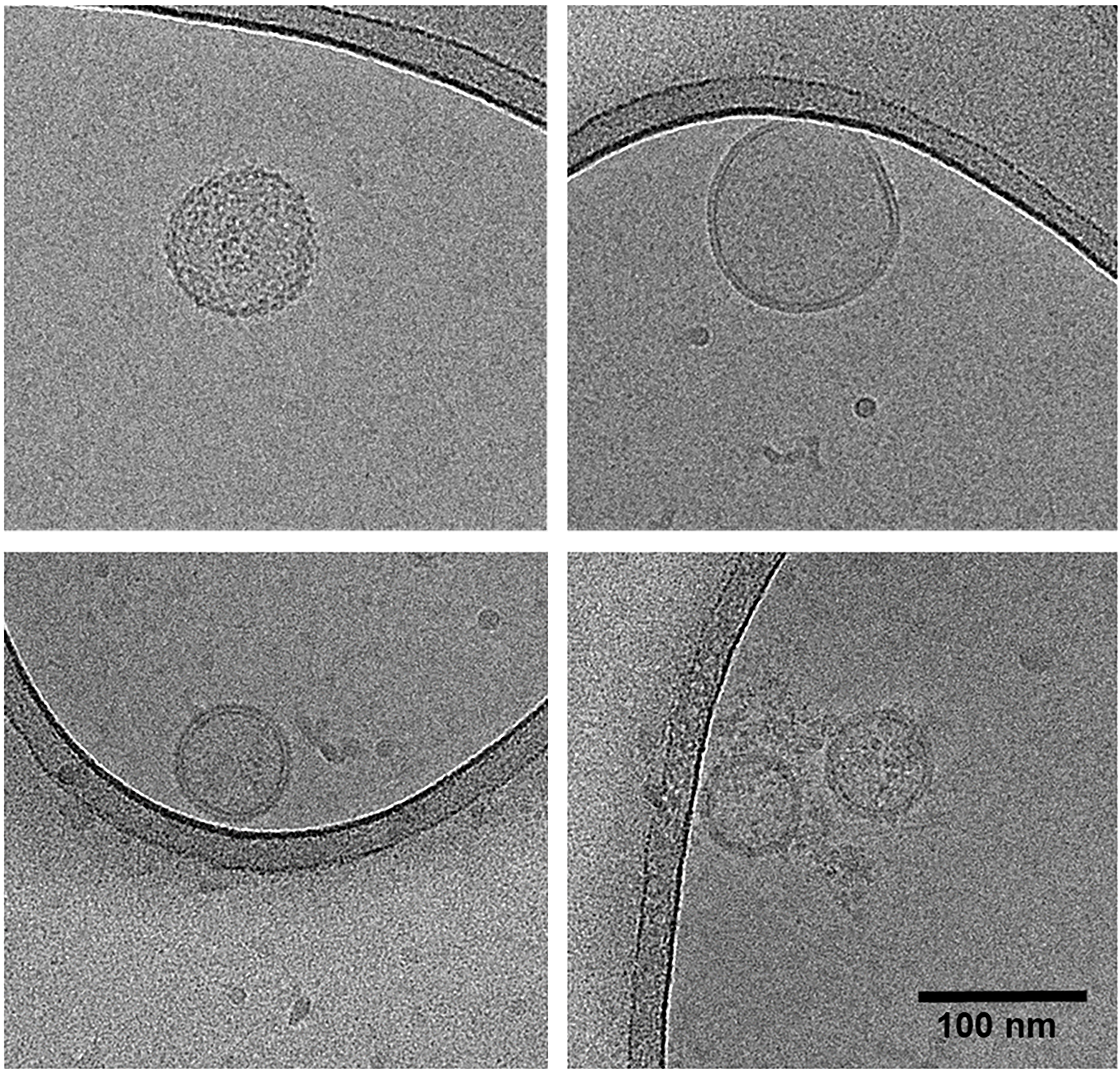
Figure 4. Cryo-transmission electron microscopy micrographs of feces-derived vesicles. Top left: Vesicle sample A1; top right: vesicle sample B1; bottom panels: vesicle sample C1. All samples originate from original sampling.


