


fig5
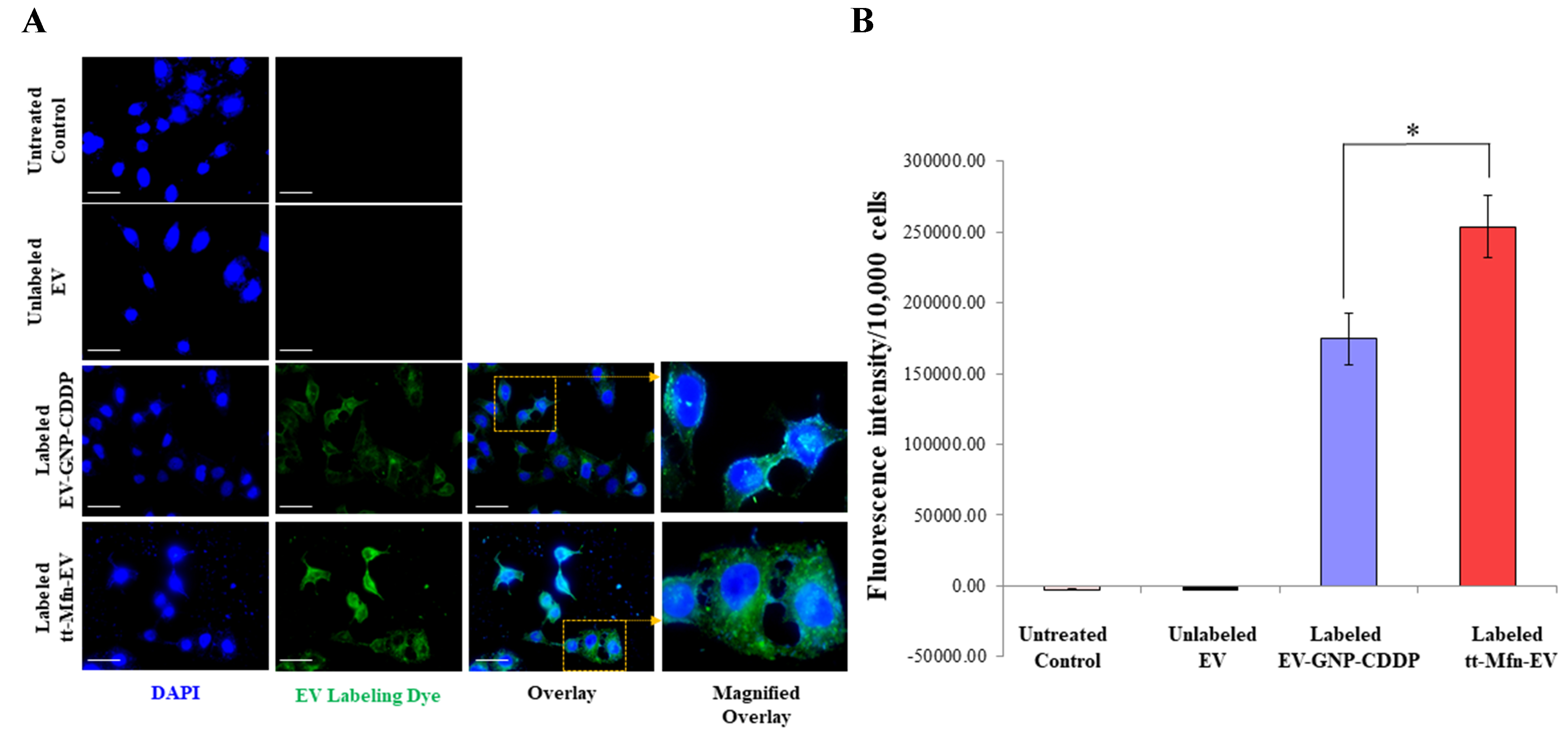
Figure 5. Measurement of tumor cell uptake of tt-Mfn-EVs. (A) Fluorescence microscopy images of A549 cells at 24 h after treatment with fluorescently labeled EV-GNP-CDDP and tt-Mfn-EVs. Untreated cells and cells treated with unlabeled EVs served as controls. Magnified overlay image showed a shift from punctate to diffuse cytoplasmic green fluorescence in A549 cells after 24 h, confirming efficient EV uptake. Fluorescein (green)-labeled EVs; DAPI (blue)-nucleus. Magnification, 60×; scale bar: 25 µm; (B) Quantification of cellular uptake based on fluorescence intensity in cells treated with labeled EV-GNP-CDDP and tt-Mfn-EVs, compared to unlabeled EVs and untreated controls. Fluorescent signal confirmed uptake in both labeled groups, with the highest uptake observed in tt-Mfn-EV-treated cells. The bar graph represents the mean ± SD from three replicates (n = 3). Statistical significance was assessed using an unpaired Student’s t-test, with P values indicated as *P < 0.05. tt-Mfn-EVs: Tumor-targeted multifunctional extracellular vesicles; GNP: gold nanoparticle; CDDP: cisplatin; DAPI: 4, 6-diamidino-2-phenylindole; SD: standard deviation.


