


fig1
From: Tumor-targeted multifunctional extracellular vesicles as drug carriers for lung cancer therapy
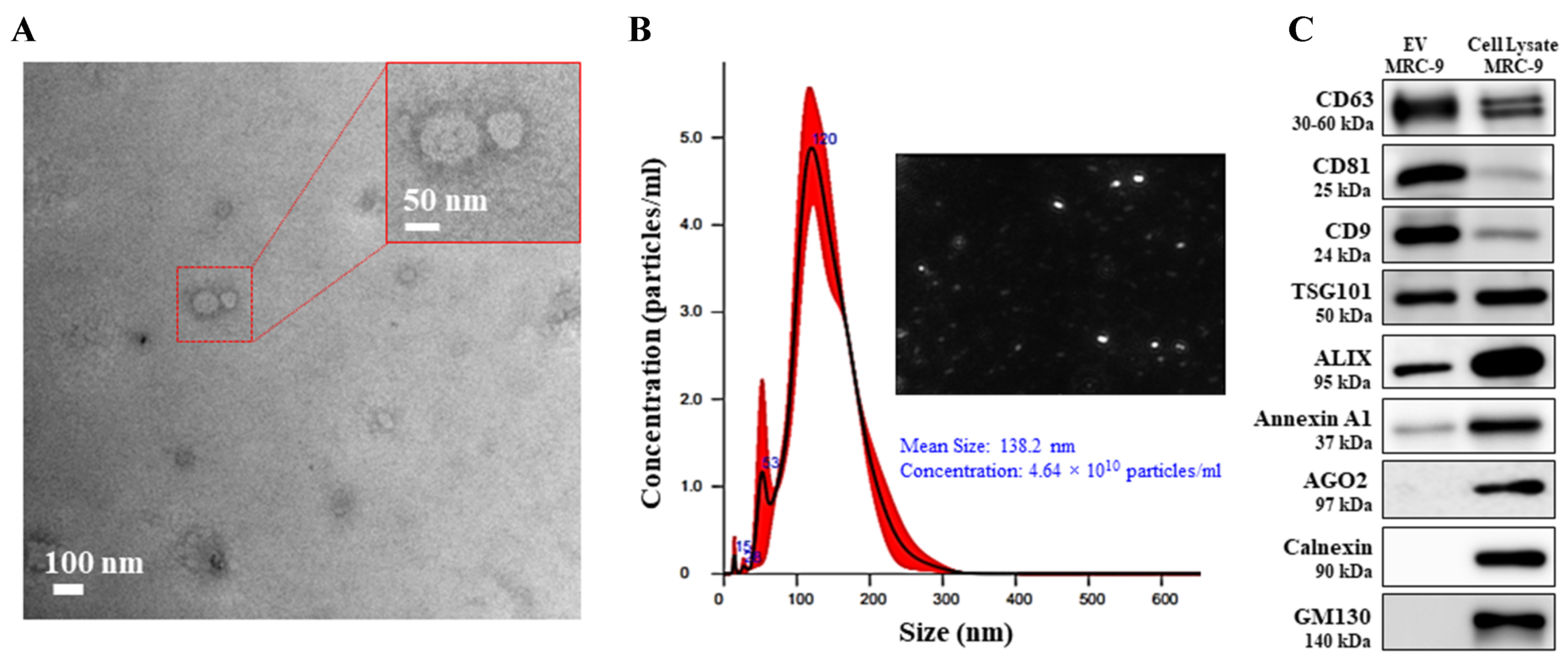
Figure 1. Characterization of EVs isolated from human lung fibroblast MRC-9 cell line. (A) TEM image (scale bar 100 nm) and the inset represents an enlarged image; (B) Histogram of NTA analysis and image of distributed EVs; (C) Western blot analysis for protein markers of EVs (CD63, CD81, CD9, TSG101, and Alix) and for markers discerning from microvesicles and non-EVs (Annexin A1, AGO2, calnexin and GM130) were compared with MRC-9 cell lysate that served as the positive reference. EVs: Extracellular vesicles; TEM: transmission electron microscopy; NTA: nanoparticle tracking analysis; TSG101: tumor susceptibility gene 101; Alix: ALG-2-interacting protein X; AGO2: argonaute 2; GM130: Golgi matrix protein 130.


