


fig5
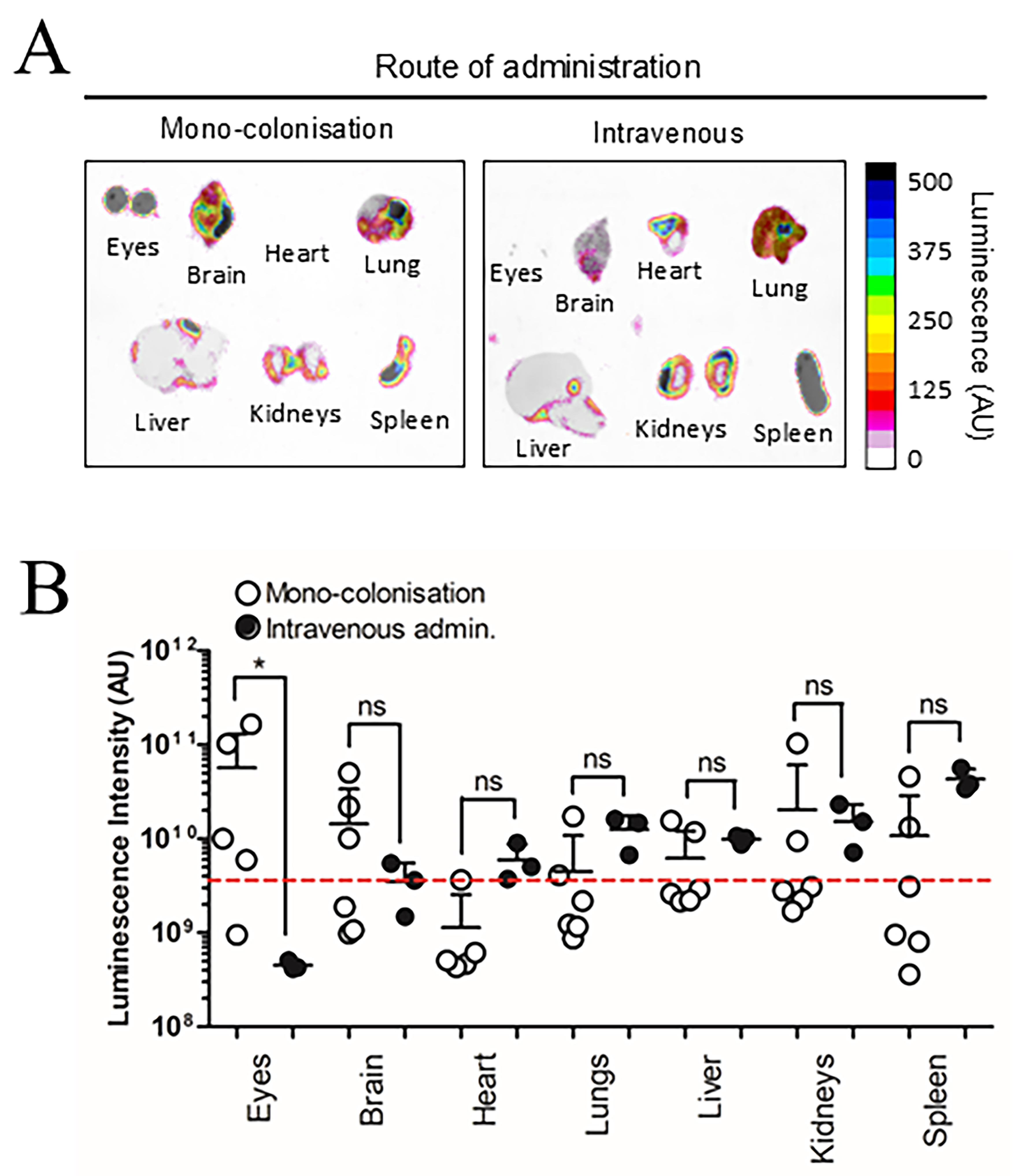
Figure 5. Route of administration alters NanoLuc-BEV biodistribution. GF mice were either monocolonised with NanoLuc-BEV-producing B. thetaiotaomicron or intravenously administered NanoLuc-BEVs (4 × 10¹⁰/mouse). (A) Organs were excised 7 days post-colonisation or 1 h post-IV administration and imaged using the Bruker In vivo Xtreme system. Substrate was injected intraperitoneally 5 min before euthanasia. Representative images from monocolonised (n = 6) or intravenously administered (n = 3) mice are shown; (B) Quantification of organ luminescence. Error bars represent mean ± SD. Red dotted line indicates LOD. *P ≤ 0.05. ns: Not significant; NanoLuc-BEV: Nanoluciferase-bacterial extracellular vesicle; GF: germ-free; B. thetaiotaomicron: Bacteroides thetaiotaomicron; IV: intravenous; SD: standard deviation; LOD: limit of detection.


