


fig3
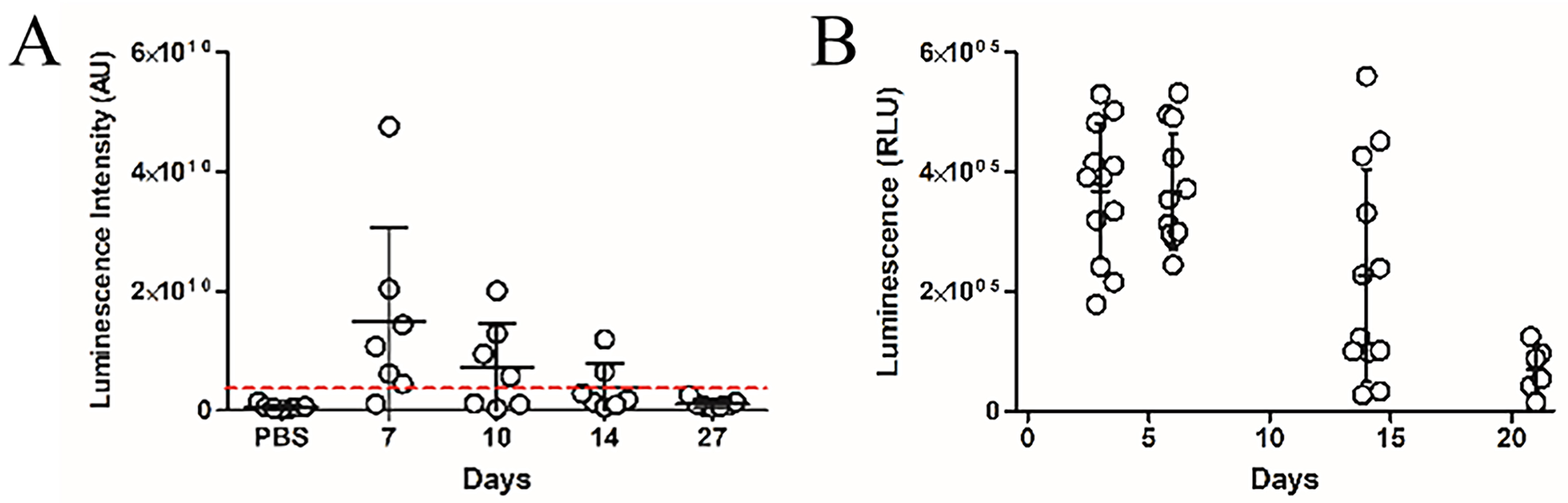
Figure 3. Time course of in vivo luminescence following colonisation with NanoLuc-BEV-producing B. thetaiotaomicron. GF mice were monocolonised at day 0. Samples were collected at 7, 10, 14, and 27 days post-colonisation. (A) Quantification of luminescence from organ tissues of animals administered PBS (n = 6) or the engineered B. thetaiotaomicron strain on day 7 (n = 6), 10 (n = 3), 14 (n = 2) and 27 (n = 4) post-colonisation. Each point represents the average luminescence for the eyes, brain, heart, lungs, liver, kidney, or spleen. Individual organs were excised for imaging using a Bruker In vivo Xtreme system. Substrate was injected intraperitoneally 5 min prior to organ excision; (B) Luminescence in the soluble fraction of faecal pellets from monocolonised GF mice. All samples were adjusted to 100 mg/mL prior to analysis (n = 12 for days 3, 6, and 14; n = 6 for day 21). Error bars represent mean ± SD. Red dotted line indicates LOD. NanoLuc-BEV: Nanoluciferase-bacterial extracellular vesicles; B. thetaiotaomicron: Bacteroides thetaiotaomicron; GF: germ-free; PBS: phosphate-buffered saline; SD: standard deviation; LOD: limit of detection.


