


fig1
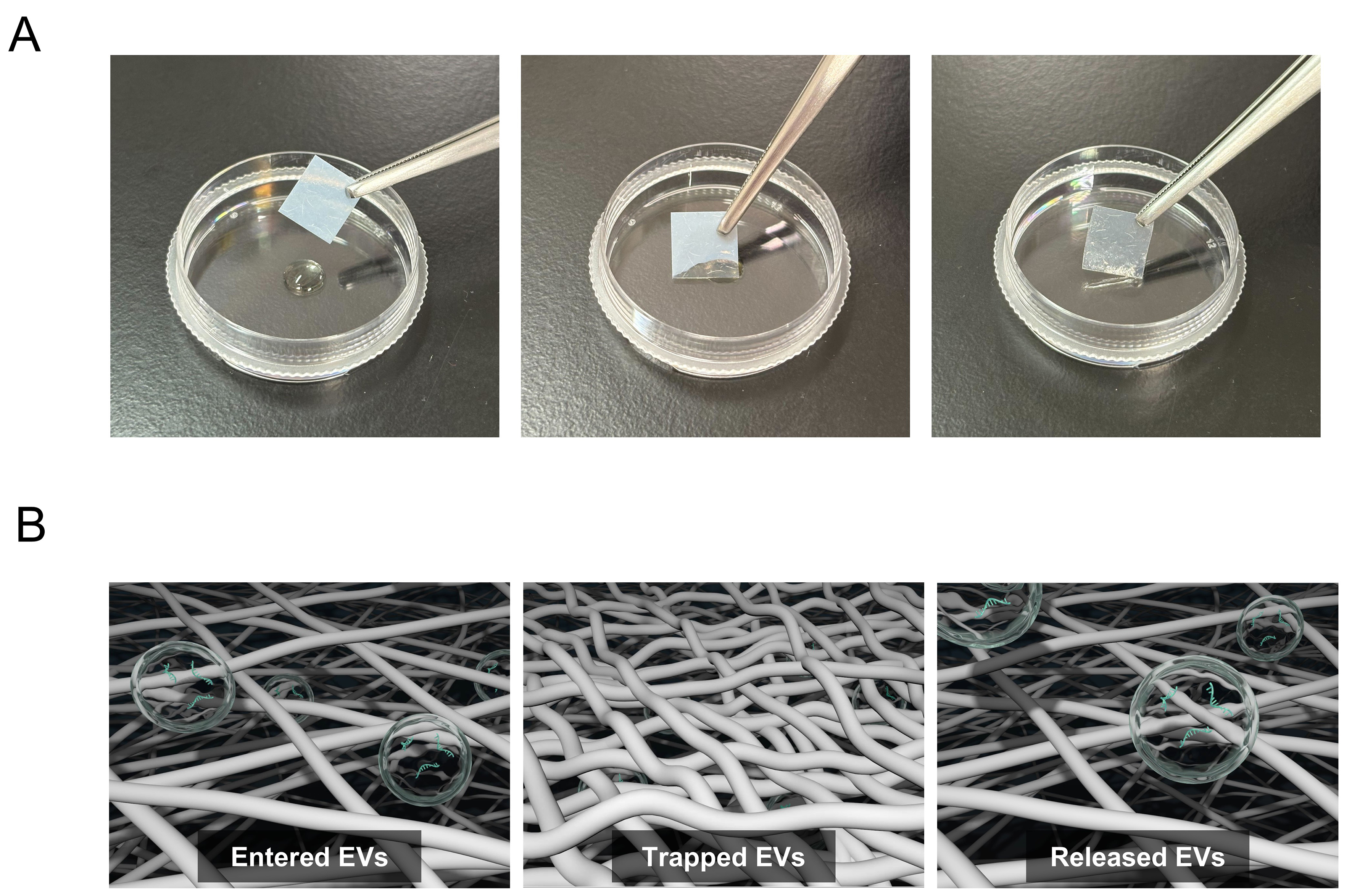
Figure 1. EV sheet, a cellulose nanofiber-based EV isolation platform. (A) Representative images of EV sheets prepared as approximately 1 cm2 square pieces. The sheets appear milky white in the dry state and become semitransparent upon water absorption. Left: dry state; Center: absorption in progress; Right: fully water-absorbed state; (B) Schematic of EV capture and release in EV sheets. Each sheet contains many small pores (< 300 nm in diameter) formed by fine cellulose nanofibers. Left: EVs are captured within the sheet. Center: Pore closure during drying retains the EVs within the pores of the sheet. Right: Soaking in PBS reopens the pores and enables the release of the retained EVs. Modified from Reference 16 based on CC BY 4.0[16]. EV: Extracellular vesicle; PBS: phosphate-buffered saline; CNF: cellulose nanofiber.


