


fig2
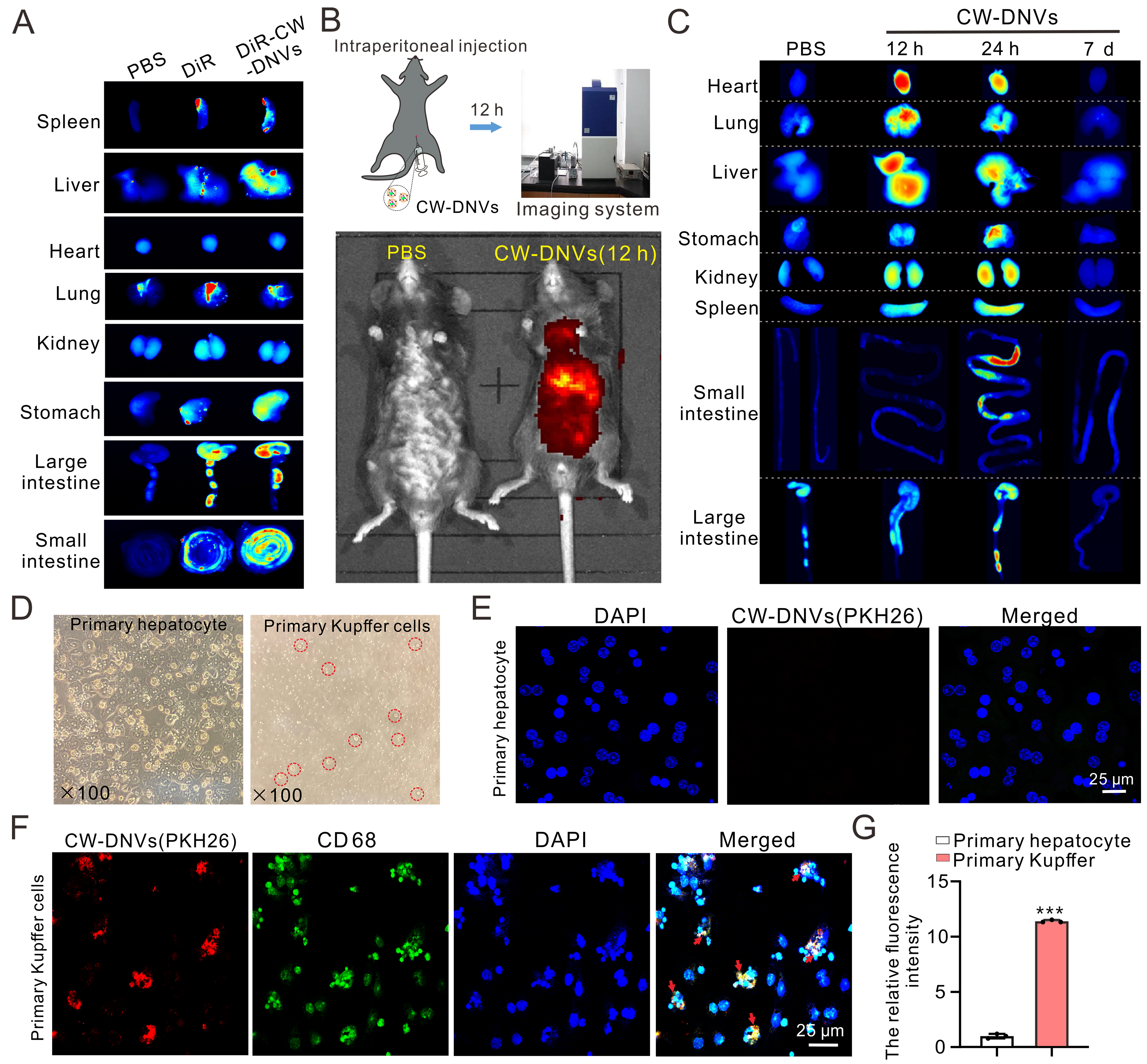
Figure 2. Accumulated delivery of CW-DNVs to liver macrophages. (A) DiR-labeled CW-DNVs were administered to mice via intraperitoneal injection, and biodistribution in organs was subsequently analyzed; (B) In vivo imaging at 12 h post-intraperitoneal injection of Dil-labeled CW-DNVs; (C) Biodistribution in organs 12 h, 24 h, and 7 day post-injection; (D) Isolated primary hepatocytes and KCs from mouse liver; (E) Uptake of PKH26-labeled CW-DNVs (red) by hepatocytes and (F) CD68+ KCs (green) 12 h post-treatment; (G) Relative fluorescence intensity of PKH26-labeled CW-DNVs in primary hepatocytes vs. KCs. Data represent the mean ± SEM (n = 3). Student’s t-test. ***P < 0.001. CW-DNVs: Curcuma wenyujin-derived nanovesicles; KCs: Kupffer cells; SEM: standard error of the mean; PBS: phosphate-buffered saline.


