


fig5
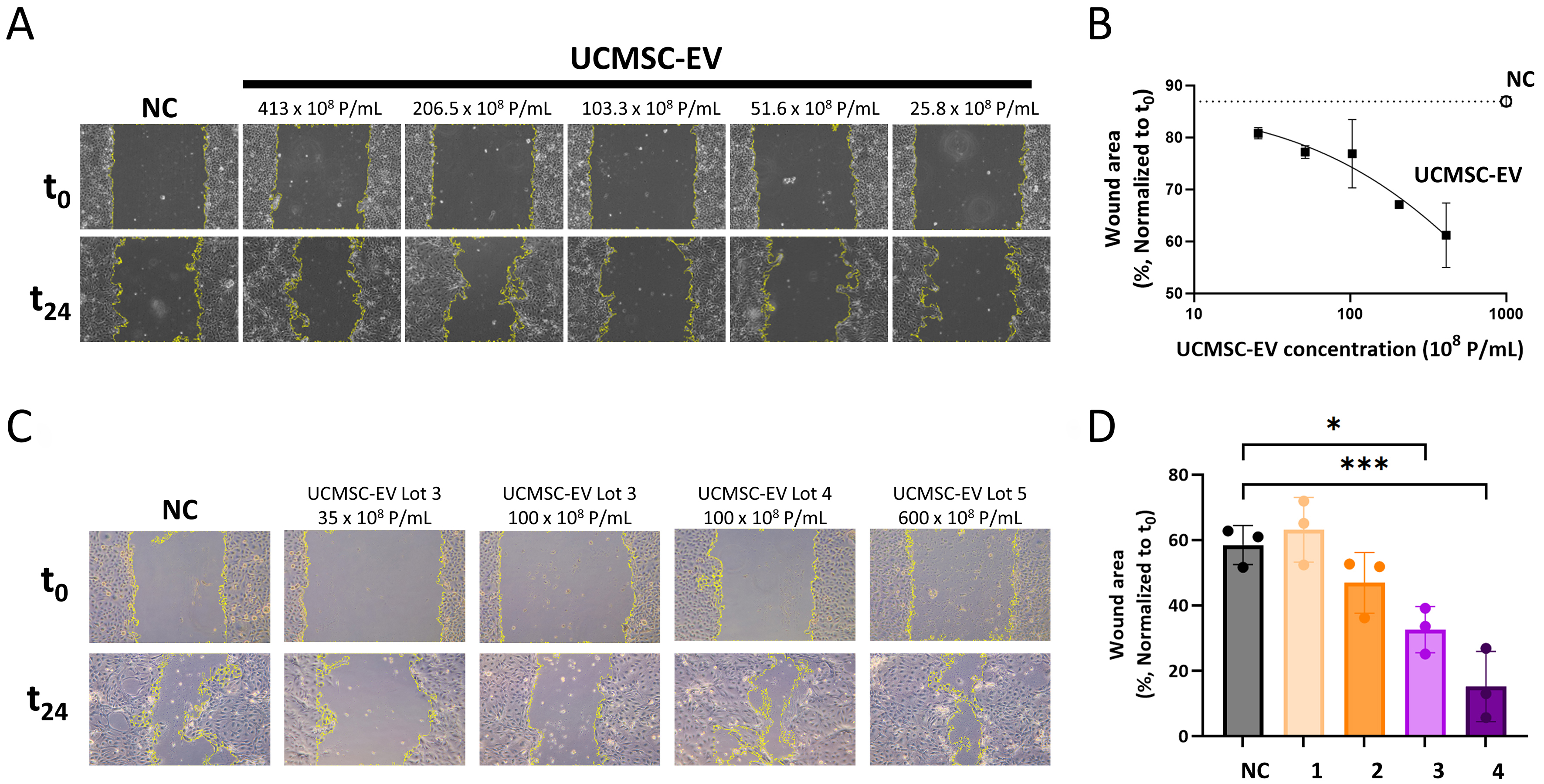
Figure 5. UCMSC-EVs promote wound healing of the human corneal epithelial cells in a dose dependent manner. (A) Bright field microscopic images show increased cell migration toward the empty area from UCMSC-EV-dosed wells after 24-h treatment; (B) Quantitative data from (A) show dose-dependent reduction of wound closure area as compared to the negative control (open circle). Data are presented as the mean ± SD, based on two independent biological replicates of each condition; (C) Bright- field microscopic images show increased cell migration across different lots of UCMSC-EV-dosed wells after 24-h treatment; (D) Quantitative data from (C) show significant reduction of wound closure area from UCMSC-EV lot 4 (Bar 3) and lot 5 (Bar 4) treatments as compared to the NC (black bar) (UCMSC-EV lot 4 100 × 108 P/mL vs. NC: *P = 0.0155, n = 3. UCMSC-EV lot 5 600 × 108 P/mL vs. NC: ***P = 0.0004, n = 3, one-way ANOVA with Dunnett’s post hoc test). Bar 1: UCMSC-EV lot 3 35 × 108 P/mL; Bar 2: UCMSC-EV lot 3 100 × 108 P/mL; Bar 3: UCMSC-EV lot 4 100 × 108 P/mL; Bar 4: UCMSC-EV lot 5 600 × 108 P/mL. NC: Negative control; UCMSC-EVs: umbilical cord mesenchymal stem cell-derived extracellular vesicles; SD: standard deviation.


