


fig1
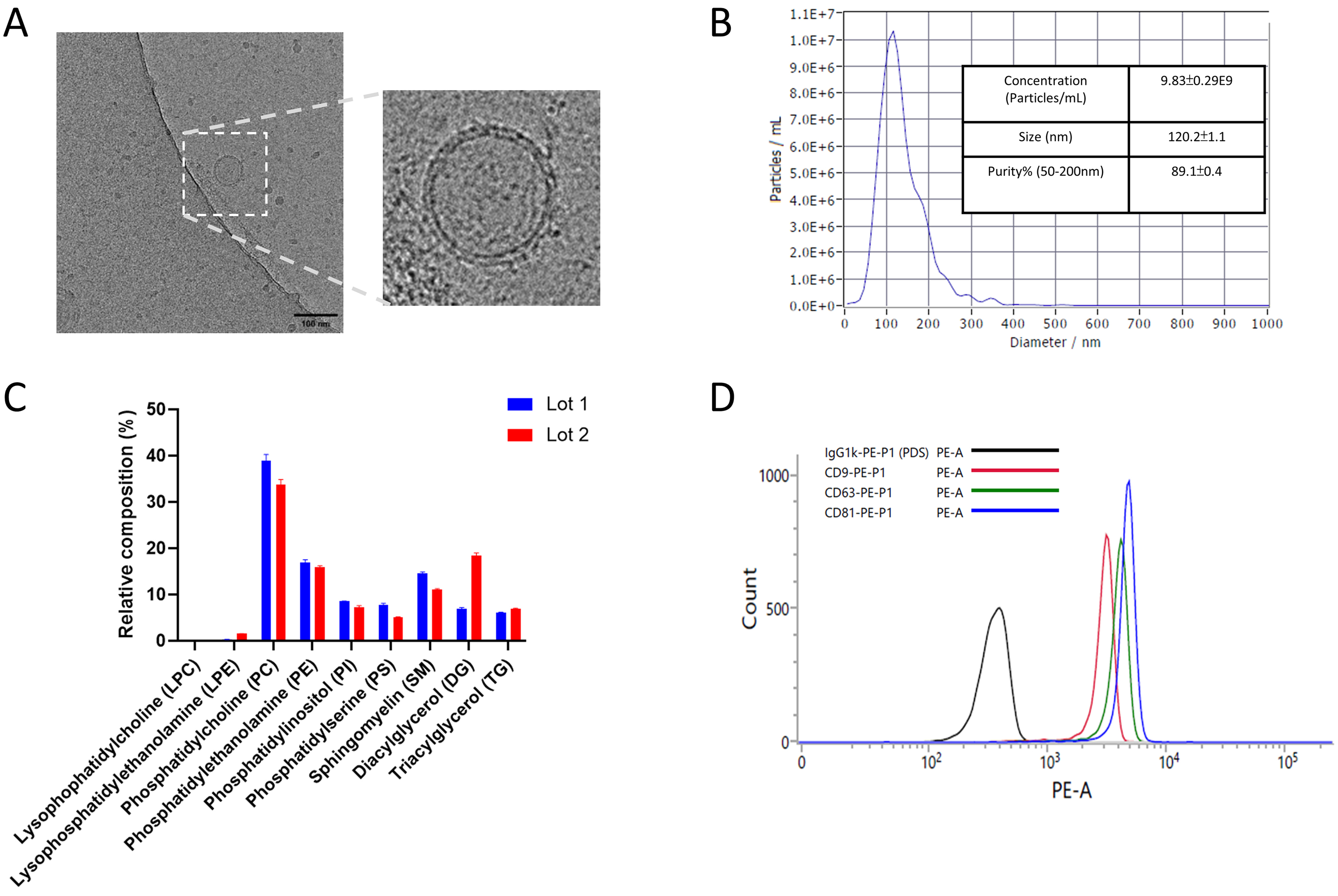
Figure 1. The physicochemical properties of the UCMSC-EVs. (A) A representative Cryo-EM image showed that UCMSC-EVs contained lipid bilayered particles. The scale bar is 100 nm; (B) NTA analysis showed that UCMSC-EVs had a median particle size around 120 nm with a purity of approximately 89%; (C) The bar plot shows similar lipid profiles of two independent UCMSC-EV lots measured by LC-MS/MS. Relative composition was calculated as (specific measured lipid/ total measured lipid) × 100%. The error bar is the standard deviation of four independent technical replicates of the same lot; (D) The anti-CD81 bead-based flow analysis shows that UCMSC-EVs stained positive for exosome markers (i.e., CD81, CD9, CD63), compared with the IgG isotype control. CD9: Cluster of differentiation 9; CD63: cluster of differentiation 63; CD81: cluster of differentiation 81; Cryo-EM: cryo-electron microscopy; IgG: immunoglobulin G; LC-MS/MS: liquid chromatography-tandem mass spectrometry; NTA: nanoparticle tracking analysis; UCMSC-EVs: umbilical cord mesenchymal stem cell-derived extracellular vesicles.


