


fig1
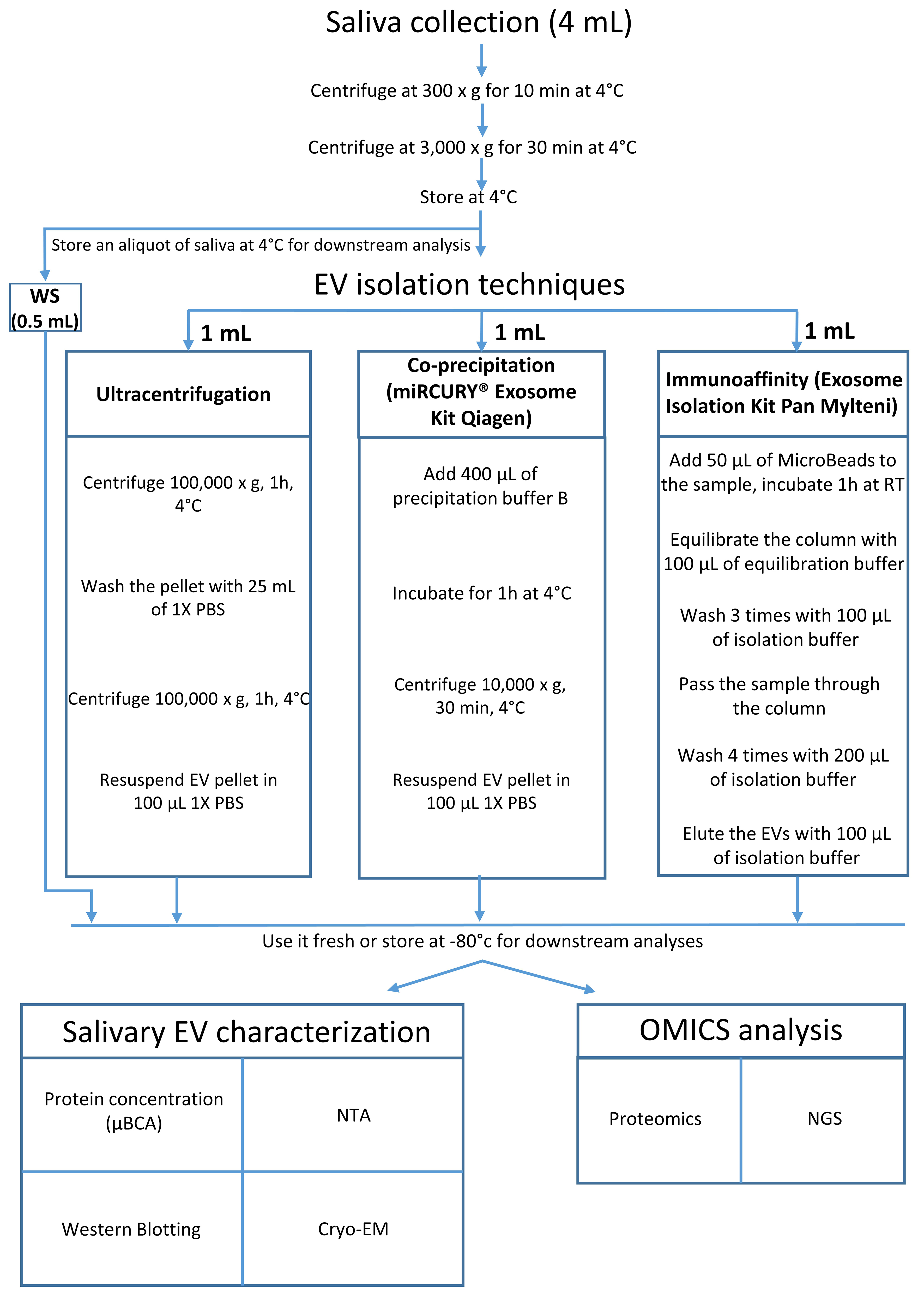
Figure 1. Schematic overview of the experimental workflow for salivary EV isolation and analysis. Unstimulated saliva (4 mL) was collected from healthy donors and sequentially centrifuged at 300 × g and 3,000 × g to remove cells, large debris, and residual contaminants. The resulting WS was divided into four aliquots: 0.5 mL was retained as the WS control, and 1 mL was allocated to each of the three EV isolation methods: UC, Q, and M. The isolated EVs were subsequently characterized and subjected to proteomic and small RNA sequencing analyses. EV: Extracellular vesicles; WS: whole saliva supernatant; UC: ultracentrifugation; Q: PEG-based co-precipitation; M: immunoaffinity capture; PBS: phosphate-buffered saline; RT: room temperature; NTA: nanoparticle tracking analysis; NGS: next-generation sequencing; EM: electron microscopy; Cryo-EM: cryogenic electron microscopy; μBCA: micro bicinchoninic acid assay.


