


fig1
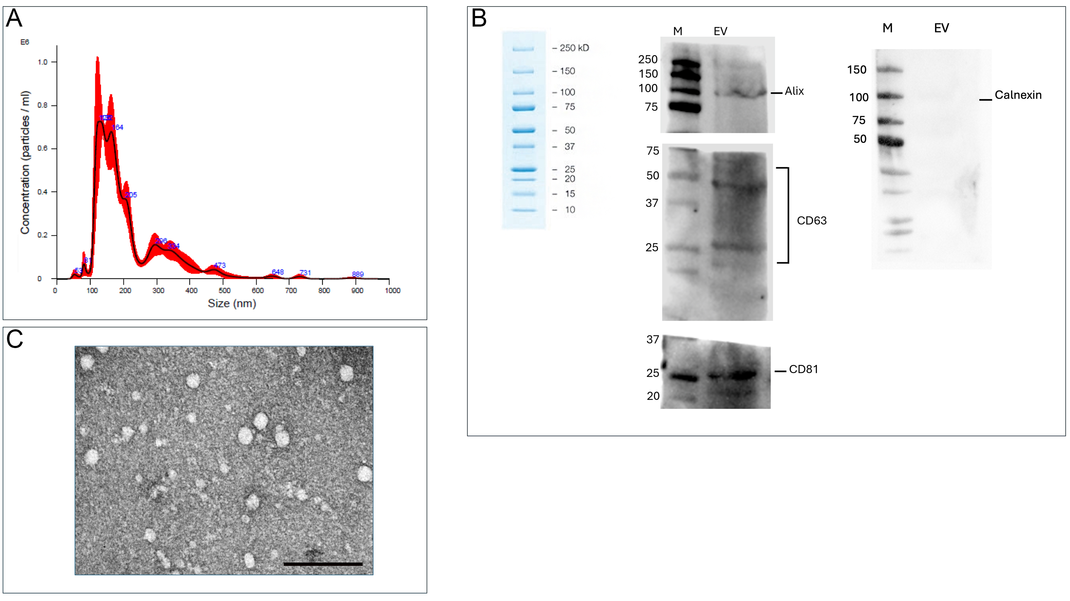
Figure 1. EV characterization. (A) NanoSight analysis of AMC-EV size distribution and concentration; (B) Western blot analysis of the EV-associated protein Alix (internal marker) and the surface markers (CD63 and CD81). Absence of Calnexin indicates lack of cellular contamination. Three biological samples were pooled together to perform Western blot analysis in technical triplicate; (C) TEM of AMC-derived EVs. Scale bar: 1 µm. AMC-EVs: Amniotic mesenchymal cell-derived extracellular vesicles; EVs: extracellular vesicles; TEM: transmission electron microscopy; CD63: cluster of differentiation 63; CD81: cluster of differentiation 81.


