


fig1
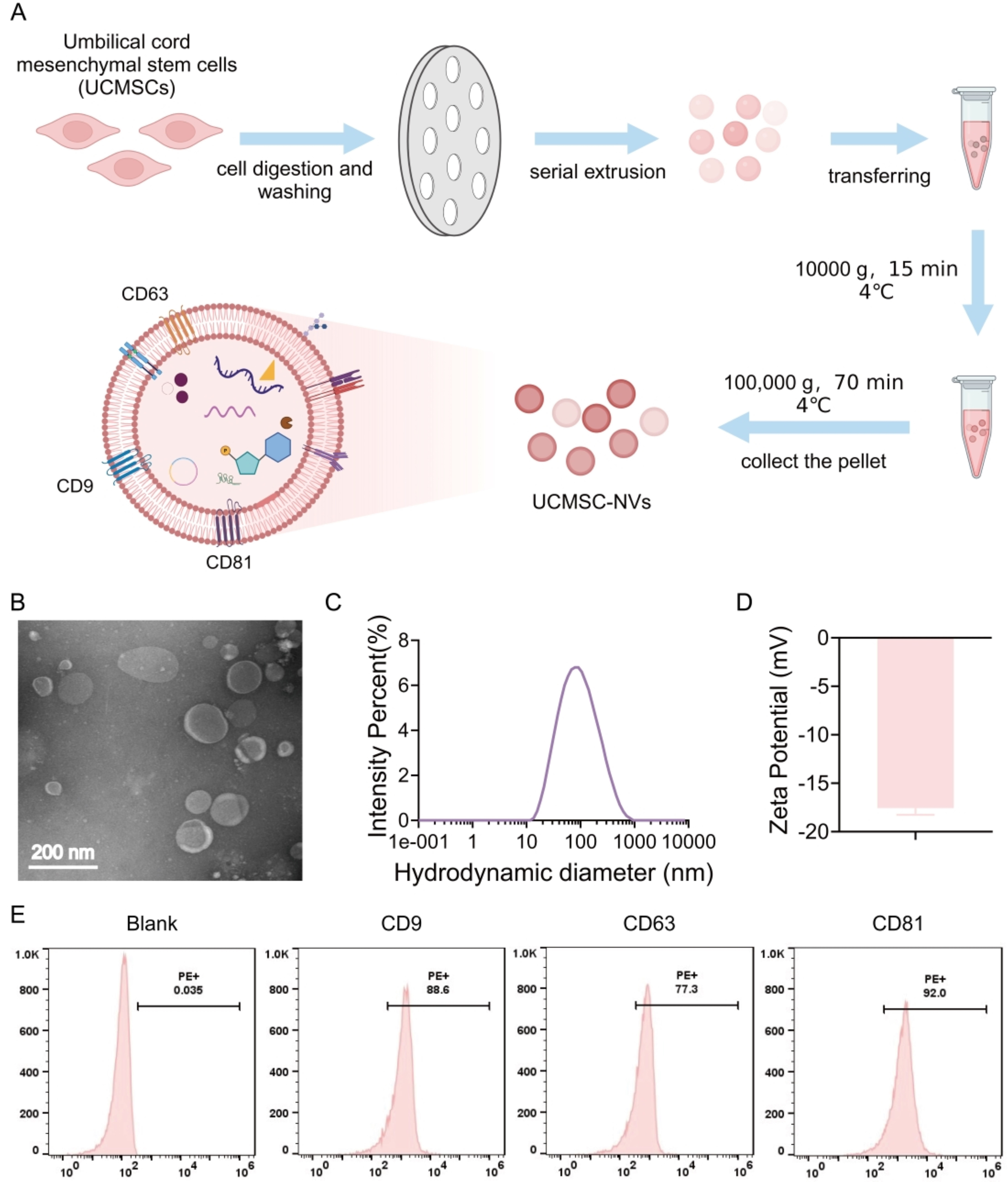
Figure 1. Preparation and Characterization of UCMSC-NVs. (A) Schematic illustration of the preparation of UCMSC-NVs through the serial extrusion of UCMSCs. Created in BioRender. Mou, X. (2026) https://app.biorender.com/illustrations/66b98605fa47a99541b0f630?slideId=72844bad-74f0-4878-9558-da4925fd93f8; (B) TEM characterization depicting the morphology and size of UCMSC-NVs; (C) DLS analysis illustrating the size distribution of UCMSC-NVs; (D) Zeta potential assessment of UCMSC-NVs; (E) Flow cytometry analysis of known. UCMSC-NVs: Nanovesicles originating from human umbilical cord mesenchymal stem cells; TEM: transmission electron microscopy; DLS: dynamic light scattering.


