


fig6
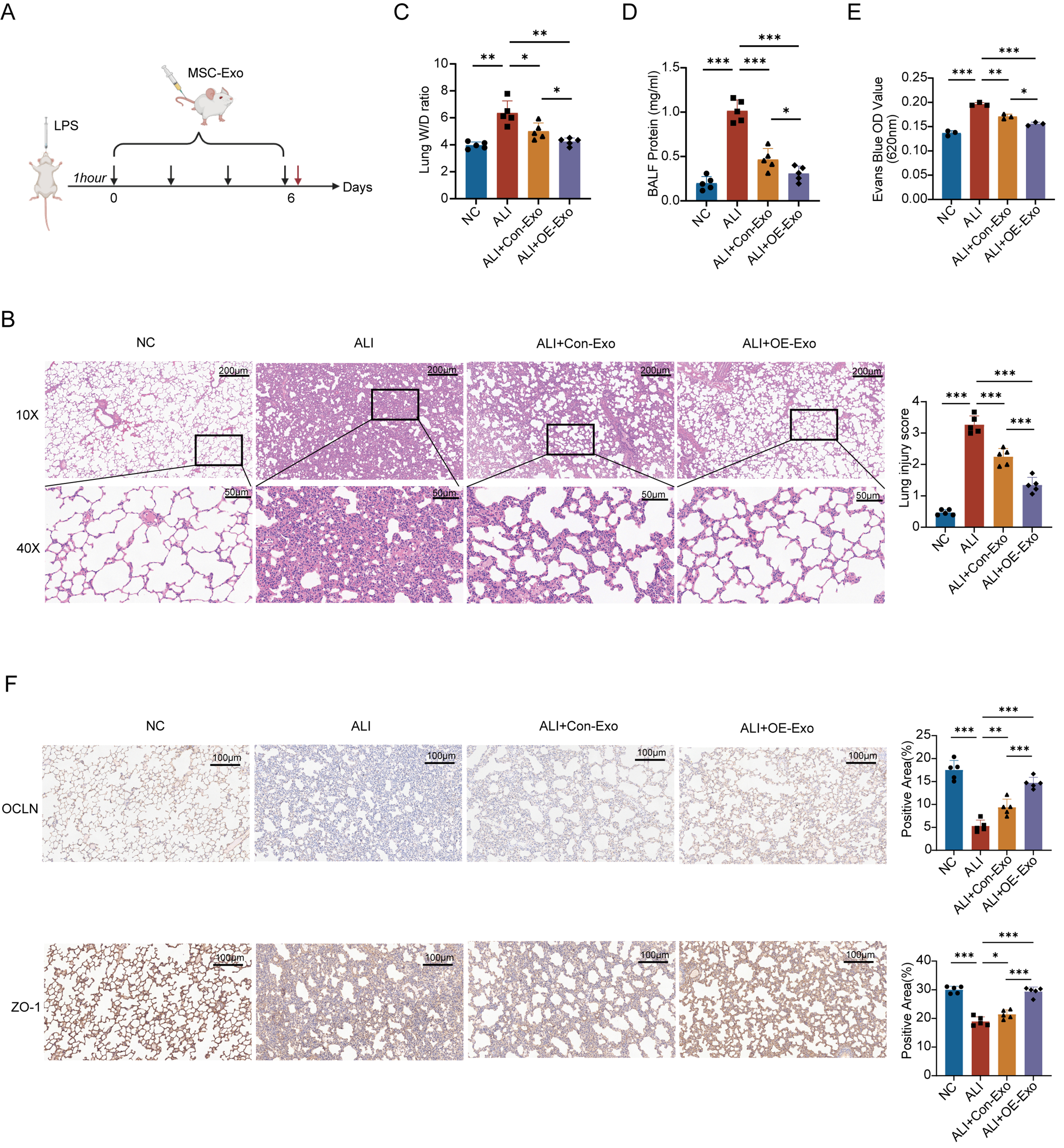
Figure 6. NRP1 can significantly alleviates pulmonary vascular hyperpermeability induced by LPS in ALI rats. (A) Establishment of rat ALI model and validation of MSC-Exo in vivo; (B) Histopathological changes and injury severity in lung tissues under different treatments were evaluated by H&E stained sections. Images reveal severe LPS-induced lung injury, while the therapeutic efficacy of OE-Exo surpasses that of conventional MSC-Exo. Quantitative analysis of ung tissue injury scores (n = 5). All experimental data were independently repeated three times; (C) The W/D weight ratio demonstrates the severity of pulmonary edema (n = 5); (D) Total protein concentration in BALF reflects lung vascular barrier integrity (n = 5); (E) Evans blue dye assay analyzes lung vascular barrier function via measurement of OD values (n = 3). All experimental data were independently repeated three times; (F) Immunohistochemical staining of OCLN and ZO-1 in lung tissue sections. Quantitative analysis of OCLN-positive immunoreactivity and ZO-1-positive immunoreactivity. All experimental data were independently repeated three times. Data are presented as mean ± SD. Statistical analysis was performed using one-way ANOVA followed by Tukey’s multiple comparisons test. *P < 0.05; **P < 0.01; ***P < 0.001. NRP1: Neuropilin-1; LPS: lipopolysaccharide; ALI: acute lung injury; MSC-Exo: mesenchymal stem cell-derived exosomes; Con-Exo: exosomes derived from control mesenchymal stem cells; OE-Exo: exosomes derived from NRP1-overexpressing mesenchymal stem cells; H&E: hematoxylin and eosin; W/D: wet-to-dry; BALF: bronchoalveolar lavage fluid; OD: optical density; OCLN: occludin; ZO-1: zonula occludens-1; NC: normal control; SD: standard deviation; ANOVA: analysis of variance.


