


fig2
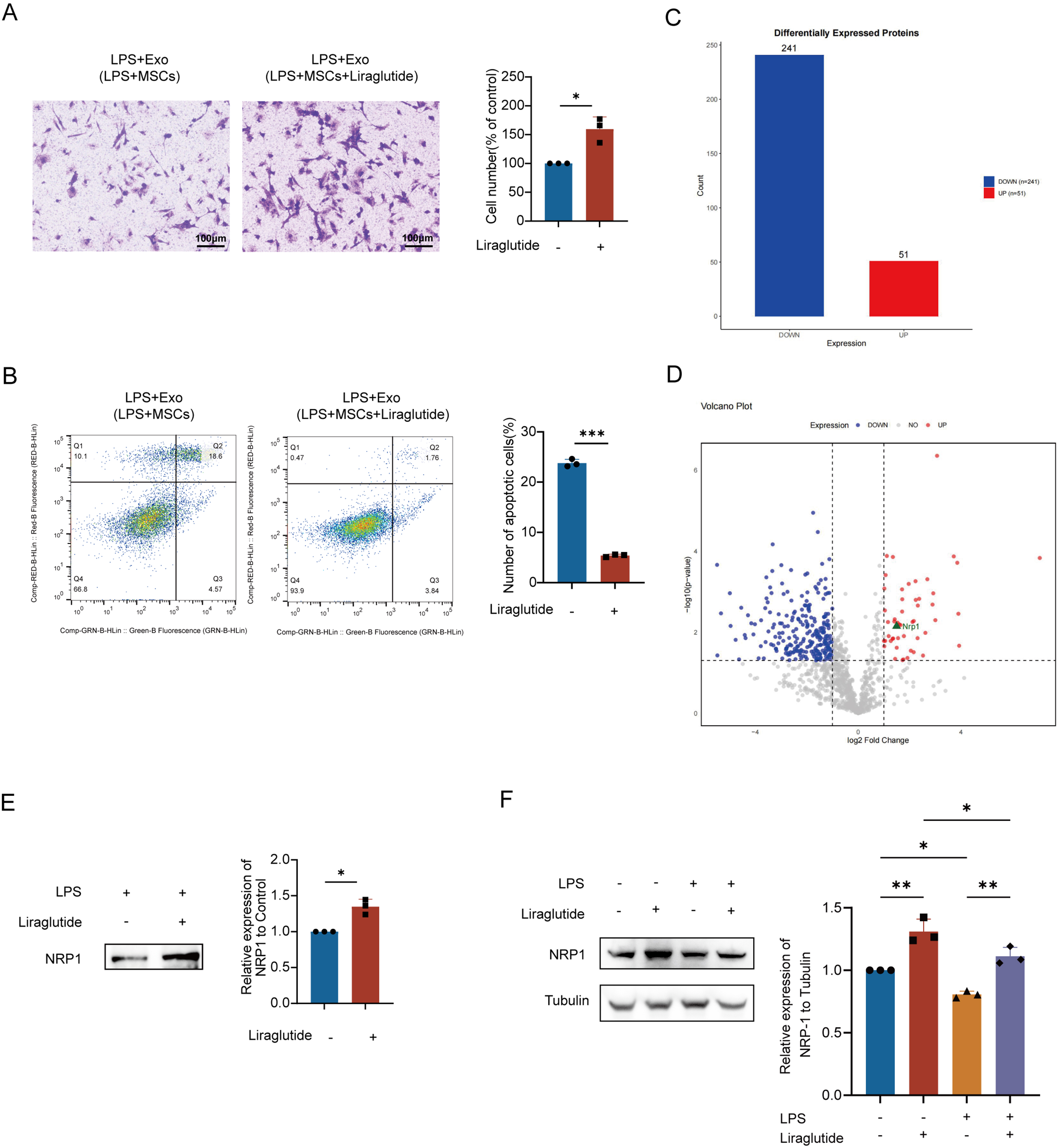
Figure 2. Liraglutide promoted the therapeutic effect of MSCs exosomes and increased the expression of NRP1 in exosomes. (A) Transwell assay demonstrates that liraglutide enhanced the LPS-pretreated MSC-Exo-mediated migration of PMVECs. Scale bar = 50 μm; (B) Annexin V-FITC/PI flow cytometry confirms that liraglutide strengthened the LPS-pretreated MSC-Exo-induced suppression of PMVEC apoptosis. All experiments were independently repeated three times; (C) Bar plot showing quantitative distribution of up-/down-regulated proteins (X-axis: regulation direction; Y-axis: protein count); (D) Volcano plot visualizing differential expression [X-axis: log2(fold change) with left/right quadrants indicating down-/up-regulation; Y-axis: -log10(P-value)]. Distance from origin correlates with statistical significance; (E) Combination treatment with liraglutide and LPS for 48 h significantly upregulated NRP1 protein levels in MSC-Exo. Equal amounts of exosomal protein (10 μg per lane) were loaded based on BCA quantification; (F) NRP1 expression in PMVECs under different treatments (Control, LPS, Liraglutide, and Liraglutide + LPS). LPS stimulation (12 h) resulted in a significant reduction in NRP1 expression, while liraglutide partially reversed this effect. Liraglutide (10 nM) was applied as indicated. All experiments were independently repeated three times. Data are presented as mean ± SD. Statistical analysis for (A, B, and E) was performed using unpaired Student’s t-test, while (F) was analyzed using one-way ANOVA followed by Tukey’s multiple comparisons test. *P < 0.05; **P < 0.01; ***P < 0.001. LPS: Lipopolysaccharide; Exo: exosome; MSCs: mesenchymal stem cells; MSC-Exo: mesenchymal stem cell-derived exosomes; Exo (LPS + MSCs): exosomes derived from lipopolysaccharide-treated mesenchymal stem cells; PMVECs: pulmonary microvascular endothelial cells; NRP1: neuropilin-1; FITC: fluorescein isothiocyanate; PI: propidium iodide; BCA: bicinchoninic acid; SD: standard deviation; ANOVA: analysis of variance.


