


fig1
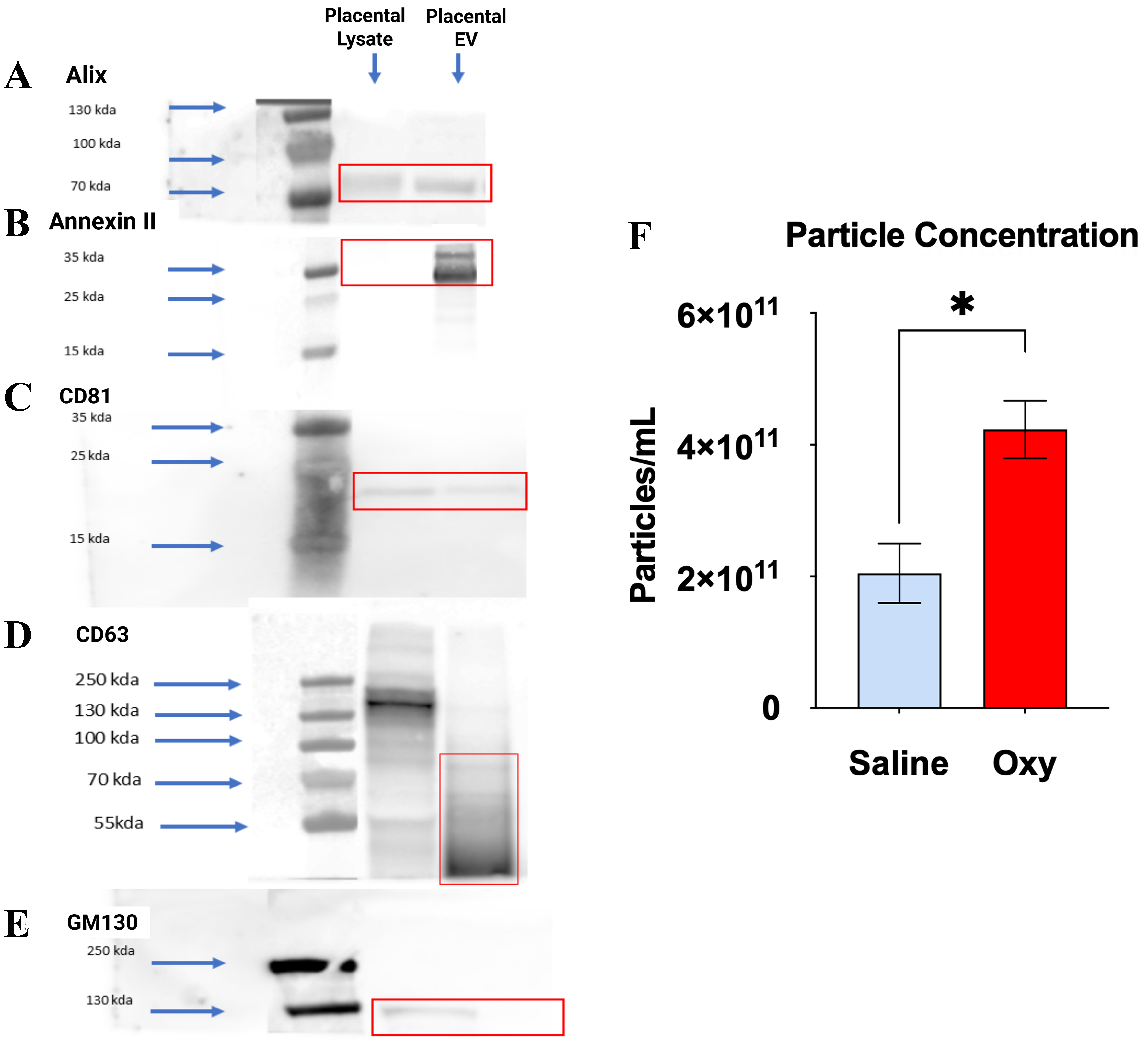
Figure 1. Characterization of placental sEVs via Western blot and particle concentration analysis. (A-D) Western blot analysis demonstrates the enrichment of canonical EV markers in placental EV fractions compared to placental lysate, confirming successful EV isolation. EV samples from both saline- and oxycodone-treated groups were probed for (A) Alix (~90-100 kDa), (B) Annexin II (~36 kDa), (C) CD81 (~25 kDa), and (D) CD63 (~30-60 kDa), all of which showed prominent bands in the EV lanes (highlighted in red boxes); (E) GM130 (~130-250 kDa), a Golgi matrix protein used as a negative control, was detected only in placental lysates and not in EV fractions, indicating minimal cellular contamination. Blue arrows mark molecular weight standards; (F) NTA shows a statistically significant increase in particle concentration in EVs from the oxycodone group compared to saline controls, indicating enhanced EV production. Values are expressed as particles/mL ± SEM. *P < 0.05. Oxycodone exposure alters the morphology and size distribution of PSEVs. An unpaired t-test followed by Welch’s test correction with P < 0.05 was performed to identify significant differences between groups (saline vs. Oxycodone). n = 3 biological replicates per group: Saline: n = 3, OXY: n = 3, [Supplementary Table 1]. sEVs: Small extracellular vesicles; EV: extracellular vesicle; NTA: nanoparticle tracking analysis; SEM: standard error of the mean; PSEVs: placenta-derived small extracellular vesicles; OXY: Oxycodone.


