


fig2
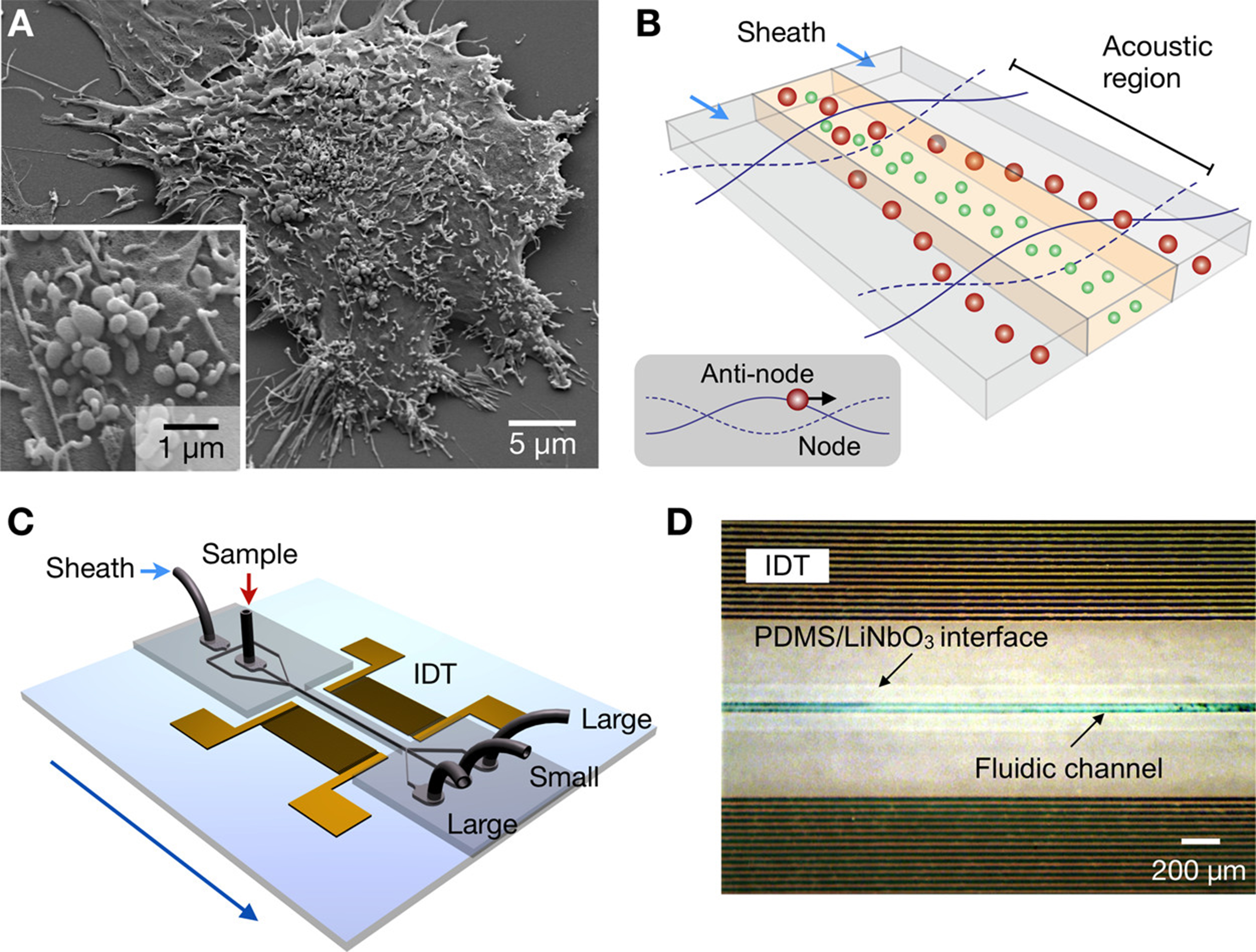
Figure 2. Acoustic nanofilter for MVs label separation. (A) Scanning electron microscopy image of MVs produced by human brain tumor cells (GBM20/3); (B) Schematic of filter operation. MVs in the acoustic region are moved to the nodes of the acoustic pressure region while under acoustic radiation (inset) pressure. As the acoustic force is related to MV volume, larger MVs travel faster and get eliminated by sheath flows positioned in the node region, whereas small MVs are retained by the center flow; (C) Schematic of the device. An acoustic wave is generated across the flow direction employing a pair of IDT electrodes. Small MVs are gathered at the center outlet, whereas large MVs are gathered at the two side outlets; (D) Micrographs showing a working prototype. A piezoelectric (LiNO3) substrate served as the pattern for IDT electrodes. The substrate and fluidic channel were irreversibly fused together. Reproduced with permission from[165]. MVs: Microvesicles; IDT: interdigitated transducer; PDMS: polydimethylsiloxane.


