


fig4
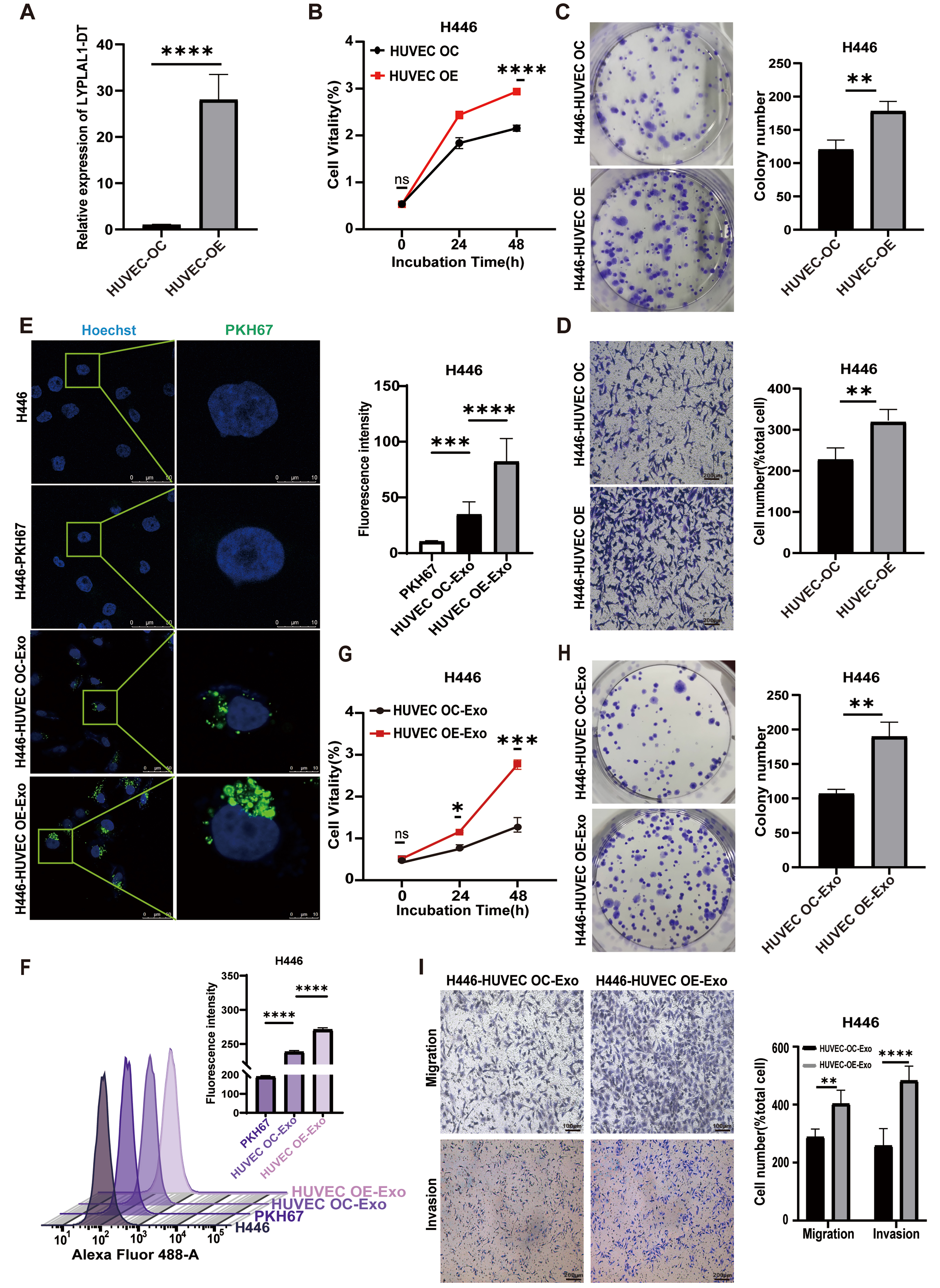
Figure 4. Enhanced proliferation, migration, and invasion of H446 cells mediated by HUVEC-derived exosomal LYPLAL1-DT. (A) The expression levels of LYPLAL1-DT were assessed in HUVECs with LYPLAL1-DT OE and OC; (B and C) When H446 cells were co-cultured with LYPLAL1-DT-OE-HUVECs, their proliferation capacity was significantly greater than that of cells co-cultured with control HUVECs; (D) The migration ability of H446 cells in the OE co-culture group was significantly greater than that in the OC group (scale bar: 200 μm); (E and F) PKH67-labeled exosomes derived from HUVECs were internalized by H446 cells (scale bars: 50 and 10 μm); (G and H) H446 cells treated with OE-Exo exhibited significantly enhanced proliferation compared to those treated with OC-Exo; (I) OE-Exo treatment significantly enhanced the migration and invasion of H446 cells compared to OC-Exo treatment (scale bar: 100 μm). Data are presented as mean ± SD. Differences between the two groups were analyzed by Student’s t-test, while comparisons among more than two groups were analyzed by one-way ANOVA. (*P < 0.05; **P < 0.01; ***P < 0.001; ****P < 0.0001). HUVEC: Human umbilical vein endothelial cells; LYPLAL1-DT: LYPLAL1 divergent transcript; OE-Exo: exosomes from overexpressing cells; OC-Exo: exosomes from control cells; SD: standard deviation; ANOVA: analysis of variance; PKH67: PKH67 Green Fluorescent Cell Linker.


