


fig2
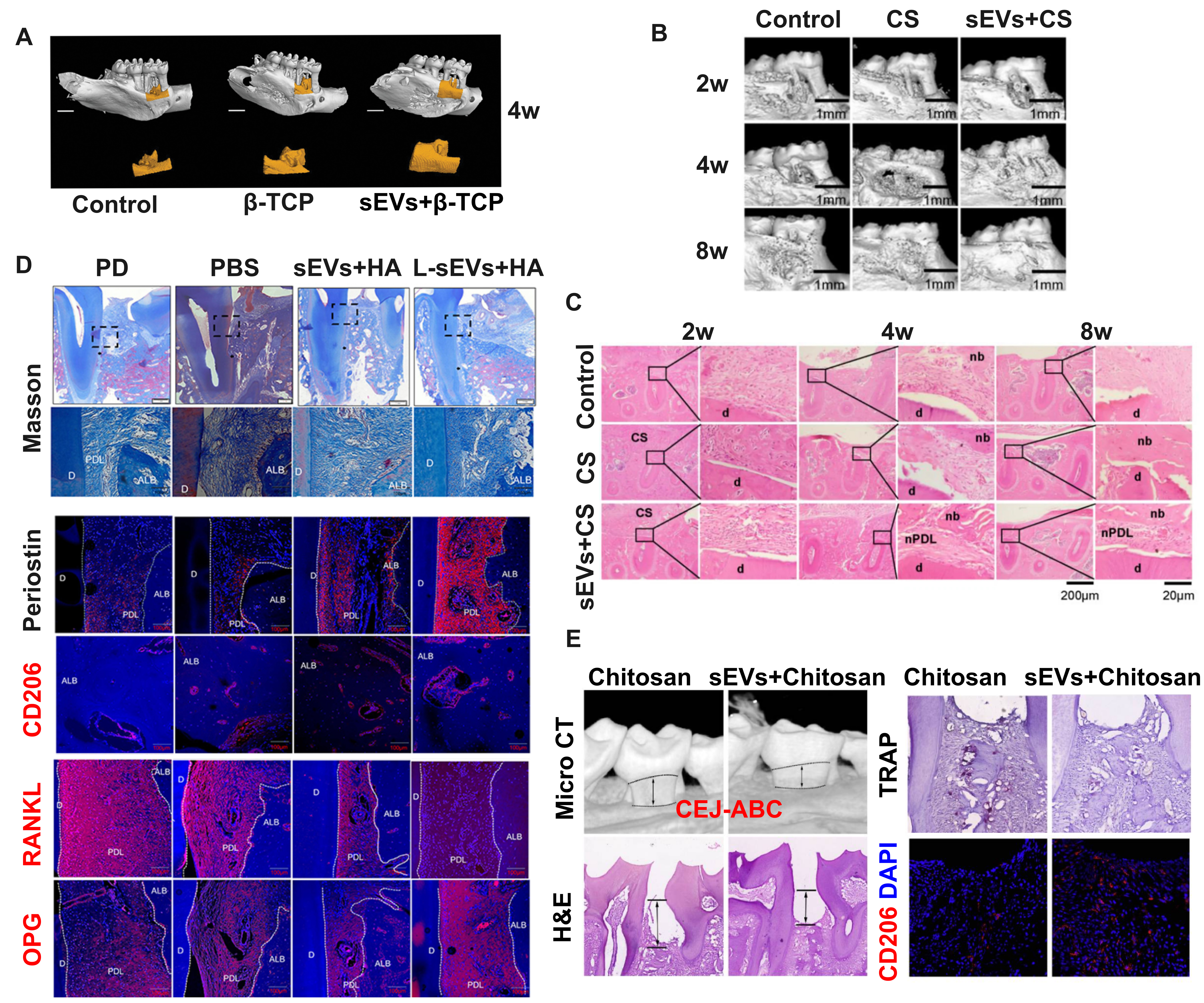
Figure 2. MSCS-sEV-laden biomaterials enhance periodontal regeneration in preclinical models. (A) Micro-CT reconstruction images demonstrating new bone formation in rat periodontal defects 4 weeks after treatment with PDLSC-sEVs loaded β-TCP scaffolds, compared to control treatments. Adapted from Ref.[19]; (B) Longitudinal micro-CT analysis showing progressive new bone formation at 2, 4, and 8 weeks post-treatment in control, CS, and DFSC-sEVs + CS groups. Scale bars = 1 mm. Adapted from Ref.[20]; (C) Histomorphological analysis using H&E staining reveals nb and nPDL formation in DFSC-sEVs + CS-treated defects. Note the organized tissue architecture compared to controls. Scale bars = 20 and 200 μm. Adapted from Ref.[20]; (D) Comparative histological analysis using Masson’s trichrome staining and immunofluorescence for Periostin, CD206, RANKL, and OPG in periodontal defects. Treatment with DFSC and L-DFSC-sEVs + HA increased periodontal ligament width, vessel density, and expression of regenerative markers (Periostin, CD206, a typical marker of anti-inflammatory M2 macrophage) while decreasing the RANKL/OPG ratio compared to controls. Scale bars = 100 μm. Adapted from Ref.[67]; (E) Therapeutic efficacy of DPSC-sEVs-loaded chitosan hydrogels in experimental periodontitis. Micro-CT reconstruction, H&E staining, TRAP staining, and CD206 immunofluorescence demonstrate that DPSC-sEVs + chitosan treatment reduces the CEJ-ABC distance and osteoclast numbers while promoting anti-inflammatory macrophage polarization (increased CD206 expression) compared to chitosan-only controls. Scale bars = 200 μm (micro-CT) and 100 μm (histology). Adapted from Ref.[63]. MSCs: Mesenchymal stem cells; sEVs: small extracellular vesicles; Micro-CT: micro-computed tomography; PDLSC-sEVs: periodontal ligament stem cell-derived small extracellular vesicles; β-TCP: beta-tricalcium phosphate; CS: collagen sponge; DFSC-sEVs: dental follicle stem cell-derived small extracellular vesicles; H&E: hematoxylin and eosin; nb: new bone; nPDL: new periodontal ligament; d: dentin; DFSC-sEVs + HA: DFSC-sEVs loaded in hyaluronic acid gel; L-DFSC-sEVs + HA: lipopolysaccharide-preconditioned DFSC-sEVs loaded in hyaluronic acid gel; ALB: alveolar bone; DPSC-sEVs: dental pulp stem cell-derived small extracellular vesicles; CEJ-ABC: distance between cementoenamel junction and alveolar bone crest; TRAP: tartrate-resistant acid phosphatase; OPG: osteoprotegerin; RANKL: receptor activator of nuclear factor kappa-B ligand; M2: M2 macrophage phenotype.


