


fig7
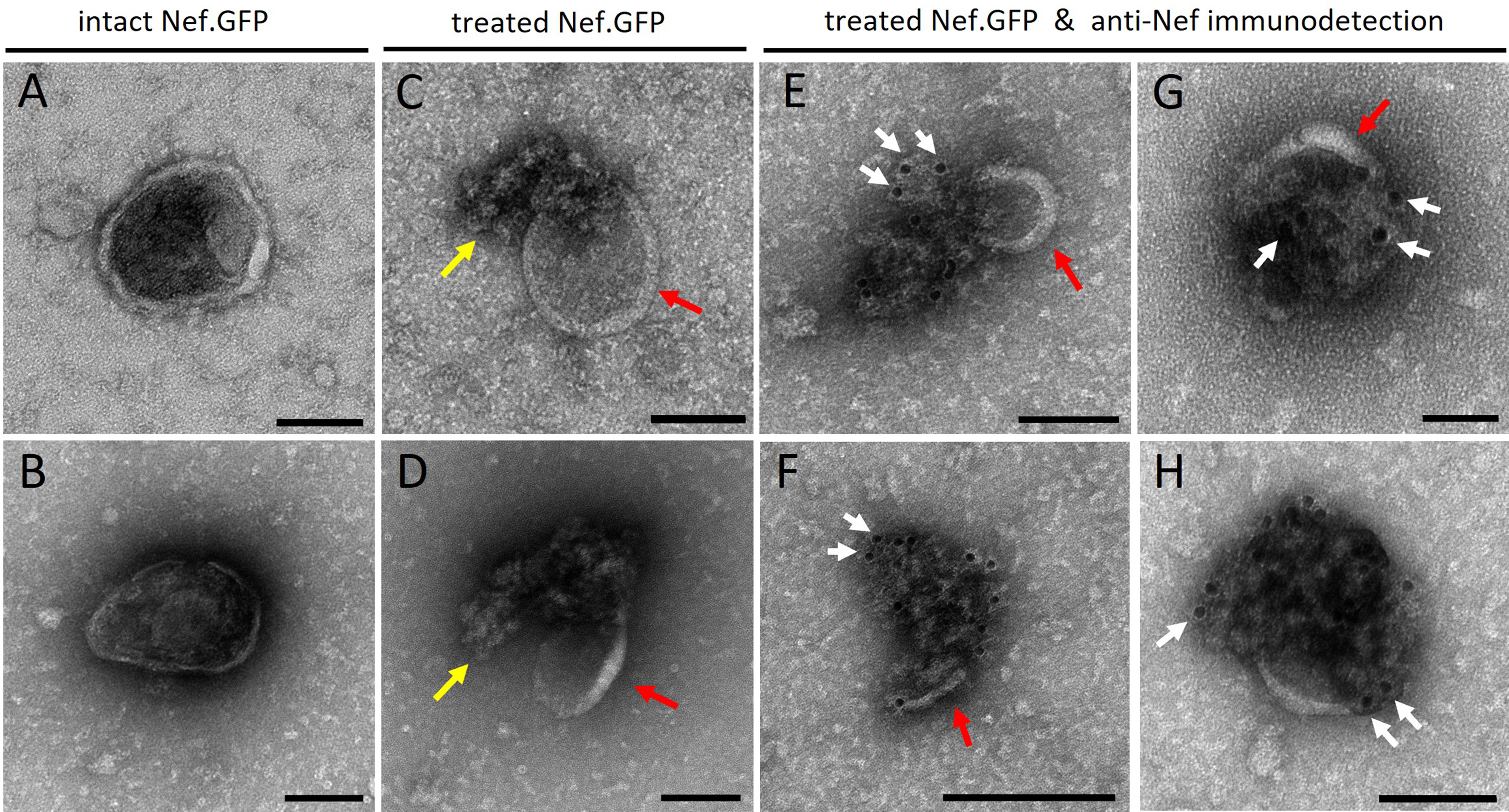
Figure 7. h-microglia expressing Nef.GFP release Nef sequestered inside the lumen of EVs. Transmission electron microscopy images of Nef.GFP EVs, isolated from pooled fractions 5-9 after iodixanol gradient separation of crude EVs and negatively stained with uranyl acetate. Nef.GFP EVs were either left intact or perforated with 0.05% Triton X-100 detergent, and then labeled with antibodies against Nef and IgGs conjugated to gold particles. (A and B) negative staining of intact EVs; (C and D) Negative staining of EVs treated with detergent; (E-H) Anti-Nef immunogold labelling of Nef.GFP EVs after detergent treatment. Arrows indicate membrane remnants (red), released cargo and/or damaged membrane (yellow), gold particles identifying Nef protein (white; at least three instances per micrograph). Scale bar: 100 nm (black line). To highlight individual EVs in the field of view and their potential colocalization with gold particles, images were cropped in Velox. During cropping, the software adjusts and redraws the scale bar according to the new field size, which was not uniform across the cropped images and therefore resulted in inconsistent scale bars. Nef.GFP: Nef green fluorescent protein; EV: extracellular vesicle; Triton X-100: Triton X-100 detergent; IgG: immunoglobulin G.


