


fig6
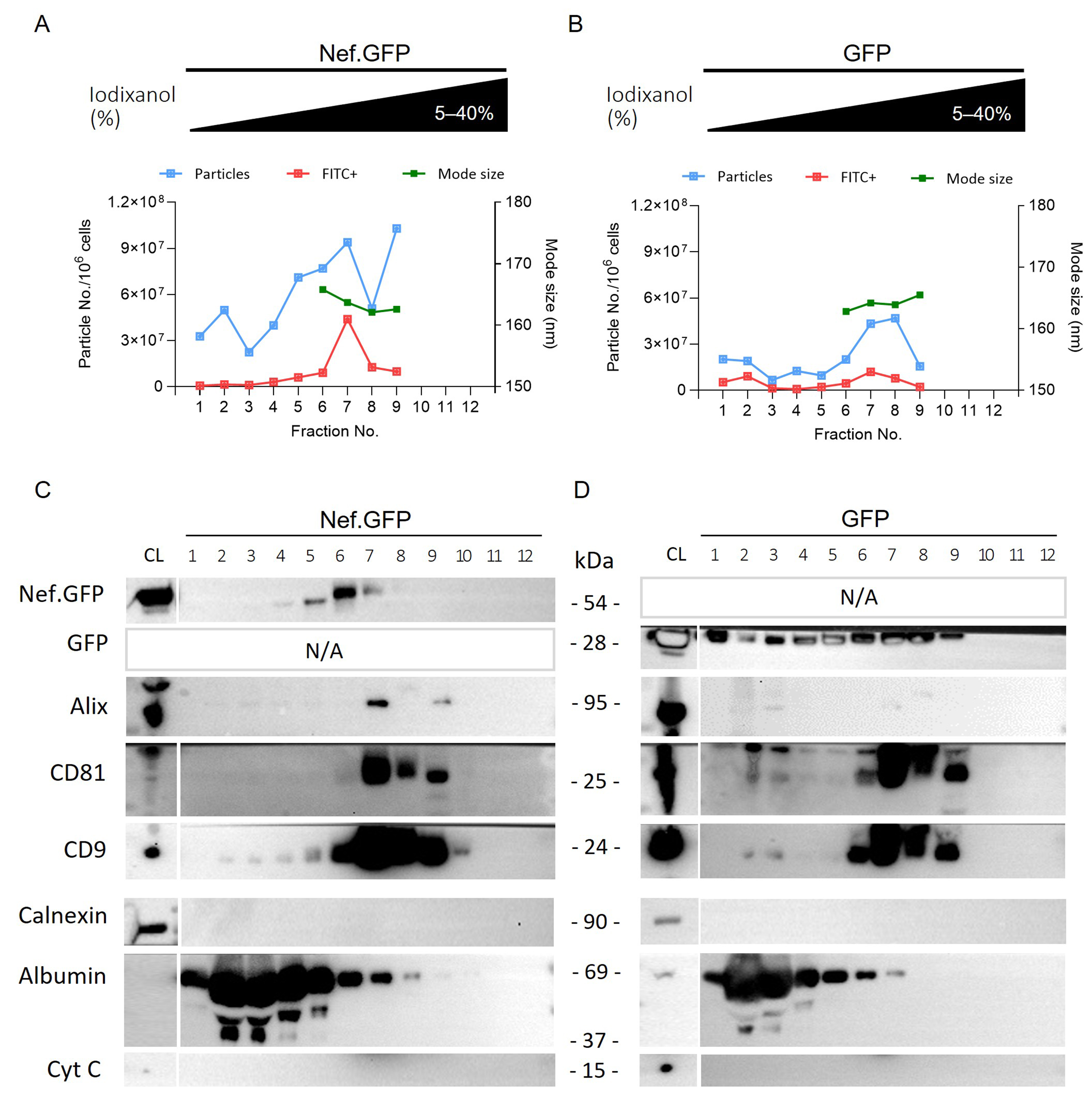
Figure 6. Inducible Nef.GFP expression in hmicroglia promotes the release of small Nef.GFP-positive EVs. (A and B) Crude EV samples enriched from the culture media of Nef.GFP and GFP expressing h-microglia, after removal of the 10,000 × g pellet, were further separated on 5%-40% iodixanol density gradient. The twelve collected fractions were analyzed by nano-flow cytometry (particles (blue) and Nef.GFP+ or GFP+ EVs (FITC+, red) per million cells), and nanoparticle tracking analysis [average mode size (nm; green) for fractions with at least 10 particles per frame]; (C and D) The same 12 fractions from Nef.GFP and GFP EV samples were also analyzed by immunoblotting with antibodies against GFP, typical EV proteins (Alix, CD81, and CD9), and impurity markers (Calnexin, Albumin and Cytochrome c). CL: Cell lysate; Nef.GFP: Nef green fluorescent protein; h-microglia: human microglia; GFP: green fluorescent protein; EV: extracellular vesicle; FITC: fluorescein isothiocyanate; Alix: ALG-2-interacting protein X; CD81: cluster of differentiation 81; CD9: cluster of differentiation 9; Calnexin: calnexin protein; Albumin: serum albumin; Cytochrome c: cytochrome c protein.


