

Gut microbiota-derived extracellular vesicles in sepsis-associated liver injury: emerging mediators of gut-liver axis dysfunction
 0
Graphical Abstract
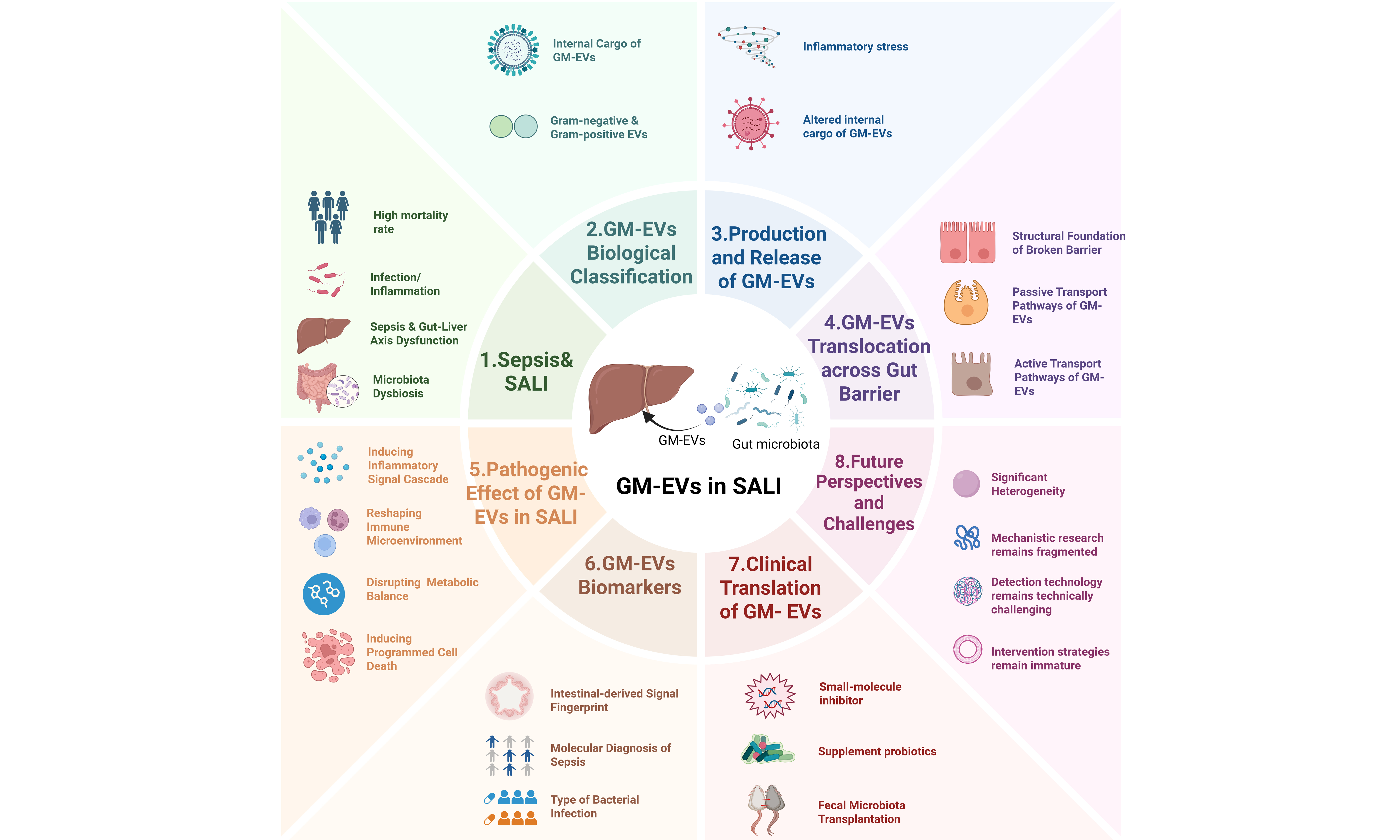
Abstract
Sepsis is a life-threatening syndrome of multiple organ dysfunction driven by a dysregulated host response to infection. Its high mortality is closely associated with disruption of gut-liver axis homeostasis. Gut microbiota-derived extracellular vesicles (GM-EVs) have emerged as important mediators of cross-kingdom communication and molecular exchange between intestinal microbes and the host. Accumulating evidence highlights their regulatory role in the progression of sepsis-associated gut-liver axis dysfunction. This review systematically summarizes the biogenesis of GM-EVs and the regulation of their release under septic conditions, as well as the mechanisms underlying their transport across the impaired intestinal epithelial barrier. We further elaborate on the pathological effects of GM-EVs in the liver, including amplification of hepatic inflammatory cascades, disruption of intrahepatic immune homeostasis, reprogramming of hepatic metabolism, and induction of hepatocellular injury and programmed cell death. Additionally, we discuss the translational potential of strategies targeting GM-EVs for the prevention and treatment of sepsis. Finally, we examine current research limitations and future directions, with a focus on the potential of GM-EVs as biomarkers and therapeutic targets for the early detection and precision management of sepsis-associated liver injury.
Keywords


 Download PDF
INTRODUCTION
Sepsis is a life-threatening syndrome of organ dysfunction caused by a dysregulated host response to infection. It represents a major public health issue in critical care medicine worldwide. Epidemiological data indicate more than 48.9 million new cases of sepsis worldwide each year, with approximately 11 million related deaths; the sepsis-associated mortality rate remains high[1]. The pathological process of sepsis involves infection-induced excessive activation of innate immunity, amplification of systemic inflammation, endothelial barrier disruption, dysregulation of the coagulation-fibrinolysis system, and an immunometabolic imbalance, ultimately leading to multiple organ dysfunction[2]. Among the forms of multiple organ dysfunction caused by sepsis, sepsis-associated liver injury (SALI) has received increasing attention. Approximately 34%-46% of patients with sepsis may experience varying degrees of liver dysfunction[3,4]. Clinically, this disease is characterized by SALI, which may present as hepatocellular injury, cholestasis, or a mixed pattern. The severity of the condition varies greatly, ranging from a slight increase in transaminase levels to severe liver dysfunction[3,5]. The development of SALI is generally associated with greater disease severity and a poor prognosis[6]. However, despite the well-established clinical significance of SALI, there are currently no specific therapeutic strategies targeting SALI, and its pathophysiological mechanisms remain to be further elucidated.
In recent years, the central role of the gut-liver axis in the pathogenesis of SALI has been increasingly recognized. The liver is a critical immunometabolic organ that links intestinal and systemic homeostasis. It clears gut-derived microbial products from the portal and systemic circulation and participates in the acute-phase response, bile acid metabolism, regulation of coagulation, detoxification, and immune surveillance[7,8]. The intestine and liver are anatomically and functionally interconnected through the portal vein, biliary system, and systemic circulation. The gut is increasingly recognized as a central driver of multiple organ dysfunction in sepsis rather than merely a passive victim of systemic inflammation[5,9]. The intestine is the largest immune organ and bacterial reservoir in the human body, and its barrier function is severely compromised during sepsis. Intestinal hypoperfusion, epithelial damage, disruption of tight junctions, impaired mucosal integrity, and gut dysbiosis may increase intestinal permeability and promote the translocation of microbial products [including lipopolysaccharide (LPS), peptidoglycans, bacterial DNA, and other pathogen-associated molecular patterns (PAMPs)][10]. These gut-derived microbial components and signaling molecules can then enter the portal circulation and reach the liver[11]. The resulting abnormal activation of Kupffer cells, dysfunction of hepatic sinusoidal endothelial cells, release of inflammatory mediators, microcirculatory damage, and metabolic reprogramming contribute to hepatocellular injury and liver dysfunction[12].
The gut-liver axis is a bidirectional regulatory network that links intestinal and hepatic functions. During sepsis, intestinal barrier disruption, gut dysbiosis, and the translocation of gut-derived PAMPs can increase the inflammatory and metabolic burden imposed on the liver, thereby aggravating hepatic dysfunction[13]. Conversely, impaired hepatic immune and metabolic functions may reduce bacterial clearance and disrupt bile acid synthesis, secretion, and enterohepatic circulation. These changes can alter the intestinal microbial ecosystem and weaken intestinal barrier integrity, thereby increasing microbial translocation and amplifying systemic inflammation. This bidirectional gut-liver interaction may establish a vicious cycle that contributes to sepsis-associated multiorgan dysfunction[5,14]. Therefore, disrupting pathological interactions along the gut-liver axis may provide new therapeutic strategies and targets for sepsis.
In recent years, gut microbiota-derived extracellular vesicles (GM-EVs) have attracted increasing attention as key mediators of microbe-host cross-talk. These include outer membrane vesicles (OMVs) derived from Gram-negative bacteria and cytoplasmic (cell) membrane vesicles (CMVs) derived from Gram-positive bacteria[15]. GM-EVs have nanoscale dimensions, a lipid bilayer structure, and a relatively robust capacity to protect their cargo. These vesicles can transport LPS, peptidoglycans, bacterial DNA and RNA, virulence-associated proteins, and metabolism-related molecules across the intestinal barrier into the circulatory system, enabling long-distance microbial-host communication[16,17]. Compared with free LPS or soluble metabolites, GM-EVs exhibit higher stability and targeted delivery capacity, enabling them to package microbial signals and precisely transport these signals to distant organs including the liver, thereby playing unique regulatory functions in gut-liver axis crosstalk. Under normal physiological conditions, GM-EVs secreted by the symbiotic microbiota contribute to maintaining intestinal microbial homeostasis, mucosal barrier integrity, and host immune tolerance. However, in sepsis, changes in microbiota composition, remodeling of the inflammatory microenvironment, and intestinal barrier disruption may jointly affect the production, release, and biological effects of GM-EVs. Increased intestinal barrier permeability may facilitate the translocation of GM-EVs into the submucosa and their subsequent entry into the circulation via the mesenteric lymph system or portal circulation, thereby conveying gut microbial signals to the liver[18]. Upon reaching the liver, GM-EVs are preferentially internalized by hepatic Kupffer cells. These vesicles can activate pattern recognition receptors, including Toll-like receptors (TLRs) and NOD-like receptors (NLRs), thereby initiating downstream signaling pathways involving nuclear factor kappa B (NF-κB) and mitogen-activated protein kinases (MAPK), inflammasome activation, and cellular oxidative stress and modulating intrahepatic inflammatory responses[19]. The resulting biological effects may include the release of pro-inflammatory cytokines, recruitment of immune cells, endothelial dysfunction, mitochondrial damage, dysregulation of bile acid and lipid metabolism, and alterations in hepatocellular death pathways[17]. Importantly, these effects are neither inherent nor universal properties of GM-EVs but are context-dependent and influenced by bacterial origin, vesicle composition and concentration, and the host environment.
Although the role of the gut-liver axis in SALI has been preliminarily recognized, and GM-EVs, as critical mediators of microbe-host crosstalk, have attracted increasing attention in the fields of metabolic diseases and chronic liver diseases, there remains a lack of a systematic synthesis and explanation of the mechanisms by which GM-EVs specifically mediate gut-liver axis communication dysfunction under septic conditions and thereby initiate or exacerbate hepatic injury. Most existing studies have focused on free microbial molecules or single cytokine networks, whereas an integrated understanding of the roles, cargo composition, uptake mechanisms, and downstream signaling pathways of GM-EVs as emerging mediators in SALI is still absent. In view of this, the present review aims to systematically summarize the potential effects of GM-EVs in SALI, with an emphasis on their mechanisms and clinical significance as emerging mediators of gut-liver axis dysfunction. This review is expected to provide a novel theoretical framework and insights for the early identification and targeted intervention of SALI.
GUT-LIVER AXIS DYSFUNCTION AND CLINICAL BURDEN IN SEPSIS
During sepsis, excessive systemic inflammation can cause intestinal microcirculation dysfunction, resulting in ischemia and hypoxia of the intestinal mucosa and pathological changes such as oxidative stress[20,21]. Dysregulated expression of tight junction proteins in intestinal epithelial cells disrupts intestinal barrier integrity, thereby increasing intestinal permeability[22]. This process facilitates the translocation of luminal bacteria and their derivatives, including PAMPs such as LPS, to the liver via the portal vein. As both a metabolic hub and immune sentinel, the liver is a major target organ in this pathological process[7,23]. Persistent inflammatory stimulation can lead to excessive activation of hepatic Kupffer cells and the release of pro-inflammatory mediators such as tumor necrosis factor-α (TNF-α) and interleukin-6 (IL-6). These mediators can exacerbate local hepatic injury and amplify systemic inflammation, thereby establishing a positive feedback loop that contributes to the progression of multiple organ dysfunction[24]. Therefore, preserving gut-liver axis homeostasis may help disrupt the pathological cycle of sepsis, whereas gut-liver axis dysfunction may contribute to the progression from local infections to systemic organ dysfunction.
The gut-liver axis may function as an important driver of multi-organ dysfunction during sepsis. The liver is the first organ exposed to gut-derived bacteria, endotoxins, and other PAMPs and damage-associated molecular patterns (DAMPs)[5,14]. During sepsis, intestinal barrier disruption and gut microbiota dysbiosis enable the translocation of these mediators into the portal vein and mesenteric lymph. Sustained systemic inflammatory insult combined with bile acid metabolic disorders induces extensive hepatocellular necrosis and cholestasis, thereby severely impairing the synthetic function of the liver[6,25].
Liver dysfunction impairs bacterial clearance, bile acid secretion, and acute-phase protein synthesis, further damaging the intestinal barrier and creating a damaging cycle between the gut and the liver[5,26]. Clinically, gastrointestinal injury is common among critically ill patients. A meta-analysis reported that acute gastrointestinal injury affected approximately 40% of critically ill patients, and the mortality rate among critically ill patients with acute gastrointestinal injury was 33%. Compared with patients without acute gastrointestinal injury, those with acute gastrointestinal injury have a higher risk of death[27]. A previous study detected SALI in 34.7% of adult patients with sepsis (cholestatic, 48.1%; hepatocellular, 21.8%; and shock liver, 30.1%)[28]. Hyperbilirubinemia is already present on intensive care unit (ICU) admission in 16.8% of patients with sepsis and independently predicts acute respiratory distress syndrome and mortality[29]. Moreover, in patients with sepsis complicated by liver dysfunction or failure, the mortality rate reaches 54%-68%, exceeding that of most other organ failures[6,8]. Importantly, the involvement of the gut-liver axis is not restricted to abdominal infections, which account for a sizeable fraction of sepsis episodes in the ICU[30]. Secondary gut injury also occurs in non-abdominal sepsis; for example, experimental Staphylococcus aureus pneumonia induces intestinal epithelial apoptosis and increased gut permeability, and gut-derived mediators carried by the mesenteric lymph have been shown to cause distant lung injury[26,31]. Collectively, these findings support the concept that the gut acts as the “motor” of multiple organ dysfunction syndrome and that, regardless of the primary infectious focus, the injured gut can initiate and propagate systemic inflammation and remote organ failure, positioning the gut–liver axis as a central node and potential therapeutic target in sepsis.[9,31].
BIOLOGICAL CHARACTERISTICS AND CLASSIFICATION OF GM-EVs
GM-EVs are key mediators of information exchange between the gut microbiota and the host. It has been confirmed that the majority of intestinal bacteria (including Gram-negative and some Gram-positive bacteria) can secrete such particles[32]. GM-EVs possess a typical lipid bilayer-encapsulated spherical structure that can effectively resist enzymatic degradation and acid-base damage in the gastrointestinal environment and protect various bioactive components such as proteins, nucleic acids, and lipids within the vesicles. This property provides a structural basis for the long-distance translocation of GM-EVs in the body, allowing them to cross tissue barriers and mediate intercellular communication across organs[33-35]. The biological effects of GM-EVs are neither intrinsic nor uniform; rather, they depend on the bacterial source, vesicle biogenesis, cargo composition, exposure dose, recipient cell, and host environment[36,37]. Bacterial species and cell envelope architecture are important determinants of vesicle biology. Gram-negative bacteria mainly release OMVs, whereas Gram-positive bacteria release membrane vesicles derived from the cytoplasmic membrane, often referred to as CMVs. Accordingly, OMVs and CMVs differ in their surface molecules and luminal cargo: OMVs commonly display LPS and may carry outer-membrane proteins, periplasmic factors, toxins, and regulatory RNAs, whereas CMVs may contain lipoteichoic acid (LTA), peptidoglycan-associated components, proteins, DNA, RNA, and metabolites[15,34,37,38]. These compositional differences can result in distinct and sometimes opposing effects on the host cells. For example, OMVs from several Gram-negative enteric pathogens can activate epithelial or immune cells through LPS- and TLR4-dependent pathways[39-42], whereas vesicles from commensal or probiotic bacteria have been reported to promote regulatory immune responses, macrophage M2-like polarization, and intestinal barrier protection in some experimental systems[43]. However, this distinction should not be considered as absolute, because the immunological activity of vesicles also depends on the specific bacterial strain, growth conditions, vesicle purification procedures, cargo abundance, recipient cell type, and host inflammatory state. Vesicle concentration is another important determinant of the observed response. Dose-dependent activation has been demonstrated for Streptococcus pneumoniae vesicles, whereas higher concentrations of bacterial vesicles may increase cellular stress or cytotoxicity in some experimental systems[44].
The internal cargo of GM-EVs is diverse and includes nucleic acids, proteins, and bacterial metabolites, which are key molecular carriers that regulate host physiological and pathological processes[45]. Nucleic acid cargo encapsulated within vesicles includes bacterial DNA and diverse RNA species. Notably, variably packaged small RNAs (sRNAs) can target host mRNAs and enhance molecular stability[46]. GM-EVs also carry a variety of protein cargoes, including enzymes, virulence factors, and signaling molecules related to bacterial metabolism[35]. For instance, Amuc_1100, a specific protein isolated from the outer membrane of Akkermansia muciniphila, retains its structural stability at pasteurization temperatures and exerts protective effects on intestinal barrier function through interaction with Toll-like receptor 2 (TLR2)[47]. Furthermore, the metabolic products within the vesicles reflect the metabolic status of the bacteria, directly acting on host cells to exert regulatory effects. These diverse substances are encapsulated within the phospholipid bilayer of the vesicles and thus can reach the target organs intact and release their contents[48-50].
The compositional characteristics of GM-EVs directly determine their immune activation abilities. During the pathological process of sepsis, GM-EVs can trigger excessive inflammatory responses in the liver, thereby leading to liver damage and organ dysfunction[19,51]. Gram-negative bacterium-derived GM-EVs primarily encapsulate LPS, whereas LTA is the major PAMP cargo released by Gram-positive bacteria. Both molecules are sensed by host pattern recognition receptors to initiate immune activation. Vesicle surface LPS stimulates canonical innate immune signaling upon specific binding to cellular Toll-like receptor 4 (TLR4), which then activates the NF-κB cascade, induces massive pro-inflammatory cytokine secretion and provokes inflammatory responses[52]. By comparison, LTA mediates immune responses predominantly through the TLR2 pathway[53]. In addition to vesicle surface antigens, intraluminal nucleic acid cargo of GM-EVs possesses strong immune-activating activity. Internalized GM-EVs undergo lysosomal degradation inside the host cells, releasing bacterial DNA for sensing by cyclic GMP-AMP synthase (cGAS). Activated cGAS further engages the stimulator of interferon genes (STING) signaling cascade, induces the production of type I interferons and inflammatory mediators and exacerbates systemic inflammatory bursts[54].
REGULATORY MECHANISMS GOVERNING GM-EV PRODUCTION AND RELEASE DURING SEPSIS
Sepsis-associated remodeling and redistribution of GM-EVs
In the early stages of sepsis, after the body is invaded by pathogenic bacteria, PAMPs and a large number of endogenous inflammatory mediators released by host immune cells jointly create a highly inflammatory systemic environment[55]. During this process, factors including intestinal hypoperfusion, ischemia and hypoxia, oxidative stress, and bile acid metabolic dysfunction contribute to the disruption of the intestinal microecological balance, significantly altering the composition and metabolic state of the gut microbiota and influencing the generation and release of GM-EVs[21,22,56]. Mechanistically, a systemic infection may create an intestinal microenvironment that is permissive to changes in bacterial vesicle release. In Gram-negative bacteria, inflammatory stress, outer membrane perturbation, antimicrobial peptide exposure, and oxidative stress can influence outer membrane remodeling and OMV formation[33,57]. Gram-positive bacteria may also release CMVs through cell wall remodeling, changes in membrane architecture, and stress response pathways[32,34]. In the context of sepsis, these bacterial responses may alter the abundance or molecular composition of selected GM-EV populations.
Nevertheless, the direction and magnitude of these changes likely depend on bacterial species, disease stage, local nutrient and inflammatory conditions, antibiotic exposure, and host-derived factors. These mechanisms provide biological plausibility for sepsis-associated GM-EV remodeling, but do not establish a uniform increase in GM-EV production. Evidence from experimental sepsis models supports the notion that sepsis remodels the intestinal GM-EV pool. In a murine cecal ligation and puncture model, extracellular vesicles (EVs) from intestinal lavage of septic mice increased during sepsis[58]. Similarly, studies in patients with sepsis and experimental sepsis models have reported changes in circulating EV abundance and associated inflammatory cargo. Circulating EV preparations generally contain vesicles from multiple sources, including platelets, endothelial cells, leukocytes, epithelial cells, and bacteria. Therefore, an increase in total circulating EVs cannot be interpreted as evidence of increased GM-EV production without bacteria-specific identification and quantitative normalization[59].
Gut microbiota imbalance and alterations in GM-EV profiles
Gut dysbiosis may alter the abundance, taxonomic profile, cargo composition, and biological activities of GM-EVs. However, this relationship is unlikely to be uniform or deterministic because vesicle production and cargo loading are influenced not only by the relative abundance of bacterial taxa, but also by bacterial growth conditions, inflammatory stress, nutrient availability, antibiotic exposure, and host-derived environmental signals. Therefore, changes in the GM-EV pool may not mirror changes in the entire bacterial community.
Experimental and translational studies support this concept. In a mouse model of diet-induced obesity, gut dysbiosis was associated with a reduction in total fecal bacterial EVs and an increased proportion of LPS-positive EVs. Proteobacteria-associated vesicles were enriched among the LPS-positive EVs and detected at higher levels in the hepatic portal vein and liver, together with increased hepatic expression of TLR4, macrophage markers, and inflammatory mediators[18]. These findings suggest that dysbiosis alters not only the quantity of GM-EVs but also the relative representation of vesicle populations with distinct inflammatory potentials. In addition, analyses of fecal microbiota-derived EVs from individuals with diarrhea, morbid obesity, or Crohn’s disease identified disease-associated differences in the taxonomic composition of EVs and showed that EV preparations from different clinical groups produced distinct effects on intestinal epithelial permeability in vitro[60]. Although these findings are not specific to sepsis, they provide experimental evidence that disease-associated changes in the intestinal ecosystem can be accompanied by the functional remodeling of the GM-EVs compartment.
In sepsis, disorders of the gut microbiota are manifested by a significant reduction in beneficial bacteria and excessive proliferation of potentially pathogenic bacteria[61]. For instance, the abundance of probiotics such as Bifidobacteria and Lactobacilli, which have anti-inflammatory and barrier-protective effects, decreases while the relative abundance of Gram-negative bacteria such as Enterobacteriaceae increases substantially[62,63]. Furthermore, the widespread use of antibiotics, a common treatment method for sepsis, can exacerbate intestinal flora imbalance, further disrupt microbial diversity, and weaken host immune defense capabilities[64,65]. Changes in the composition and ecological state of the intestinal microbiota may be accompanied by alterations in the abundance, taxonomic profile, cargo composition, and biological activity of GM-EVs produced by different bacterial taxa. However, the direction and magnitude of these changes are influenced by bacterial physiology, environmental conditions, and host factors and do not necessarily mirror changes in the whole microbiota[18,60,66,67].
In sepsis, dysbiosis, intestinal hypoperfusion, inflammatory stress, altered nutrient availability, and antibiotic exposure alter the production and composition of GM-EVs. Increased intestinal permeability may facilitate the translocation of selected vesicle populations to the portal circulation and the liver. Nevertheless, whether these changes promote hepatic injury depends on the bacterial sources of the vesicles, their molecular cargo, vesicle abundance and concentration, route and duration of exposure, hepatic clearance capacity, and the inflammatory and metabolic states of the host. Furthermore, direct experimental evidence linking specific sepsis-associated bacterial taxa to defined GM-EV populations and subsequent liver injury remains limited. Therefore, GM-EV remodeling should be considered a plausible mechanism linking dysbiosis with gut-liver signaling, rather than a uniform or inevitable consequence of microbiota disruption.
PATHWAYS AND MECHANISMS BY WHICH GM-EVs CROSS THE DAMAGED INTESTINAL BARRIER IN SEPSIS
Passive transport pathway of GM-EVs across the intestinal barrier
The monolayer of intestinal columnar epithelial cells, together with the apical junctional complex, constitutes the principal physical barrier of the intestine, serving as the first line of defense that restricts the translocation of luminal microorganisms, toxins, and their derivatives into host tissues[68]. Under physiological conditions, tight junctions are assembled by occludin, claudin family proteins, junctional adhesion molecule-A (JAM-A), and intracellular scaffolding proteins, including zonula occludens-1 (ZO-1) and zonula occludens-2 (ZO-2). These junction proteins are anchored to the peripheral actin cytoskeleton to form a continuous circumferential sealing structure. These structural features block the paracellular passive diffusion of macromolecules, microbial products, and vesicle-like substances, thereby preserving epithelial cell polarity and the homeostatic state of low intestinal permeability[68-70]. Following the onset of sepsis, the massive release of pro-inflammatory mediators triggers a systemic inflammatory response that invades the intestinal mucosa, resulting in augmented intestinal epithelial apoptosis, disorganized tight junction architecture, and elevated barrier permeability. Sepsis-associated inflammatory cascades, neutrophil extracellular trap formation, endoplasmic reticulum (ER) stress, and mitochondrial injury collectively facilitate intestinal epithelial cell death, which in turn leads to villous epithelial shedding and disruption of mucosal integrity, ultimately leading to structural defects in the intestinal barrier[71-73]. Additionally, inflammatory signals cause changes in the expression and subcellular localization of tight junction proteins. TNF-α triggers the endocytic internalization of occludin, thereby directly destabilizing the structural integrity of tight junctions. Studies on animal models of sepsis have documented divergent alterations in the expression of multiple junctional proteins, including occludin, claudin isoforms, ZO-1, and JAM-A. These molecular aberrations lead to the aberrant opening of paracellular spaces and markedly elevated intestinal permeability, culminating in the pathological hallmark of intestinal leakage[74-76]. Advanced sepsis is accompanied by systemic hypoperfusion and intestinal ischemia/reperfusion (I/R) injury, which exacerbates epithelial apoptosis, tight junction disruption, and mucosal inflammatory responses. Data obtained from human intestinal I/R models have demonstrated that the small intestine shows greater epithelial damage, higher pro-inflammatory cytokine expression, and greater neutrophil infiltration than the colon[77]. Such barrier disruption may increase the vulnerability of the intestinal mucosa to additional injury from pathogen-derived products, including GM-EVs. In this context, experimental evidence from an enteric bacterial model provides a mechanistic example of how GM-EV cargo aggravates epithelial barrier disruption. OMVs secreted by Campylobacter jejuni carry proteolytic activity associated with the serine protease HtrA (together with Cj0511 and Cj1365c), which cleaves the adherens junction protein E-cadherin and the tight junction protein occludin in intestinal epithelial cells, thereby impairing intestinal barrier integrity[78]. In one study, GM-EVs derived from E. coli were fluorescently tagged with Cy7 near-infrared dye. In vivo imaging revealed widespread systemic distribution of Cy7 signals in mice. Although signal intensity gradually decreased over time, prominent retention was observed in the peritoneal cavity and liver 24 h after administration[79]. This evidence demonstrates that GM-EVs compromise intestinal barrier integrity and increase epithelial permeability, which promotes the translocation of bacteria and their metabolites into the systemic circulation and subsequently causes injury to distant organs. Such barrier disruption and increased intestinal permeability provide the anatomical and pathophysiological basis for the translocation of GM-EVs and other microbial derivatives across the mucosa into the submucosa and subsequently into the systemic circulation.
The paracellular pathway is a crucial passive route for the transepithelial transport of GM-EVs upon pathological disruption of the intestinal barrier during sepsis [Figure 1A]. This transport pathway is primarily established by two types of impairments in the intestinal physical barrier: epithelial denudation or mucosal erosion resulting from enterocyte apoptosis and necrosis and elevated paracellular permeability induced by inflammatory modulation of tight junction complexes[68,69]. The first involves mucosal defects caused by apoptosis, necrosis, and shedding of intestinal epithelial cells. Under conditions of sepsis and critical illness, enterocyte apoptosis is augmented, whereas the proliferative and migratory repair capacity of the epithelium is compromised, eventually disrupting intestinal mucosal continuity[80]. Open defects emerge in the originally intact epithelial barrier when erosion, ulceration, or focal exfoliation occurs on the intestinal epithelial surface. Under such circumstances, the transmural transport of substances is no longer predominantly governed by the “on-off” state of tight junctions. Instead, luminal contents can directly infiltrate the deep mucosal layers through these ruptured gaps. Large molecules, microbial components, and intact viable bacteria can traverse the damaged epithelium and infiltrate the lamina propria[68]. GM-EVs are spherical nanostructures enclosed by a lipid bilayer, with diameters ranging from 20 to 300 nm[81]. Accordingly, in regions with severe intestinal mucosal erosion or epithelial exfoliation, GM-EVs tend to undergo non-selective passive leakage across mucosal defects driven by concentration gradients and local interstitial fluid flow rather than being internalized via receptor-specific recognition or energy-dependent transcytosis. The second impairment is attributed to widened intercellular spaces induced by the dissociation and remodeling of tight junction complexes. Physiologically, intact tight junctions are only permeable to small molecules such as water molecules and inorganic ions[82]. Sepsis triggers the internalization, degradation, and abnormal membranous localization of tight junction proteins, which widens intercellular gaps enough to permit the passage of most GM-EVs[69,70].
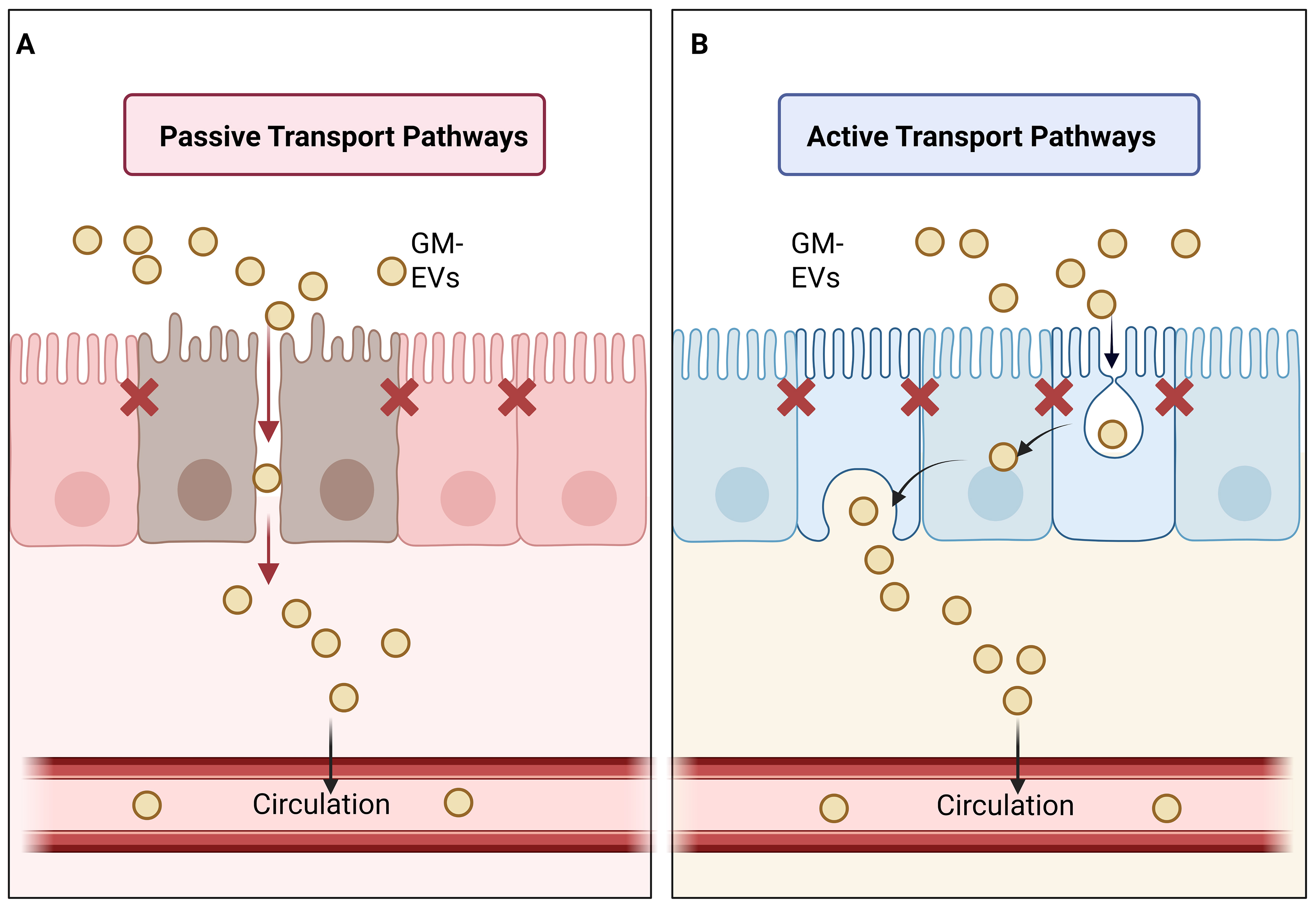
Figure 1. Putative transepithelial translocation pathways of GM-EVs in sepsis. (A) Sepsis-induced barrier dysfunction may disrupt tight junctions and widen intercellular gaps, permitting increased paracellular passage of GM-EVs into the submucosa; (B) When the barrier is only partially impaired, GM-EVs are proposed to cross the epithelium mainly via receptor-mediated endocytosis-dependent transcellular transport. The relative contribution of each route in sepsis requires further validation. Created in BioRender. yuan, Y. (2026) https://BioRender.com/r4i6yno. GM-EVs: Gut microbiota-derived extracellular vesicles.
Active transport pathways of GM-EVs across the intestinal barrier
When the intestinal epithelial barrier remains structurally intact, or tight junction injury is too mild to permit massive paracellular translocation of intact GM-EVs, GM-EVs can achieve transepithelial transport via the transcytotic pathway mediated by endocytosis-exocytosis cascades [Figure 1B]. Distinct from simple passive leakage, this process relies on active internalization by the epithelial plasma membrane and intracellular vesicular trafficking; the process is moderately energy-dependent and is not fully constrained by the extent of tight-junction opening. Intestinal epithelial cells abundantly express diverse pattern recognition receptors such as TLR2, TLR4, TLR7, and NOD1[81,83]. GM-EVs carry microbe-associated molecular patterns, including LPS, peptidoglycan fragments, bacterial RNA, DNA, and virulence-associated proteins, either on their membrane surface or within the vesicular lumen, which can be recognized by epithelial cells and subsequently trigger endocytic uptake. Accumulating evidence indicates that bacterial extracellular vesicles (BEVs) enter epithelial cells via clathrin-dependent endocytosis, dynamin-dependent endocytosis, lipid raft/caveolae-associated internalization, and other non-canonical endocytic pathways. These internalized vesicles are transported intracellularly within early endosomes, late endosomes, or lysosome-associated compartments[84-87]. Thus, the transcytotic pathway is a vital complementary mechanism by which GM-EVs traverse the intestinal epithelial barrier. This pathway explains how GM-EVs can interact with the host mucosal immune system, even when the barrier remains intact without complete collapse, or the paracellular route is restricted.
CORE PATHOGENIC EFFECT OF GM-EVs IN MEDIATING LIVER INJURY IN SEPSIS
Inflammatory cascade trigger
SALI is a multifactorial process involving systemic inflammatory activation, Kupffer-cell and neutrophil-mediated injury, hepatic sinusoidal endothelial dysfunction, microcirculatory disturbance, tissue hypoxia, cholestatic and metabolic dysregulation, mitochondrial injury, oxidative stress, and regulated cell death[88,89]. GM-EVs may function as gut-derived amplifiers of hepatic inflammation [Figure 2]. For example, PAMPs and DAMPs can activate hepatic immune cells and endothelial cells, whereas cytokine release, oxidative stress, impaired sinusoidal perfusion, and mitochondrial dysfunction may further aggravate hepatocyte injury and disrupt hepatic homeostasis. Within this established pathophysiological framework, GM-EVs should be considered emerging gut-derived mediators that may amplify, rather than independently account for, hepatic inflammation in sepsis. Experimental evidence has supported the biological plausibility of this concept. Jain et al. demonstrated in a diet-induced obese mouse model that LPS-containing bacterial EVs could be detected along the gut-hepatic portal vein-liver axis and accumulated in the hepatic tissue. Hepatic exposure to these vesicles is associated with increased TLR4 expression, altered macrophage marker expression, and elevated inflammatory chemokine expression[18]. These findings suggest that gut-derived bacterial EVs can reach the liver and engage in the established PAMP-sensing pathways. However, because this study was not performed in a sepsis model, it should be interpreted as evidence for portal delivery and potential hepatic immunostimulation rather than as direct proof that LPS-containing EVs act as quantitatively dominant drivers of SALI.
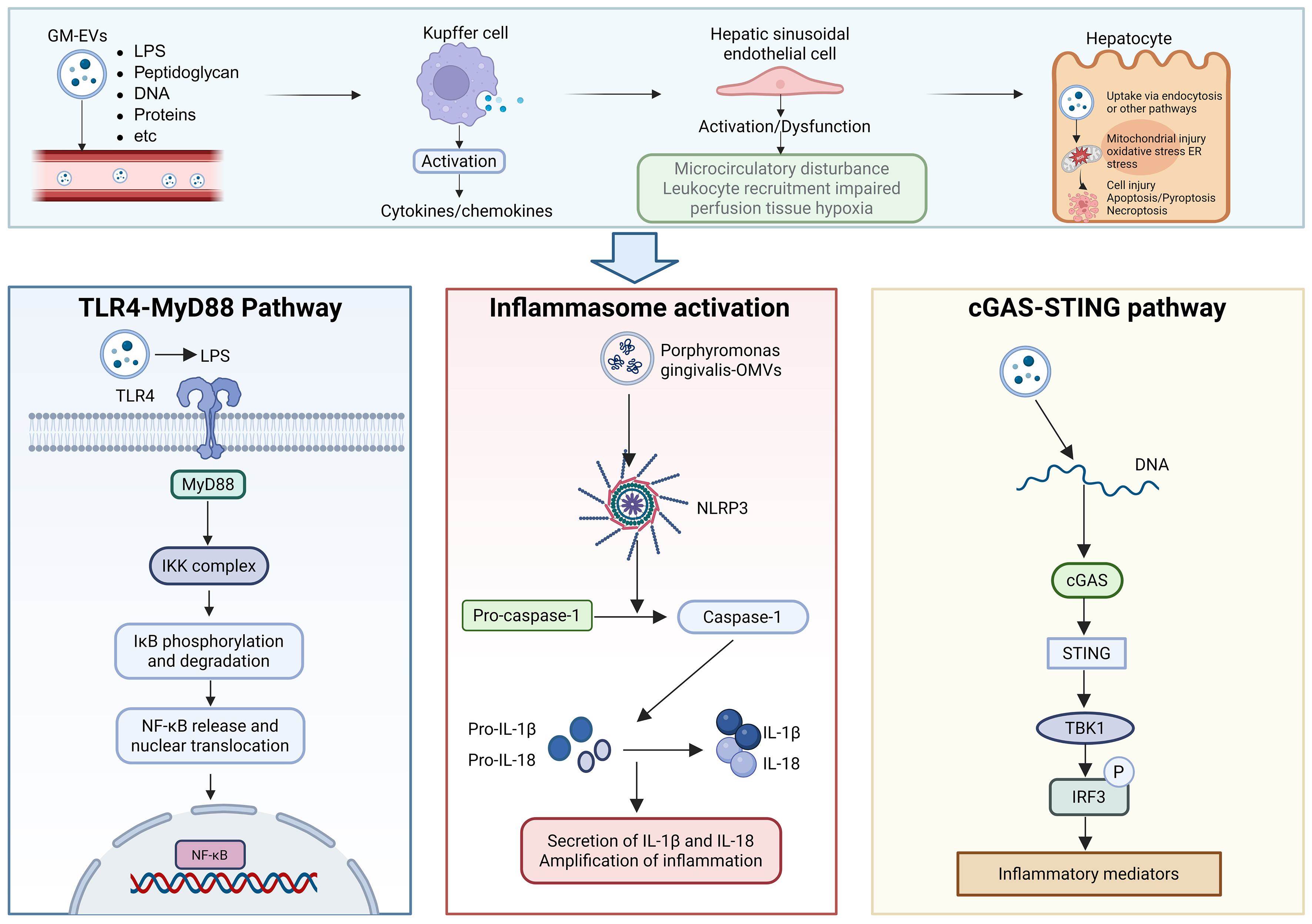
Figure 2. Potential inflammatory pathways of GM-EVs in sepsis-associated liver injury. Created in BioRender. yuan, Y. (2026) https://BioRender.com/wnng3vs. LPS: Lipopolysaccharide; TLR4: toll-like receptor 4; IKK: IκB kinase; IκB: inhibitor of NF-κB; NLRP3: NOD-like receptor family, pyrin domain containing 3; NF-κB: nuclear factor kappa B; IL-1β: interleukin-1 beta; IL-18: interleukin-18; cGAS: cyclic GMP-AMP synthase; STING: stimulator of interferon genes; TBK1: TANK-binding kinase 1; IRF3: interferon regulatory factor 3.
Evidence from host-derived EVs provides complementary support for the broader concept of EV-mediated inflammatory amplification but should be distinguished from evidence concerning GM-EVs. In an experimental study, LPS-stimulated macrophages loaded high-mobility group box 1 (HMGB1) into EVs via a receptor for advanced glycation end products (RAGE)-dependent mechanism. Host macrophage-derived HMGB1-positive EVs were internalized by hepatocytes via transferrin-mediated endocytosis and activated the NOD-like receptor protein 3 (NLRP3) inflammasome, leading to hepatocyte pyroptosis. In addition, serum levels of HMGB1-positive EVs were positively associated with liver injury biomarkers in patients with sepsis[90]. This study demonstrated that EV-mediated transfer of inflammatory DAMPs could contribute to hepatocyte injury during sepsis; however, the EVs examined were primarily host macrophage-derived rather than gut microbiota-derived. Therefore, these findings should not be interpreted as direct evidence that GM-EVs carry HMGB1 or induce the same pathway. After uptake by hepatic immune or parenchymal cells[91-93], GM-EVs may activate different signaling pathways depending on their cargo composition. Their downstream signaling may be mediated via the TLR4/myeloid differentiation primary response 88 (MyD88)-dependent pathway, which triggers the activation of the inhibitor of NF-κB (IκB) kinase complex. This process further induces the phosphorylation and subsequent degradation of IκB, thereby releasing NF-κB from cytoplasmic sequestration. The released NF-κB then translocates to the nucleus and initiates the transcription of pro-inflammatory genes[94-96]. Specific bacterial OMVs have been shown to activate inflammasome signaling in experimental models, although the available evidence is not specific to SALI. OMVs produced by Porphyromonas gingivalis increase NLRP3 inflammasome expression, facilitate the cleavage of pro-caspase-1 and pro-interleukin-1β (IL-1β) into their mature forms (caspase-1 and IL-1β), and ultimately promote the secretion of mature IL-1β and interleukin-18 (IL-18)[97]. Reciprocal regulation among these pathways and their concurrent activation synergistically boost the production of intrahepatic pro-inflammatory cytokines, the maturation of inflammasome effector molecules, and the release of chemotactic signals, thereby establishing an inflammatory response network amplified through multi-node cascades[98-100]. Although these findings are not specific to sepsis, they provide experimental evidence of bacterial OMV-mediated inflammasome activation.
In addition to LPS- and TLR-associated signaling, bacterial DNA transported by GM-EVs may provide an additional route for innate immune activation via cytosolic DNA-sensing pathways. Experimental studies have shown that microbial DNA-containing EVs activate the cGAS-STING pathway, resulting in the production of inflammatory mediators[54,92]. However, most available evidence for this mechanism has been obtained from models other than human SALI. Therefore, cGAS-STING signaling should be considered as a complementary and cargo-dependent pathway through which selected GM-EV populations may influence hepatic or systemic inflammation, rather than a universally activated pathway in all patients with sepsis. These GM-EV-associated pathways may interact with and potentially amplify other mechanisms of SALI. Cytokines and chemokines released by the activated macrophages may promote hepatic sinusoidal endothelial activation, leukocyte recruitment, oxidative stress, and microcirculatory dysfunction. These changes may impair hepatic clearance of gut-derived microbial products and prolong the exposure of hepatic cells to circulating GM-EVs. Conversely, persistent intestinal barrier disruption, dysbiosis, and increased vesicle release may increase the delivery of selected GM-EVs to the portal and systemic circulation. This reciprocal relationship may contribute to a positive feedback loop involving intestinal barrier dysfunction, hepatic accumulation of microbial signals, and progressive liver injury.
Reshaping the immune microenvironment
GM-EVs may do more than activate a single pro-inflammatory pathway. As bidirectional immunomodulators, they may reshape cellular functions and immune-response patterns within the hepatic immune microenvironment, thereby driving the dynamic transition of the sepsis course from an early phase of excessive inflammation to a later stage characterized by immune suppression and immune tolerance. Because hepatic sinusoids receive high blood flow and filter circulating material, Kupffer cells, hepatic sinusoidal endothelial cells, and dendritic cells (DCs) serve as the predominant cell populations responsive to circulating GM-EVs[23]. The LPS, lipoproteins, and peptidoglycan fragments carried by these vesicles are capable of activating the NF-κB, MAPK, and inflammasome-associated signaling pathways via pattern recognition receptors. Moreover, small bacterial RNAs and other nucleic acid components encapsulated within these vesicles may modulate the expression of host immune genes through cross-kingdom regulation. Driven by the aforementioned signaling cascades, macrophages undergo a phenotypic shift from a homeostatic surveillance and immune-tolerant phenotype toward a pro-inflammatory effector phenotype, accompanied by elevated expression of pro-inflammatory cytokines and chemokines[101,102].
During the acute inflammatory stage of sepsis, pathogenic bacteria-derived GM-EVs can activate a potent innate immune response through the pattern recognition receptor pathway and mediate a cascade that amplifies the response from innate to adaptive immunity. Their immunological effects are initially manifested through the activation of pattern recognition receptors. Specifically, LPS, lipoproteins, and nucleic acids contained within Gram-negative bacterium-derived GM-EVs are recognized by TLR4, TLR2, and endosomal TLRs, respectively. This recognition drives the maturation of antigen-presenting cells, upregulates the expression of MHC-II and co-stimulatory molecules, and facilitates the transmission of innate immune signals to the adaptive immune system[103]. The homeostatic functions of Kupffer cells, including scavenging portal vein-derived microbial products, apoptotic cells, and cellular debris as well as sustaining hepatic immune tolerance, may be impaired. Accordingly, Kupffer cells undergo a phenotypic shift from their homeostatic role as critical guardians of the hepatic immune barrier to becoming central hubs driving local immune injury.
GM-EVs do not exclusively exert pro-inflammatory effects; their immunophenotypes are governed by their bacterial origin, encapsulated cargo profile, and host barrier function. Commensal bacteria stimulate interleukin-10 (IL-10) secretion by DCs, facilitate regulatory T-cell responses, and sustain mucosal immune homeostasis. EVs released by Bacteroides thetaiotaomicron act on monocytes and macrophages via TLR2 signaling to elevate IL-10 levels, while restraining pro-inflammatory cytokine production in a dose-dependent manner. Moreover, persistent H3K4me1 epigenetic remodeling is induced to build long-term innate immune tolerance[104]. Furthermore, other studies have reported that EVs secreted by Bacteroides thetaiotaomicron can stimulate intestinal mucosal and peripheral DCs to concurrently produce pro-inflammatory IL-6 and anti-inflammatory IL-10 in healthy human subjects, thereby maintaining intestinal immune homeostasis[105]. DCs sense Bacteroides fragilis OMV-associated polysaccharide A (PSA) via TLR2 in a Gadd45α-dependent manner, which enhances the generation of regulatory T cells and anti-inflammatory cytokines[106]. Studies of the immunomodulatory effects of PSA derived from Bacteroides fragilis have demonstrated that PSA suppresses IL-17/Th17-mediated inflammatory responses and sustains immune homeostasis through IL-10-producing CD4+ T cells[107]. EVs secreted by Lactobacillus plantarum drive human THP-1 monocytic cells toward an anti-inflammatory M2 phenotype (predominantly the M2b subtype) by inducing the biased expression of cell surface markers and cytokines characteristic of M2 macrophages[108]. Accordingly, the immunomodulatory functions of GM-EVs are not limited to their pro-inflammatory effects. Instead, GM-EVs recalibrate the balance between innate immune sensing, antigen presentation, and T-cell responses in response to the microbial community composition, vesicle cargo profiles, barrier integrity, and overall immune status of the host.
Lipid metabolism disorders
During sepsis, the liver is subjected to inflammatory insults and metabolic reprogramming, with dysregulated lipid metabolism being one of the most prominent manifestations. Massive lipid droplet accumulation, defective fatty acid oxidation, triglyceride deposition, and intensified lipid peroxidation in the liver are vital pathological drivers of SALI progression from reversible liver injury to hepatic failure. Upon hepatic delivery of LPS carried by GM-EVs, LPS activates Kupffer cells via TLR4 and stimulates the secretion of inflammatory factors. These mediators directly trigger hepatocyte apoptosis and disrupt fatty acid oxidation by repressing the PPARα pathway. Specifically, robust suppression of hepatic PPARα signaling compromises fatty acid β-oxidation, resulting in substantial accumulation of circulating free fatty acids and their ectopic deposition in the liver and kidneys. Oxidative stress and reactive oxygen species, together with excess lipids, promote lipid peroxidation. This process produces lipotoxic metabolites, including malondialdehyde and 4-hydroxynonenal, which eventually induce parenchymal cell apoptosis in the liver and kidneys, causing multi-organ damage[109,110]. Abrupt intracellular accumulation of free fatty acids disturbs the lipid bilayer composition of the ER membrane and directly provokes severe ER stress, accompanied by persistent activation of three major transmembrane receptor pathways, namely PERK, IRE1α, and ATF6[111]. As the central organelle responsible for hepatic lipid synthesis and protein folding, the ER exerts compensatory protective effects against mild ER stress during the early stages of sepsis. Nevertheless, severe lipotoxicity induced by sustained lipid overload switches the output of the unfolded protein response (UPR) from a pro-survival to a pro-apoptotic phenotype, thereby triggering hepatocyte death[112] and aggravating the progression of hepatic failure.
Induction of programmed cell death
In addition to metabolic disturbances, GM-EVs elicit parenchymal liver injury by triggering programmed cell death in hepatocytes. The pro-inflammatory and cytotoxic effects of GM-EVs on immune cells have been well characterized. Previous studies demonstrated that EVs derived from Salmonella typhimurium deliver flagellin into the cytoplasm of bone marrow-derived macrophages (BMDMs), which activate the canonical NLRC4 inflammasome. This cascade drives caspase-1 activation and the secretion of mature IL-1β. LPS encapsulated within EVs initiates GSDMD-dependent pyroptosis via the caspase-11 signaling pathway[113]. Deo et al. reported that macrophages exposed to OMVs secreted by Neisseria gonorrhoeae, uropathogenic Escherichia coli and Pseudomonas aeruginosa undergo mitochondrial apoptosis and NLRP3 inflammasome activation. OMVs and their cargo toxins cause mitochondrial dysfunction and suppress host protein synthesis, consequently depleting MCL-1, an unstable anti-apoptotic member of the BCL-2 protein family, and ultimately driving BAK-dependent mitochondrial apoptosis[114]. Vanaja et al. demonstrated that treatment of BMDMs with EVs induced proteolytic maturation of caspase-1 and IL-1β as well as IL-1β secretion. Importantly, EV stimulation induced cell death in a dose-dependent manner[115]. Fusobacterium nucleatum secreted EVs loaded with diverse molecules. In a macrophage/Caco-2 co-culture system, Fusobacterium nucleatum-derived EVs markedly disrupted the epithelial barrier and induced oxidative stress; this pathological phenomenon was attributed to epithelial necrotic cell death triggered by the activation of receptor-interacting protein kinase 1 (RIPK1) and receptor-interacting protein kinase 3 (RIPK3)[116]. OMVs derived from Desulfovibrio fairfieldensis disrupt epithelial barrier integrity and trigger inflammatory responses and pyroptosis in macrophages[117]. Collectively, the above studies demonstrate that bacteria-derived EVs can activate canonical and non-canonical programmed cell death pathways via multiple signaling cascades; these effects have defined molecular mechanisms underlying cellular injury and pro-inflammatory responses.
POTENTIAL AND CHALLENGES OF GM-EVs AS BIOMARKERS FOR SALI
Compared with conventional hepatic injury biomarkers, GM-EVs can provide mechanistic information about early pathological events in the disease. Clinically, routine indicators including alanine transaminase, aspartate transaminase, bilirubin, and coagulation profiles can indicate hepatocellular injury, cholestasis, and impaired hepatic synthetic function. Nevertheless, these biomarkers suffer from insufficient specificity and poor performance in early warning systems, making it difficult to discriminate between hepatic injuries induced by heterogeneous insults, such as sepsis, pharmaceutical agents, ischemia-hypoxia, and underlying chronic liver diseases. In contrast, the microbial profiles of GM-EVs, together with their surface PAMPs and encapsulated nucleic acid and protein cargo, may provide a gut-derived signature for SALI. Previous studies have suggested that GM-EVs circulating in bodily fluids are promising candidate biomarkers for the diagnosis of infectious diseases[118]. OMVs, which originate from Gram-negative bacteria and retain the characteristic signatures of parental bacteria, are regarded as promising candidates for the molecular diagnosis of Gram-negative sepsis[119]. Recent research using a fluorescent polymyxin B probe to label LPS-containing OMVs further verified that circulating bacterial OMVs were detectable in the early phase of infection and were capable of distinguishing bacterial infection phenotypes, providing methodological implications for employing GM-EVs to enable early detection of sepsis-associated organ injury[120].
Nevertheless, the clinical translation of GM-EVs into bona fide biomarkers of SALI faces several limitations. First, circulating GM-EVs are present in relatively low abundance and heavily contaminated with host-derived EVs, lipoproteins, protein aggregates, and cell-free nucleic acids. Current isolation approaches fail to meet requirements for purity, throughput, cost-effectiveness, and clinical practicality[121]. Second, GM-EVs exhibit pronounced heterogeneity and their molecular compositions are modulated by multiple variables, including bacterial species and strains, infection sites, antibiotic exposure, nutritional status, pre-existing hepatic diseases, and sampling time windows. Accordingly, a single molecular biomarker does not consistently reflect the complex pathological process underlying gut-liver axis injury. Third, robust direct clinical evidence substantiating the causal linkage between GM-EVs and SALI remains scarce, and most relevant conclusions are merely drawn from indirect inferences derived from separate investigations concerning bacterial EVs, gut-liver axis homeostasis, and GM-EV biology in sepsis.
TRANSLATIONAL POTENTIAL OF GM-EVs
Considering the potential hazards posed by GM-EVs in sepsis-induced gut-liver axis dysfunction, intervention in the biogenesis and release of GM-EVs at the source represents a highly promising therapeutic strategy. Small-molecule inhibitors that specifically block GM-EV biogenesis without compromising bacterial viability have been developed. EV biogenesis is governed by sophisticated mechanisms, including the regulation of periplasmic enzymes. Precise targeting of these pathways is expected to selectively eliminate detrimental GM-EVs secreted by pathogenic or opportunistic bacteria while preserving overall gut microbiota homeostasis. For instance, the release of OMVs in Gram-negative bacteria is tightly associated with dynamic remodeling between the periplasmic space and the bacterial outer membrane[32,122]. High-throughput screening or structure-based drug design can identify compounds capable of interfering with the activities of key proteins involved in vesicle budding (such as periplasmic enzymes and outer membrane proteins), thereby inhibiting the biogenesis of pro-inflammatory EVs at the molecular level[123]. Compared to broad-spectrum antibiotics, such targeted interventions can better preserve the symbiotic functions of the gut microbiota and avert secondary injury resulting from overall dysbiosis of the microbiota, offering a more refined regulatory strategy for the treatment of sepsis.
However, restoring gut microbial balance to indirectly remodel the release profile of GM-EVs constitutes another vital intervention approach. Supplementation with specific probiotics (e.g., Lactobacillus and Bifidobacterium) stimulates these microbes to secrete EVs that exert anti-inflammatory and tissue-protective effects, consequently reshaping the gut microbial community and alleviating systemic inflammatory responses. For example, EVs derived from Lactiplantibacillus plantarum have been reported to alleviate acute lung injury[124]. EVs derived from Bifidobacterium longum AO44 exert anti-inflammatory properties and induce IL-10 secretion in a co-culture system consisting of splenocytes, DCs, and CD4+ T cells[125]. EVs secreted by Bifidobacterium longum NSP001 play a key role in maintaining host immune homeostasis by inhibiting the STAT3 signaling pathway[126]. Fecal microbiota transplantation (FMT) is a robust strategy for comprehensive gut microbiota remodeling. Previous studies have demonstrated that FMT reduces hepatic aminotransferase activity, serum total bilirubin, and hepatic pro-inflammatory cytokine levels; ameliorates hepatic apoptosis; decreases cleaved caspase-3 expression; and alleviates pathological lesions in liver tissue[127]. Therefore, the combined application of these approaches to regulate intestinal microbiota homeostasis is expected to serve as an effective strategy for modulating GM-EVs and ameliorating sepsis-induced gut-liver axis dysfunction.
FUTURE PERSPECTIVES AND CHALLENGES
Although the roles of GM-EVs in microbe-host interactions and multiple organ injuries during sepsis have been widely validated, numerous research limitations and barriers to their clinical translation remain. Current research limitations are reflected in four aspects. First, GM-EVs exhibit heterogeneity. The composition and biological functions of the GM-EVs secreted by different bacterial strains under various pathological conditions differ significantly. The regulatory mechanisms underlying this heterogeneity and their correlation with sepsis severity have not been elucidated, and standardized systems for accurate typing and functional identification are still lacking. Second, mechanistic investigations of GM-EV-mediated injury to multiple organs are fragmented. Most studies have focused on individual organs or isolated signaling pathways, whereas the core mechanisms by which GM-EVs integrate multi-organ damage signals and mediate synergistic injuries across organs remain to be elucidated. Third, prevailing detection techniques for GM-EVs are cumbersome, costly, and insufficiently sensitive, and fail to meet the requirements for rapid clinical diagnosis and dynamic monitoring of disease progression. Fourth, therapeutic strategies targeting GM-EVs are far from mature. Pharmacologically targeted interventions tend to disturb the normal physiological functions of bacteria. The preparation of specific antibodies faces great technical challenges, and the clinical safety and efficacy of engineered GM-EVs await further systematic verification.
Future studies should focus on several key priorities to address these limitations. By adopting cutting-edge technologies, such as single-cell sequencing and nanotechnology, researchers can dissect the compositional and functional signatures of diverse GM-EV subtypes and clarify the regulatory mechanisms governing GM-EV heterogeneity, together with their correlations with sepsis severity and patient prognosis. Using research platforms such as multi-organ chips and animal models, researchers should investigate the integrated signaling networks and holistic effects responsible for GM-EV-mediated synergistic multi-organ injury to identify novel therapeutic targets. Moreover, detection methodologies should be refined to develop low-cost, highly sensitive, rapid, and user-friendly assays that facilitate precise clinical diagnosis and treatment. It is also necessary to refine targeted intervention strategies and GM-EV engineering and modification techniques, validate the safety and efficacy of the relevant therapeutic regimens, and accelerate their translation into clinical practice. Furthermore, exploration of the crosstalk between GM-EVs and other pathogenic mediators (such as host-derived EVs and inflammatory cytokines) is required to construct a comprehensive regulatory network of microbe-host interplay, thereby enriching the overall pathological framework of sepsis.
Conclusion
Current evidence supports an emerging and testable model in which sepsis-associated dysbiosis, intestinal barrier dysfunction, and altered bacterial extracellular vesicle biology facilitate the release, translocation, and hepatic exposure of selected GM-EV populations. These vesicles may subsequently amplify hepatic inflammatory, endothelial, oxidative, metabolic, and cell death pathways, thereby contributing to gut-liver axis dysfunction and SALI. The relative contribution of GM-EVs has not been fully quantified; however, their pathophysiological significance lies in serving as upstream, transportable, and potentially targetable mediators that connect intestinal dysbiosis to hepatocyte injury. Therefore, we believe that GM-EVs represent a complementary and mechanistically interrelated pathway within the gut-liver axis, rather than an isolated or dominant mechanism in sepsis-related liver injury. Clarifying the precise contribution of GM-EVs relative to other determinants and their hierarchical relationships warrants dedicated experimental studies. Notably, direct clinical evidence linking GM-EVs to SALI remains limited, and several steps of the proposed pathway warrant validation in sepsis-specific models and clinical settings. Several critical challenges remain, including unclear regulatory mechanisms of GM-EVs, suboptimal clinical detection systems, incomplete targeted therapeutic strategies, and substantial obstacles in clinical translation. Future investigations should further elucidate the complex pathogenic mechanisms of GM-EVs, optimize detection and intervention techniques, and address bottlenecks in translational research. These efforts will facilitate the translation of findings on GM-EVs into diagnostic and therapeutic applications for SALI, thereby improving hepatic outcomes and reducing the disease burden in patients with sepsis.
DECLARATIONS
Acknowledgments
We thank BioRender for providing the illustration platform used to prepare the figures in this article. The Graphical Abstract was created with BioRender.com. (2026) https://BioRender.com/1m1qykw.
Authors’ contributions
Conceptualization: Sun T, Yuan Y
Literature collection and collation: Ge Y, Ji J
Visualization: Zhang X, Guo R
Drafting of the manuscript: Yuan Y, Liang H
Revision of the manuscript: Zhang Z, Zhang R
All authors have approved the final version of the manuscript for publication.
Availability of data and materials
Not applicable.
AI and AI-assisted tools statement
Not applicable.
Financial support and sponsorship
This study was supported by the Noncommunicable Chronic Diseases-National Science and Technology Major Project (Grant No. 2023ZD0506504), Medical Science and Technology Tackling Plan Provincial and Ministerial Major Projects of Henan Province (Grant No. SBGJ202501004), Scientific Research and Innovation Excellence Team of First Affiliated Hospital of Zhengzhou University (Grant No. ZYCXTD2023006), and the Key Project of the Medical Science and Technology Tackling Program of Henan Province (Provincial and Ministerial Co-construction) (Project No. SBGJ202302034).
Conflicts of interest
All authors declared that there are no conflicts of interest.
Ethical approval and consent to participate
Not applicable.
Consent for publication
Not applicable.
Copyright
© The Author(s) 2026.
REFERENCES
1. Rudd KE, Johnson SC, Agesa KM, et al. Global, regional, and national sepsis incidence and mortality, 1990-2017: analysis for the Global Burden of Disease Study. The Lancet 2020;395:200-11.
2. Zhang W, Jiang H, Wu G, et al. The pathogenesis and potential therapeutic targets in sepsis. MedComm 2023;4:e418.
3. Katsounas A, Tsochatzis E, Rello J. Sepsis-associated liver injury: from liver-test risk signals to immunobiology-guided precision medicine. Intensive Care Med 2026.
4. Guo Y, Guo W, Chen H, Sun J, Yin Y. Mechanisms of sepsis-induced acute liver injury: a comprehensive review. Front Cell Infect Microbiol 2025;15:1504223.
5. Sun J, Zhang J, Wang X, et al. Gut-liver crosstalk in sepsis-induced liver injury. Crit Care 2020;24:3327.
6. Xu X, Yang T, An J, Li B, Dou Z. Liver injury in sepsis: manifestations, mechanisms and emerging therapeutic strategies. Front Immunol 2025;16:1575554.
7. Strnad P, Tacke F, Koch A, Trautwein C. Liver - guardian, modifier and target of sepsis. Nat Rev Gastroenterol Hepatol 2017;14:55-66.
9. Klingensmith NJ, Coopersmith CM. The gut as the motor of multiple organ dysfunction in critical illness. Crit Care Clin 2016;32:203-12.
10. Zheng L, Jia T, Li Y, Zhang Z, Su H, Zhang R. The interplay between gastrointestinal dysfunction and gut microbiota dynamics in sepsis. Front Cell Infect Microbiol 2026;16:1761536.
11. Shahid A, Chambers S, Scott-Thomas A, Bhatia M. Gut microbiota and liver dysfunction in sepsis: the role of inflammatory mediators and therapeutic approaches. Int J Mol Sci 2024;25:13415.
12. Murao A, Jha A, Kato T, et al. Gut-primed neutrophils activate Kupffer cells to promote hepatic injury in mouse sepsis. Nat Commun 2025;16:65572.
13. Pabst O, Hornef MW, Schaap FG, Cerovic V, Clavel T, Bruns T. Gut-liver axis: barriers and functional circuits. Nat Rev Gastroenterol Hepatol 2023;20:447-61.
14. Bauer M. The liver-gut-axis: initiator and responder to sepsis. Curr Opin Crit Care 2022;28:216-20.
15. Margutti P, D’Ambrosio A, Zamboni S. Microbiota-derived extracellular vesicle as emerging actors in host interactions. Int J Mol Sci 2024;25:8722.
16. Ma K, Zhang Q, Jin Z, et al. Gut microbiota-derived extracellular vesicles: bridging microbial-host crosstalk in metabolic disorders. Cell Commun Signal 2026;24:3034.
17. Li Y, Li J, Dullaart R, Moshage H. Bacterial extracellular vesicles in the pathogenesis and treatment of liver diseases. J Gastroenterol Hepatol 2026;41:880-94.
18. Jain H, Kumar A, Almousa S, et al. Characterisation of LPS+ bacterial extracellular vesicles along the gut-hepatic portal vein-liver axis. J Extracell Vesicles 2024;13:e12474.
19. Effah CY, Ding X, Drokow EK, Li X, Tong R, Sun T. Bacteria-derived extracellular vesicles: endogenous roles, therapeutic potentials and their biomimetics for the treatment and prevention of sepsis. Front Immunol 2024;15:1296061.
20. Singer M, Deutschman CS, Seymour CW, et al. The third international consensus definitions for sepsis and septic shock (Sepsis-3). JAMA 2016;315:801.
21. Turner JR. Intestinal mucosal barrier function in health and disease. Nat Rev Immunol 2009;9:799-809.
22. Haussner F, Chakraborty S, Halbgebauer R, Huber-Lang M. Challenge to the intestinal mucosa during sepsis. Front Immunol 2019;10:891.
24. Dixon LJ, Barnes M, Tang H, Pritchard MT, Nagy LE. Kupffer cells in the liver. Compr Physiol 2013;3:785-97.
25. Beyer D, Hoff J, Sommerfeld O, Zipprich A, Gaßler N, Press AT. The liver in sepsis: molecular mechanism of liver failure and their potential for clinical translation. Mol Med 2022;28:510.
26. Zhang X, Liu H, Hashimoto K, Yuan S, Zhang J. The gut-liver axis in sepsis: interaction mechanisms and therapeutic potential. Crit Care 2022;26:4090.
27. Zhang D, Li Y, Ding L, Fu Y, Dong X, Li H. Prevalence and outcome of acute gastrointestinal injury in critically ill patients: a systematic review and meta-analysis. Medicine 2018;97:e12970.
28. Kobashi H, Toshimori J, Yamamoto K. Sepsis-associated liver injury: Incidence, classification and the clinical significance. Hepatol Res 2013;43:255-66.
29. Zhai R, Sheu CC, Su L, et al. Serum bilirubin levels on ICU admission are associated with ARDS development and mortality in sepsis. Thorax 2009;64:784-90.
30. Blot S, Antonelli M, Arvaniti K, et al. ; the Abdominal Sepsis Study (AbSeS) group on behalf of the Trials Group of the European Society of Intensive Care Medicine. Epidemiology of intra-abdominal infection and sepsis in critically ill patients: “AbSeS”, a multinational observational cohort study and ESICM Trials Group Project. Intensive Care Med 2019;45:1703-17.
31. Meng M, Klingensmith NJ, Coopersmith CM. New insights into the gut as the driver of critical illness and organ failure. Curr Opin Crit Care 2017;23:143-8.
32. Toyofuku M, Nomura N, Eberl L. Types and origins of bacterial membrane vesicles. Nat Rev Microbiol 2019;17:13-24.
33. Schwechheimer C, Kuehn MJ. Outer-membrane vesicles from Gram-negative bacteria: biogenesis and functions. Nat Rev Microbiol 2015;13:605-19.
34. Brown L, Wolf JM, Prados-Rosales R, Casadevall A. Through the wall: extracellular vesicles in Gram-positive bacteria, mycobacteria and fungi. Nat Rev Microbiol 2015;13:620-30.
35. Kulp A, Kuehn MJ. Biological functions and biogenesis of secreted bacterial outer membrane vesicles. Annu Rev Microbiol 2010;64:163-84.
36. Liu BD, Akbar R, Oliverio A, Thapa K, Wang X, Fan G. Bacterial extracellular vesicles in the regulation of inflammatory response and host-microbe interactions. Shock 2024;61:175-88.
37. Gilmore WJ, Johnston EL, Zavan L, Bitto NJ, Kaparakis-Liaskos M. Immunomodulatory roles and novel applications of bacterial membrane vesicles. Mol Immunol 2021;134:72-85.
38. Cao Y, Lin H. Characterization and function of membrane vesicles in Gram-positive bacteria. Appl Microbiol Biotechnol 2021;105:1795-801.
39. Feuillet V, Medjane S, Mondor I, et al. Involvement of Toll-like receptor 5 in the recognition of flagellated bacteria. Proc Natl Acad Sci U S A 2006;103:12487-92.
40. Poltorak A, He X, Smirnova I, et al. Defective LPS signaling in C3H/HeJ and C57BL/10ScCr mice: mutations in Tlr4 gene. Science 1998;282:2085-8.
41. Charpentier LA, Dolben EF, Hendricks MR, Hogan DA, Bomberger JM, Stanton BA. Bacterial outer membrane vesicles and immune modulation of the host. Membranes 2023;13:752.
42. Thapa HB, Kohl P, Zingl FG, et al. Characterization of the inflammatory response evoked by bacterial membrane vesicles in intestinal cells reveals an RIPK2-dependent activation by enterotoxigenic Escherichia coli vesicles. Microbiol Spectr 2023;11:e01115-23.
43. Liang L, Yang C, Liu L, et al. Commensal bacteria-derived extracellular vesicles suppress ulcerative colitis through regulating the macrophages polarization and remodeling the gut microbiota. Microb Cell Fact 2022;21:1812.
44. Yerneni SS, Werner S, Azambuja JH, et al. Pneumococcal extracellular vesicles modulate host immunity. mBio 2021;12:e01657-21.
45. Toyofuku M, Schild S, Kaparakis-Liaskos M, Eberl L. Composition and functions of bacterial membrane vesicles. Nat Rev Microbiol 2023;21:415-30.
46. Koeppen K, Hampton TH, Jarek M, et al. A novel mechanism of host-pathogen interaction through sRNA in bacterial outer membrane vesicles. PLoS Pathog 2016;12:e1005672.
47. Plovier H, Everard A, Druart C, et al. A purified membrane protein from Akkermansia muciniphila or the pasteurized bacterium improves metabolism in obese and diabetic mice. Nat Med 2017;23:107-13.
48. Mottawea W, Yousuf B, Sultan S, et al. Multi-level analysis of gut microbiome extracellular vesicles-host interaction reveals a connection to gut-brain axis signaling. Microbiol Spectr 2025;13:e01368-24.
49. Huang J, Yu Y, Feng Z, et al. Cross-Kingdom dialogue of microbial messengers: multi-target regulatory mechanisms and therapeutic strategies of gut microbiota-derived extracellular vesicles in metabolic diseases. Int J Nanomedicine 2025;20:12573-91.
50. Jan AT. Outer membrane vesicles (OMVs) of Gram-negative bacteria: a perspective update. Front Microbiol 2017;8:1053.
51. Zhou Y, Sun Y, Yin P, Zuo S, Li H, Cao K. Bacterial extracellular vesicles: emerging mediators of gut-liver axis crosstalk in hepatic diseases. Front Cell Infect Microbiol 2025;15:1620829.
52. Kaparakis-Liaskos M, Ferrero RL. Immune modulation by bacterial outer membrane vesicles. Nat Rev Immunol 2015;15:375-87.
53. Takeuchi O, Hoshino K, Kawai T, et al. Differential roles of TLR2 and TLR4 in recognition of gram-negative and gram-positive bacterial cell wall components. Immunity 1999;11:443-51.
54. Erttmann SF, Swacha P, Aung KM, et al. The gut microbiota prime systemic antiviral immunity via the cGAS-STING-IFN-I axis. Immunity 2022;55:847-61.e10.
55. Hotchkiss RS, Moldawer LL, Opal SM, Reinhart K, Turnbull IR, Vincent J. Sepsis and septic shock. Nat Rev Dis Primers 2016;2:BFnrdp201645.
56. Tulkens J, Vergauwen G, Van Deun J, et al. Increased levels of systemic LPS-positive bacterial extracellular vesicles in patients with intestinal barrier dysfunction. Gut 2020;69:191-3.
57. McBroom AJ, Kuehn MJ. Release of outer membrane vesicles by Gram-negative bacteria is a novel envelope stress response. Mol Microbiol 2007;63:545-58.
58. Appiah MG, Park EJ, Darkwah S, et al. Intestinal epithelium-derived luminally released extracellular vesicles in sepsis exhibit the ability to suppress TNF-α and IL-17A expression in mucosal inflammation. Int J Mol Sci 2020;21:8445.
59. Li P, Wu Y, Goodwin AJ, et al. Circulating extracellular vesicles are associated with the clinical outcomes of sepsis. Front Immunol 2023;14:1150564.
60. Rodríguez-Díaz C, Martín-Reyes F, Taminiau B, et al. The metagenomic composition and effects of fecal-microbe-derived extracellular vesicles on intestinal permeability depend on the patient’s disease. Int J Mol Sci 2023;24:4971.
61. Lankelma JM, van Vught LA, Belzer C, et al. Critically ill patients demonstrate large interpersonal variation in intestinal microbiota dysregulation: a pilot study. Intensive Care Med 2017;43:59-68.
62. McDonald D, Ackermann G, Khailova L, et al. Extreme dysbiosis of the microbiome in critical illness. mSphere 2016;1:e00199-16.
63. Liang H, Song H, Zhang X, et al. Metformin attenuated sepsis-related liver injury by modulating gut microbiota. Emerg Microbes Infect 2022;11:815-28.
64. Becattini S, Taur Y, Pamer EG. Antibiotic-induced changes in the intestinal microbiota and disease. Trends Mol Med 2016;22:458-78.
65. Dethlefsen L, Relman DA. Incomplete recovery and individualized responses of the human distal gut microbiota to repeated antibiotic perturbation. Proc Natl Acad Sci U S A 2011;108:4554-61.
66. Lee E, Choi D, Kim K, Gho YS. Proteomics in gram-negative bacterial outer membrane vesicles. Mass Spectrom Rev 2008;27:535-55.
67. Stentz R, Jones E, Juodeikis R, et al. The proteome of extracellular vesicles produced by the human gut bacteria Bacteroides thetaiotaomicron in vivo is influenced by environmental and host-derived factors. Appl Environ Microbiol 2022;88:e00533-22.
68. Odenwald MA, Turner JR. The intestinal epithelial barrier: a therapeutic target? Nat Rev Gastroenterol Hepatol 2017;14:9-21.
69. Oami T, Shimazui T, Yumoto T, Otani S, Hayashi Y, Coopersmith CM. Gut integrity in intensive care: alterations in host permeability and the microbiome as potential therapeutic targets. J intensive care 2025;13:786.
70. Marchiando AM, Shen L, Graham WV, et al. Caveolin-1-dependent occludin endocytosis is required for TNF-induced tight junction regulation in vivo. J Cell Biol 2010;189:111-26.
71. Sun S, Duan Z, Wang X, et al. Neutrophil extracellular traps impair intestinal barrier functions in sepsis by regulating TLR9-mediated endoplasmic reticulum stress pathway. Cell Death Dis 2021;12:3896.
72. Cao Y, Zhang Y, Gerile W, et al. PLK1 protects intestinal barrier function during sepsis by targeting mitochondrial dynamics through TANK-NF-κB signalling. Mol Med 2022;28:597.
73. Chu C, Wang X, Chen F, et al. Neutrophil extracellular traps aggravate intestinal epithelial necroptosis in ischaemia-reperfusion by regulating TLR4/RIPK3/FUNDC1 -required mitophagy. Cell Prolif 2024;57:e13538.
74. Zuo L, Kuo W, Turner JR. Tight junctions as targets and effectors of mucosal immune homeostasis. Cell Mol Gastroenterol Hepatol 2020;10:327-40.
75. Oami T, Abtahi S, Shimazui T, et al. Claudin-2 upregulation enhances intestinal permeability, immune activation, dysbiosis, and mortality in sepsis. Proc Natl Acad Sci U S A 2024;121:e2217877121.
76. Yoseph BP, Klingensmith NJ, Liang Z, et al. Mechanisms of intestinal barrier dysfunction in sepsis. Shock 2016;46:52-9.
77. Hundscheid IH, Grootjans J, Lenaerts K, et al. The human colon is more resistant to ischemia-reperfusion-induced tissue damage than the small intestine: an observational study. Ann Surg 2015;262:304-11.
78. Elmi A, Nasher F, Jagatia H, et al. Campylobacter jejuni outer membrane vesicle-associated proteolytic activity promotes bacterial invasion by mediating cleavage of intestinal epithelial cell E-cadherin and occludin. Cell Microbiol 2016;18:561-72.
79. Jang SC, Kim SR, Yoon YJ, et al. In vivo kinetic biodistribution of nano-sized outer membrane vesicles derived from bacteria. Small 2015;11:456-61.
80. Hu Q, Ren H, Li G, et al. STING-mediated intestinal barrier dysfunction contributes to lethal sepsis. EBioMedicine 2019;41:497-508.
81. Díaz-Garrido N, Badia J, Baldomà L. Microbiota-derived extracellular vesicles in interkingdom communication in the gut. J Extracell Vesicles 2021;10:e12161.
82. Zihni C, Mills C, Matter K, Balda MS. Tight junctions: from simple barriers to multifunctional molecular gates. Nat Rev Mol Cell Biol 2016;17:564-80.
83. Gilmore WJ, Johnston EL, Bitto NJ, et al. Bacteroides fragilis outer membrane vesicles preferentially activate innate immune receptors compared to their parent bacteria. Front Immunol 2022;13:970725.
84. O’Donoghue EJ, Sirisaengtaksin N, Browning DF, et al. Lipopolysaccharide structure impacts the entry kinetics of bacterial outer membrane vesicles into host cells. PLoS Pathog 2017;13:e1006760.
85. Olofsson A, Nygård Skalman L, Obi I, Lundmark R, Arnqvist A. Uptake of Helicobacter pylori vesicles is facilitated by clathrin-dependent and clathrin-independent endocytic pathways. mBio 2014;5:e00979-14.
86. Rubio APD, Martínez J, Palavecino M, et al. Transcytosis of Bacillus subtilis extracellular vesicles through an in vitro intestinal epithelial cell model. Sci Rep 2020;10:3120.
87. O’Donoghue EJ, Krachler AM. Mechanisms of outer membrane vesicle entry into host cells: microReview - OMV entry into host cells. Cell Microbiol 2016;18:1508-17.
89. Pool R, Gomez H, Kellum JA. Mechanisms of organ dysfunction in sepsis. Crit Care Clin 2018;34:63-80.
90. Wang G, Jin S, Huang W, et al. LPS-induced macrophage HMGB1-loaded extracellular vesicles trigger hepatocyte pyroptosis by activating the NLRP3 inflammasome. Cell Death Discov 2021;7:729.
91. Luo Z, Ji Y, Gao H, et al. CRIg+ macrophages prevent gut microbial DNA-containing extracellular vesicle-induced tissue inflammation and insulin resistance. Gastroenterology 2021;160:863-74.
92. Luo Z, Ji Y, Zhang D, et al. Microbial DNA enrichment promotes liver steatosis and fibrosis in the course of non-alcoholic steatohepatitis. Acta Physiol 2022;235:e13827.
93. Qin X, Wang S, Yan Z, Zhao N, Yao J. The role of bacterial outer membrane vesicles in inflammatory response of acute-on-chronic liver failure. Front Microbiol 2025;16:1608137.
94. Gong Y, Jin L, Duan L, et al. Extracellular vesicles derived from Streptococcus anginosus aggravate lupus nephritis by triggering TLR2-MyD88-NF-κB signalling in NK cells. J Extracell Vesicles 2025;14:e70134.
95. Engevik MA, Danhof HA, Ruan W, et al. Fusobacterium nucleatum secretes outer membrane vesicles and promotes intestinal inflammation. mBio 2021;12:e02706-20.
97. Fleetwood AJ, Lee MKS, Singleton W, et al. Metabolic remodeling, inflammasome activation, and pyroptosis in macrophages stimulated by Porphyromonas gingivalis and its outer membrane vesicles. Front Cell Infect Microbiol 2017;7:351.
98. Guo J, Friedman SL. Toll-like receptor 4 signaling in liver injury and hepatic fibrogenesis. Fibrogenesis Tissue Repair 2010;3:36.
99. Fizanne L, Villard A, Benabbou N, et al. Faeces-derived extracellular vesicles participate in the onset of barrier dysfunction leading to liver diseases. J Extracell Vesicles 2023;12:12303.
101. Han E, Choi S, Lee Y, Park J, Hong S, Lee H. Extracellular RNAs in periodontopathogenic outer membrane vesicles promote TNF-α production in human macrophages and cross the blood-brain barrier in mice. FASEB J 2019;33:13412-22.
102. Guangzhang C, Fangfang F, Siqian D, et al. Outer membrane vesicles from Escherichia coli are efficiently internalized by macrophage cells and alter their inflammatory response. Microb Pathog 2023;175:105965.
103. Kawai T, Akira S. The role of pattern-recognition receptors in innate immunity: update on Toll-like receptors. Nat Immunol 2010;11:373-84.
104. Fonseca S, Carvalho AL, Miquel-Clopés A, et al. Extracellular vesicles produced by the human gut commensal bacterium Bacteroides thetaiotaomicron elicit anti-inflammatory responses from innate immune cells. Front Microbiol 2022;13:1050271.
105. Durant L, Stentz R, Noble A, et al. Bacteroides thetaiotaomicron-derived outer membrane vesicles promote regulatory dendritic cell responses in health but not in inflammatory bowel disease. Microbiome 2020;8:868.
106. Shen Y, Torchia M, Lawson G, Karp C, Ashwell J, Mazmanian S. Outer membrane vesicles of a human commensal mediate immune regulation and disease protection. Cell Host Microbe 2012;12:509-20.
107. Mazmanian SK, Round JL, Kasper DL. A microbial symbiosis factor prevents intestinal inflammatory disease. Nature 2008;453:620-5.
108. Kim W, Lee EJ, Bae I, et al. Lactobacillus plantarum -derived extracellular vesicles induce anti-inflammatory M2 macrophage polarization in vitro. J Extracell Vesicles 2020;9:1793514.
109. Feng Y, Luo S, Fang C, et al. ANGPTL8 deficiency attenuates lipopolysaccharide-induced liver injury by improving lipid metabolic dysregulation. J Lipid Res 2024;65:100595.
110. Van Wyngene L, Vanderhaeghen T, Timmermans S, et al. Hepatic PPARα function and lipid metabolic pathways are dysregulated in polymicrobial sepsis. EMBO Mol Med 2020;12:BFEMMM201911319.
111. Akazawa Y, Nakao K. Lipotoxicity pathways intersect in hepatocytes: Endoplasmic reticulum stress, c-Jun N-terminal kinase-1, and death receptors. Hepatol Res 2016;46:977-84.
112. Karagöz GE, Acosta-Alvear D, Walter P. The unfolded protein response: detecting and responding to fluctuations in the protein-folding capacity of the Endoplasmic reticulum. Cold Spring Harb Perspect Biol 2019;11:a033886.
113. Yang J, Hwang I, Lee E, et al. Bacterial outer membrane vesicle-mediated cytosolic delivery of flagellin triggers host NLRC4 canonical inflammasome signaling. Front Immunol 2020;11:581165.
114. Deo P, Chow SH, Han M, et al. Mitochondrial dysfunction caused by outer membrane vesicles from Gram-negative bacteria activates intrinsic apoptosis and inflammation. Nat Microbiol 2020;5:1418-27.
115. Vanaja S, Russo A, Behl B, et al. Bacterial outer membrane vesicles mediate cytosolic localization of LPS and caspase-11 activation. Cell 2016;165:1106-19.
116. Liu L, Liang L, Yang C, Zhou Y, Chen Y. Extracellular vesicles of Fusobacterium nucleatum compromise intestinal barrier through targeting RIPK1-mediated cell death pathway. Gut Microbes 2021;13:1-20.
117. Nie Y, Xie X, Zhou L, et al. Desulfovibrio fairfieldensis-derived outer membrane vesicles damage epithelial barrier and induce inflammation and pyroptosis in macrophages. Cells 2023;12:89.
118. Su K, Koh Kok J, Chua Y, et al. Bacterial extracellular vesicles in biofluids as potential diagnostic biomarkers. Expert Rev Mol Diagn 2022;22:1057-62.
119. Michel LV, Gaborski T. Outer membrane vesicles as molecular biomarkers for Gram-negative sepsis: Taking advantage of nature’s perfect packages. J Biol Chem 2022;298:102483.
120. Li Q, Ou Z, Lin J, et al. Specific labeling of outer membrane vesicles with antibiotic-conjugated probe reveals early bacterial infections in blood. Nat Commun 2025;16:58676.
121. Welsh JA, Goberdhan DCI, O’Driscoll L, et al. ; MISEV Consortium. Minimal information for studies of extracellular vesicles (MISEV2023): from basic to advanced approaches. J Extracell Vesicles 2024;13:e12404.
122. Schwechheimer C, Kulp A, Kuehn MJ. Modulation of bacterial outer membrane vesicle production by envelope structure and content. BMC Microbiol 2014;14:324.
123. Steenhuis M, ten Hagen-jongman CM, van Ulsen P, Luirink J. Stress-based high-throughput screening assays to identify inhibitors of cell envelope biogenesis. Antibiotics 2020;9:808.
124. Zhao Q, Lai J, Jiang Y, et al. Lactiplantibacillus plantarum -derived extracellular vesicles alleviate acute lung injury by inhibiting ferroptosis of macrophages. J Nanobiotechnol 2025;23:3405.
125. Mandelbaum N, Zhang L, Carasso S, et al. Extracellular vesicles of the Gram-positive gut symbiont Bifidobacterium longum induce immune-modulatory, anti-inflammatory effects. npj Biofilms Microbiomes 2023;9:400.
126. Nie X, Li Q, Ji H, et al. Bifidobacterium longum NSP001-derived extracellular vesicles ameliorate ulcerative colitis by modulating T cell responses in gut microbiota-(in)dependent manners. npj Biofilms Microbiomes 2025;11:27.
Cite This Article
 Open Access
How to Cite
Yuan Y, Zhang Z, Zhang X, Ge Y, Ji J, Guo R, Zhang R, Liang H, Sun T. Gut microbiota-derived extracellular vesicles in sepsis-associated liver injury: emerging mediators of gut-liver axis dysfunction. Extracell Vesicles Circ Nucleic Acids. 2026;7:2068-88. https://dx.doi.org/10.20517/evcna.168
Download Citation
If you have the appropriate software installed, you can download article citation data to the citation manager of your choice. Simply select your manager software from the list below and click on download.
Export Citation File
Type of Import
Tips on Downloading Citation
Citation Manager File Format
Type of Import
Direct Import: When the Direct Import option is selected (the default state), a dialogue box will give you the option to Save or Open the downloaded citation data. Choosing Open will either launch your citation manager or give you a choice of applications with which to use the metadata. The Save option saves the file locally for later use.
Indirect Import: When the Indirect Import option is selected, the metadata is displayed and may be copied and pasted as needed.
About This Article
Special Topic
Copyright

Data & Comments
Data
0










Comments
Comments must be written in English. Spam, offensive content, impersonation, and private information will not be permitted. If any comment is reported and identified as inappropriate content by OAE staff, the comment will be removed without notice. If you have any queries or need any help, please contact us at [email protected].