
fig1
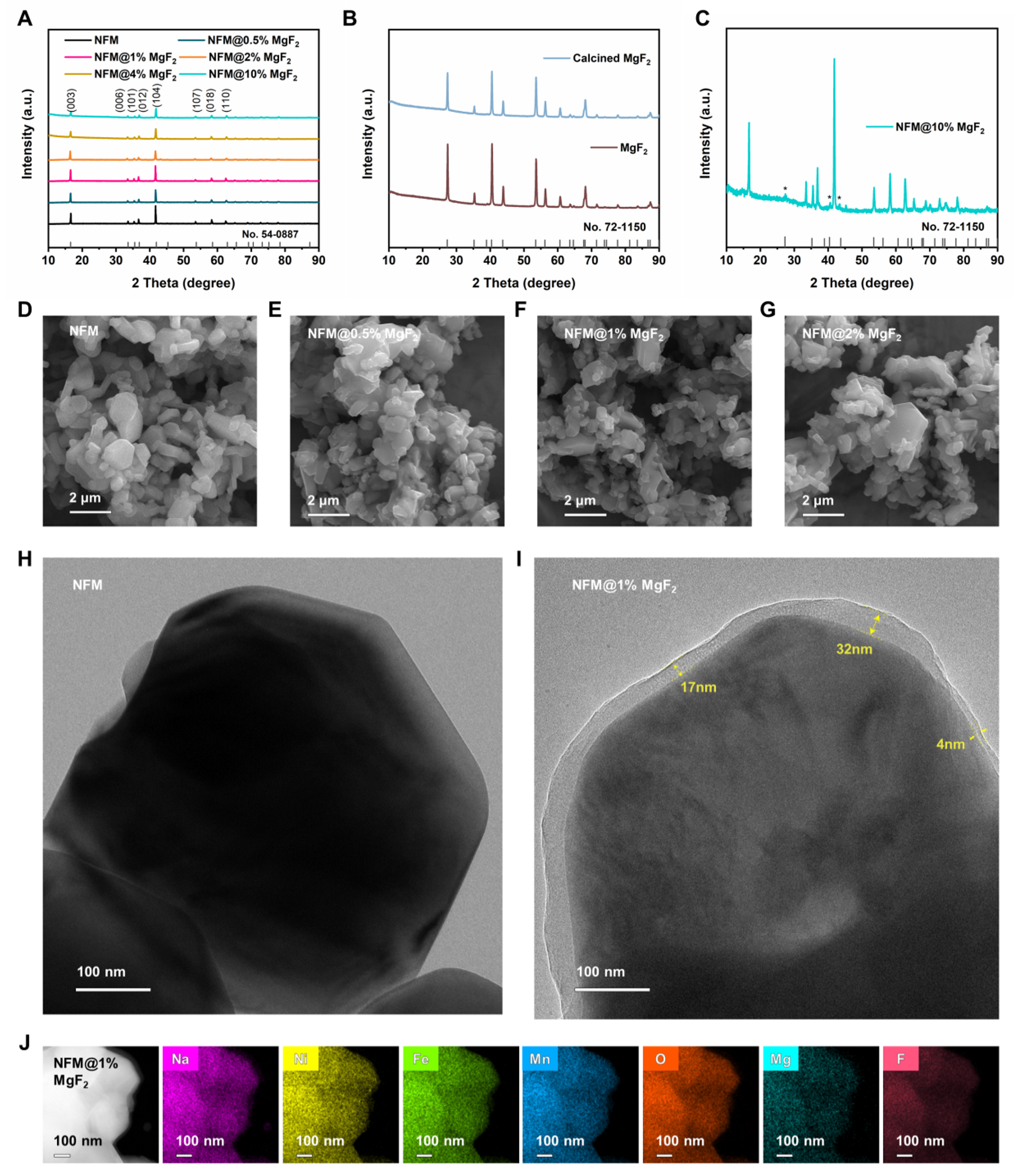
Figure 1. Morphological and structural characterization of the prepared samples. (A) Powder XRD patterns of pristine NFM and MgF2-loaded at different loading amounts, (B) XRD patterns of MgF2 before and after calcination, (C) XRD patterns of NFM@10%MgF2 sample. (D-G) SEM images of pristine NFM,