

fig5
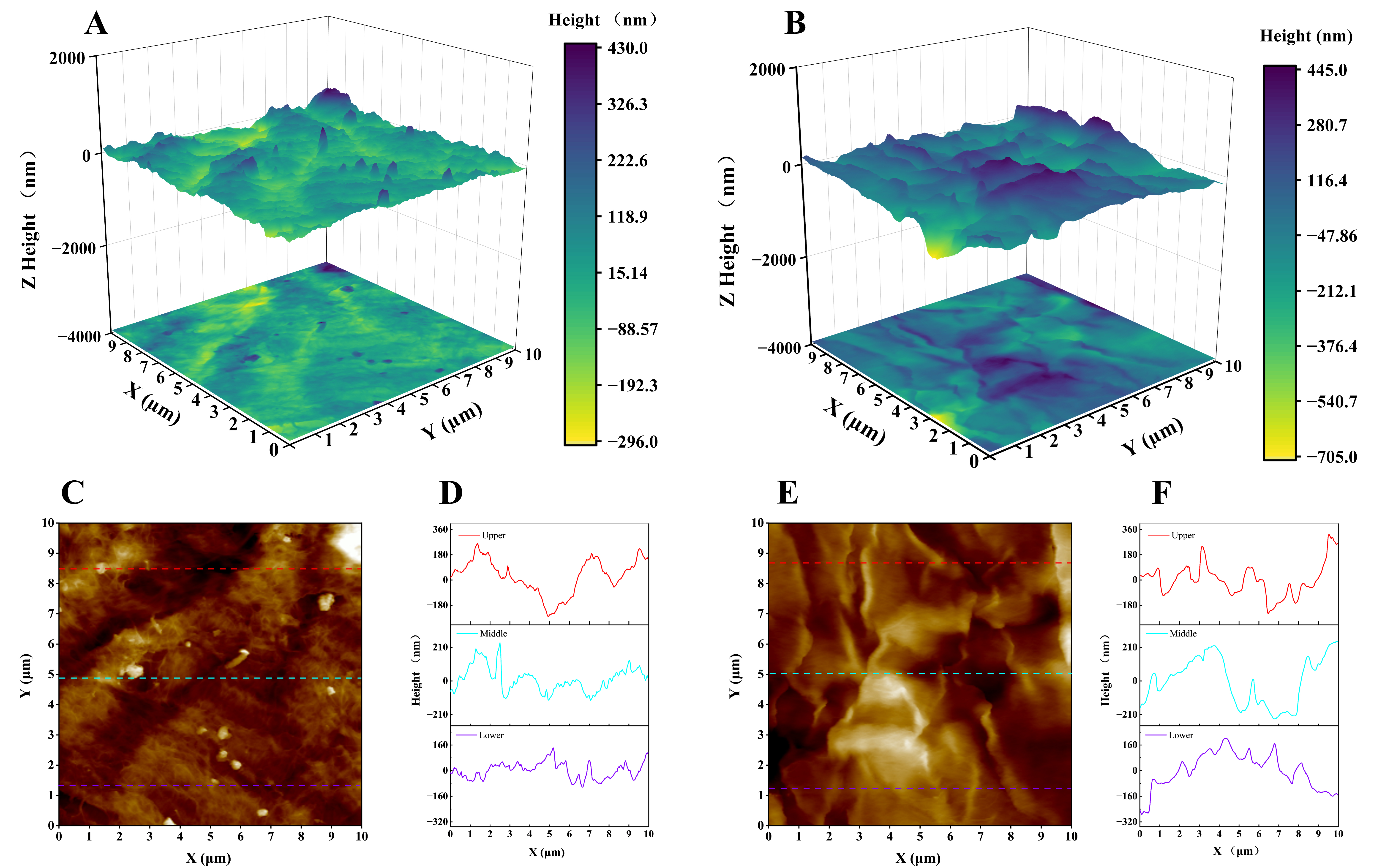
Figure 5. AFM characterization of cellulose fibers before and after bio-etching. (A) 3D surface morphology of CFs; (B) 3D surface morphology of m-CFs; (C) 2D height map of CFs; (D) The corresponding cross-sectional height profile of the original CFs; (E) 2D height map of m-CFs; (F) corresponding cross-sectional height profile of the m-CFs. AFM: Atomic force microscopy; m-CFs: modified cellulose fibers.