
fig2
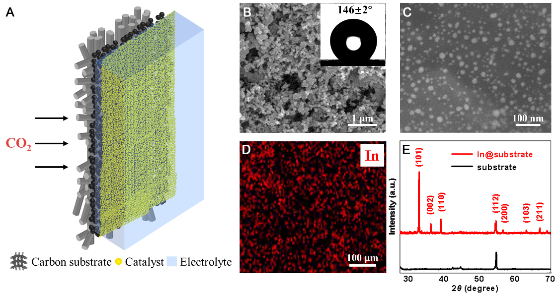
Figure 2. (A) Schematic illustration of the catalyst-integrated, superhydrophobic carbon-based TPB architecture; (B) Field emission scanning electron microscope (FE-SEM) of the superhydrophobic carbon substrate. The inset shows a water droplet placed on the substrate with a water contact angle of approximately 146 ± 2°; (C) FE-SEM of the In nanoparticles immobilized on the carbon substrate; (D) EDS elemental maps of In element; (E) XRD patterns of carbon substrate and In@substrate. EDS: Energy-dispersive X-ray spectroscopy; TPB: triphase boundary; XRD: X-ray diffraction; SEM: scanning electron microscope.