

fig3
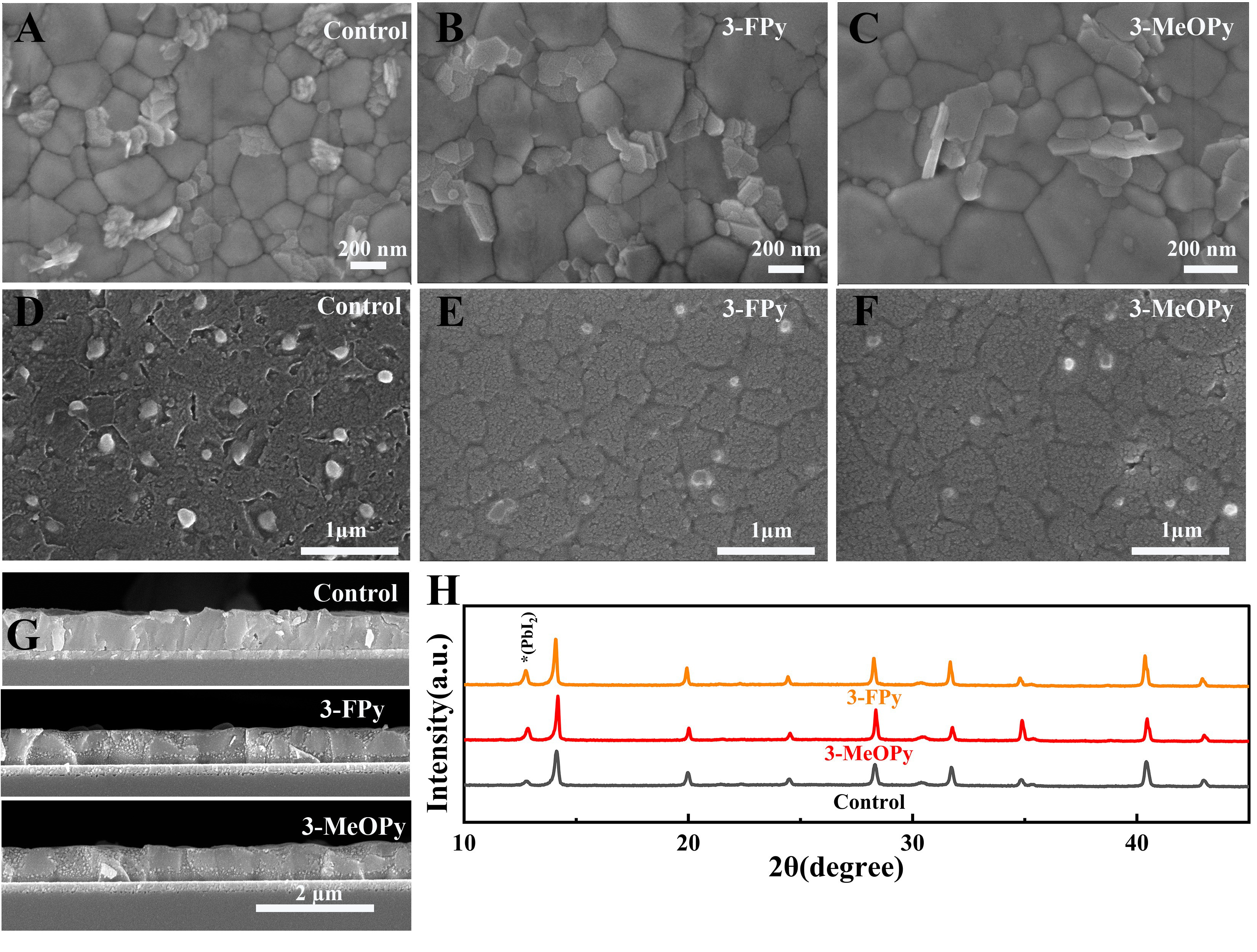
Figure 3. (A-C) Top-view SEM images of perovskite films deposited on (A) the control SAM, (B) the 3-FPy-treated SAM, and (C) the 3-MeOPy-treated SAM; (D-F) Corresponding bottom-view SEM images of the buried perovskite/SAM interfaces; (G) Cross-sectional SEM images of the complete perovskite films on the three substrates; (H) The XRD patterns for perovskite films prepared on different substrates. 3-FPy: 3-fluoropyridine; 3-MeOPy: 3-methoxypyridine; SEM: scanning electron microscopy; SAM: self-assembled monolayer; XRD: X-ray diffraction.