

fig2
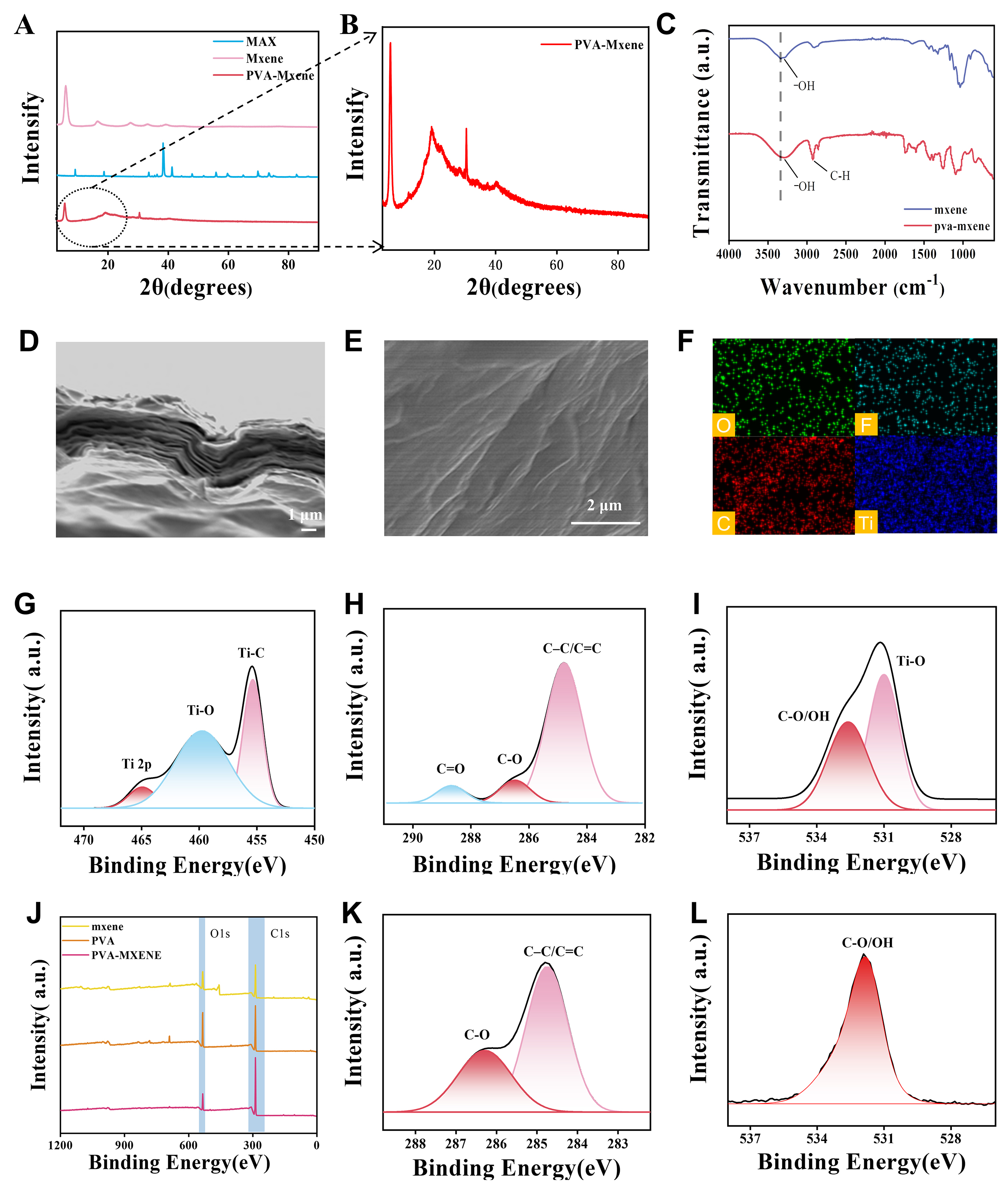
Figure 2. Structural, morphological, and compositional characterization of MXene, PVA, and PVA/MXene composite films. (A and B) X-ray diffraction (XRD) patterns of pure MXene, MAX phase, and the PVA/MXene composite film. (C) Fourier-transform infrared (FTIR) spectra comparing pure MXene and the PVA/MXene composite film, highlighting the chemical functional groups. (D and E) Surface and cross-sectional field-emission scanning electron microscope (FE-SEM) images of the MXene film at different magnifications. (F) Energy-dispersive X-ray spectroscopy (EDS) elemental mapping results showing the spatial distribution of characteristic elements within the composite. (G–I) High-resolution X-ray photoelectron spectroscopy (XPS) core-level spectra of (G) Ti 2p, (H) C 1s, and (I) O 1s of pure MXene. (J) XPS survey spectra of pristine MXene, pristine PVA, and the PVA/MXene composite film. (K and L) High-resolution XPS spectra of (K) C 1s and (L) O 1s of the PVA/MXene composite film.