


fig2
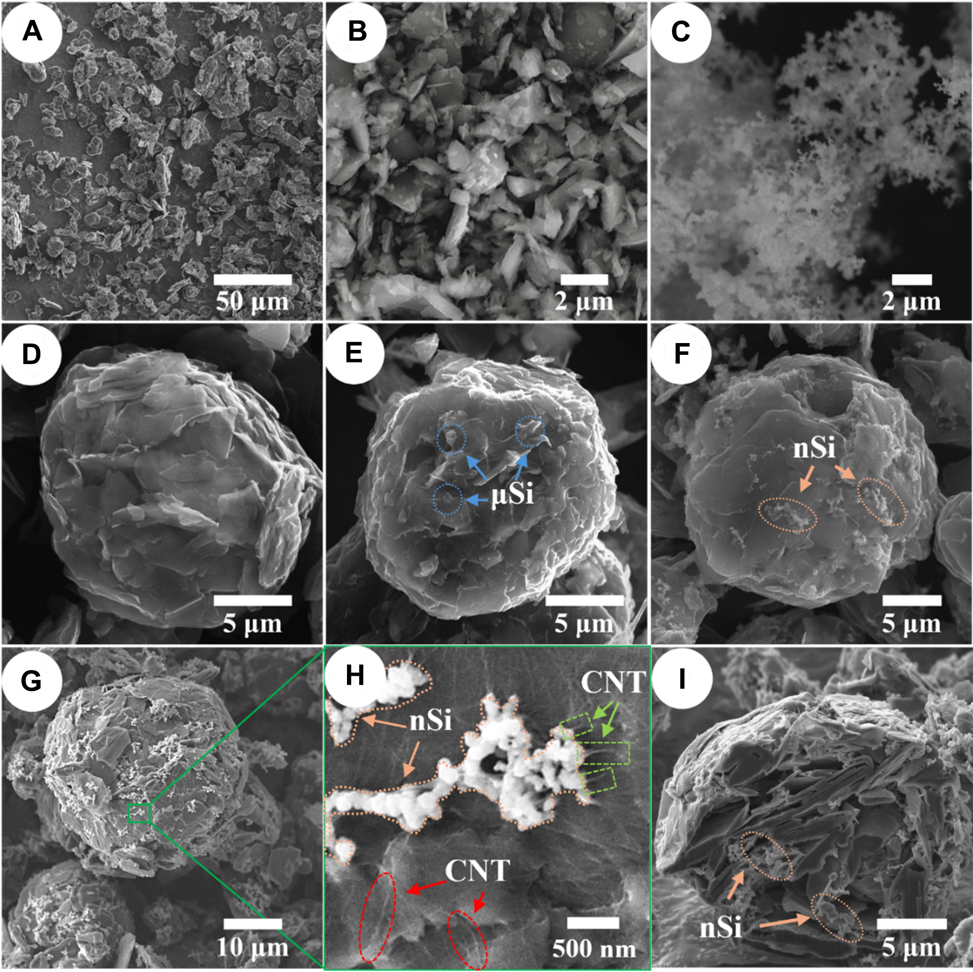
Figure 2. SEM images of (A) fNG, (B) µSi, (C) nSi, (D) GA, (E) G-9% µSi, (F) G-9% nSi, (G) G-15% nSi-CNT (lower magnification), (H) G-15% nSi-CNT (higher magnification), and (I) FIB cross-section of G-15% nSi-CNT. SEM: Scanning electron microscopy; fNG: natural graphite fines; µSi: micro-silicon; nSi: nano-silicon; GA: graphite agglomerate; CNT: carbon nanotube; FIB: focused ion beam.


