


fig2
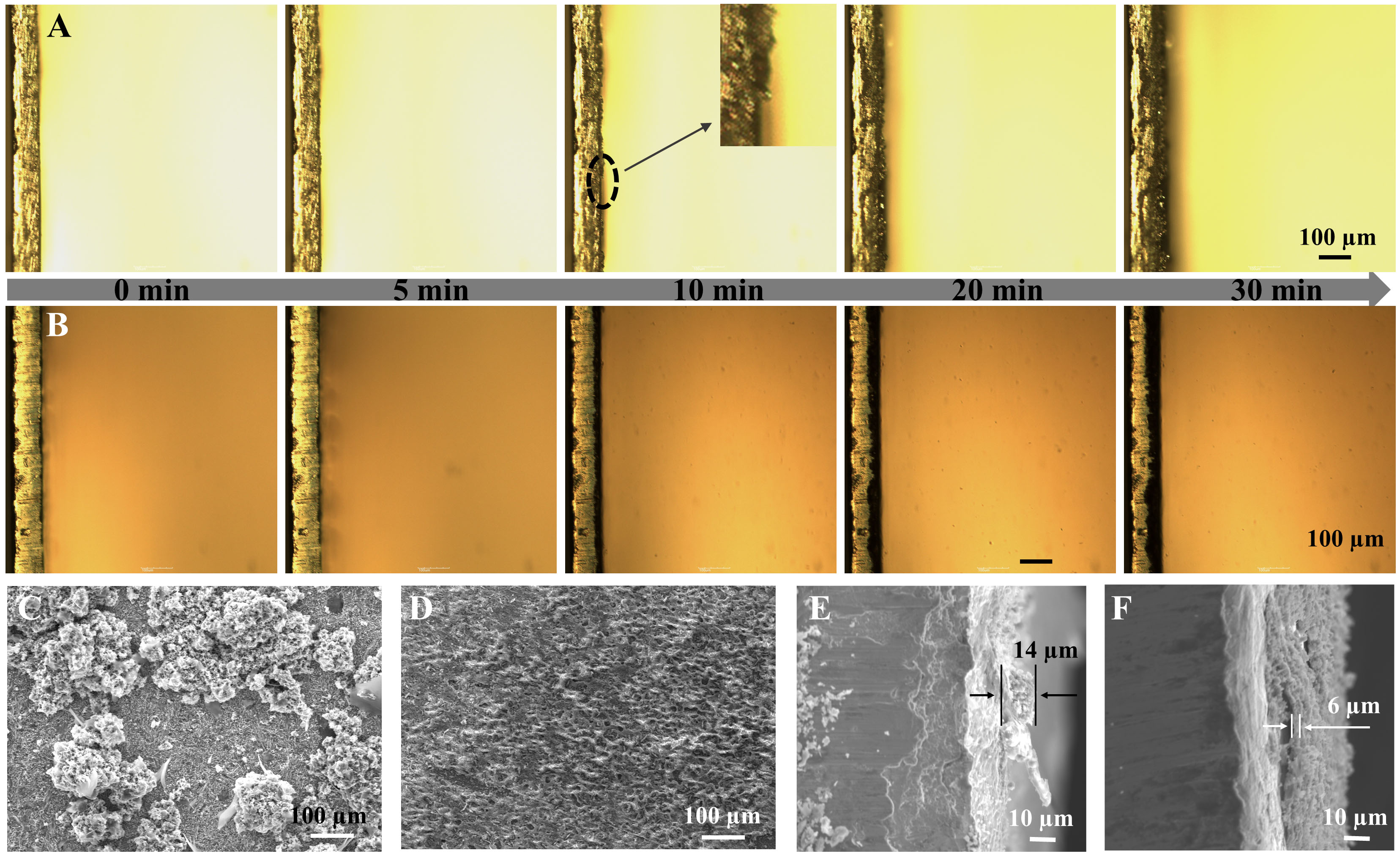
Figure 2. In situ optical microscopy images showing deposited Zn without (A) and with aniline (B) at 10 mA cm-2; Top-view and cross-section SEM of Zn anodes after 5 cycles without (C and E) and with (D and F) aniline at 10 mA cm-2. SEM: Scanning electron microscopy.


