fig1
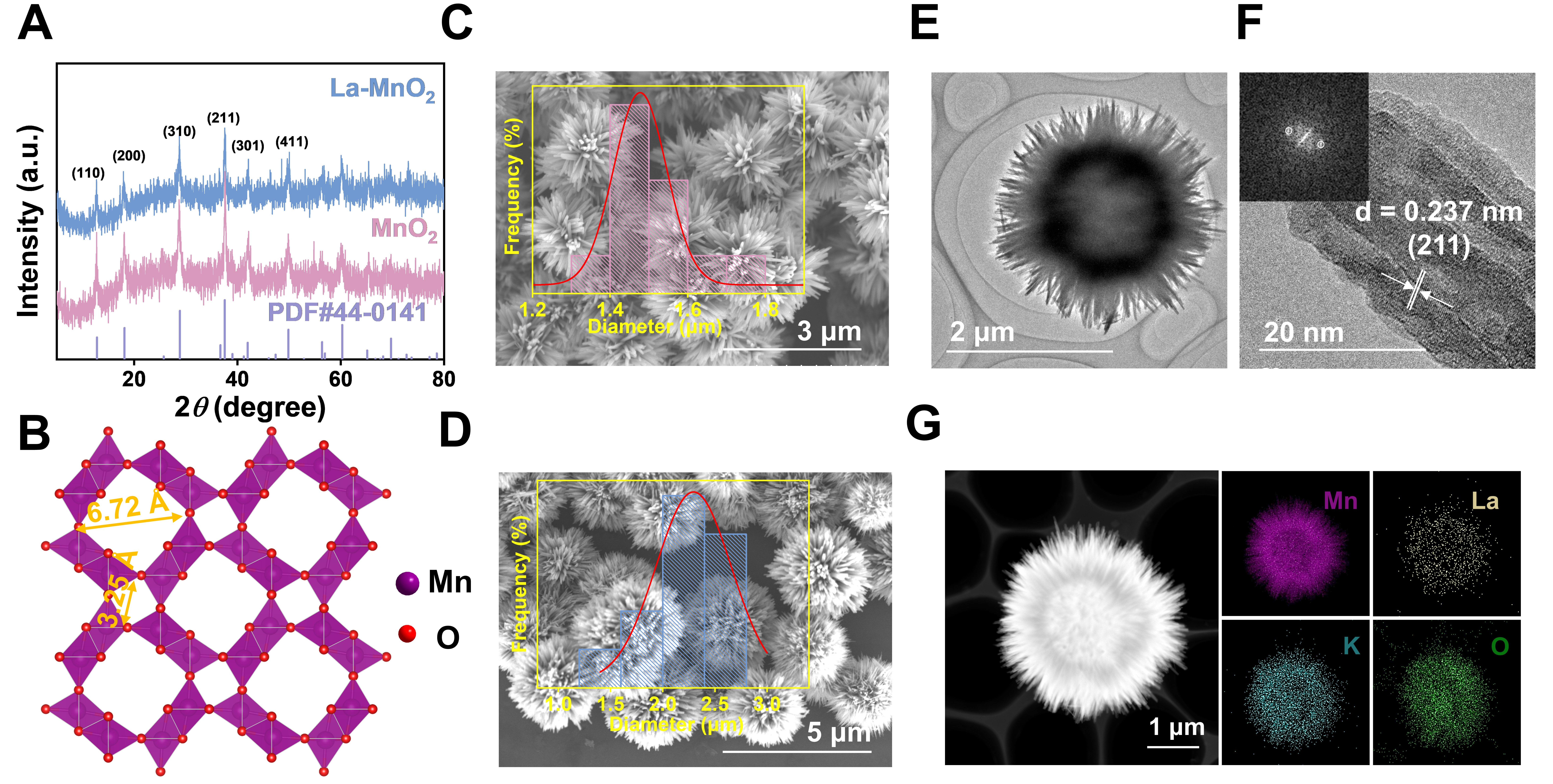
Figure 1. (A) The XRD patterns of La-doped MnO2 and MnO2; (B) The crystal structure of α-MnO2; The SEM images of (C) the sea urchin-like MnO2 and (D) La-doped MnO2 (The inset shows the particle size distributions); (E) The TEM and (F) HR-TEM images of


