

An exquisite symphony: protein post-translational modifications govern mitochondrial fate
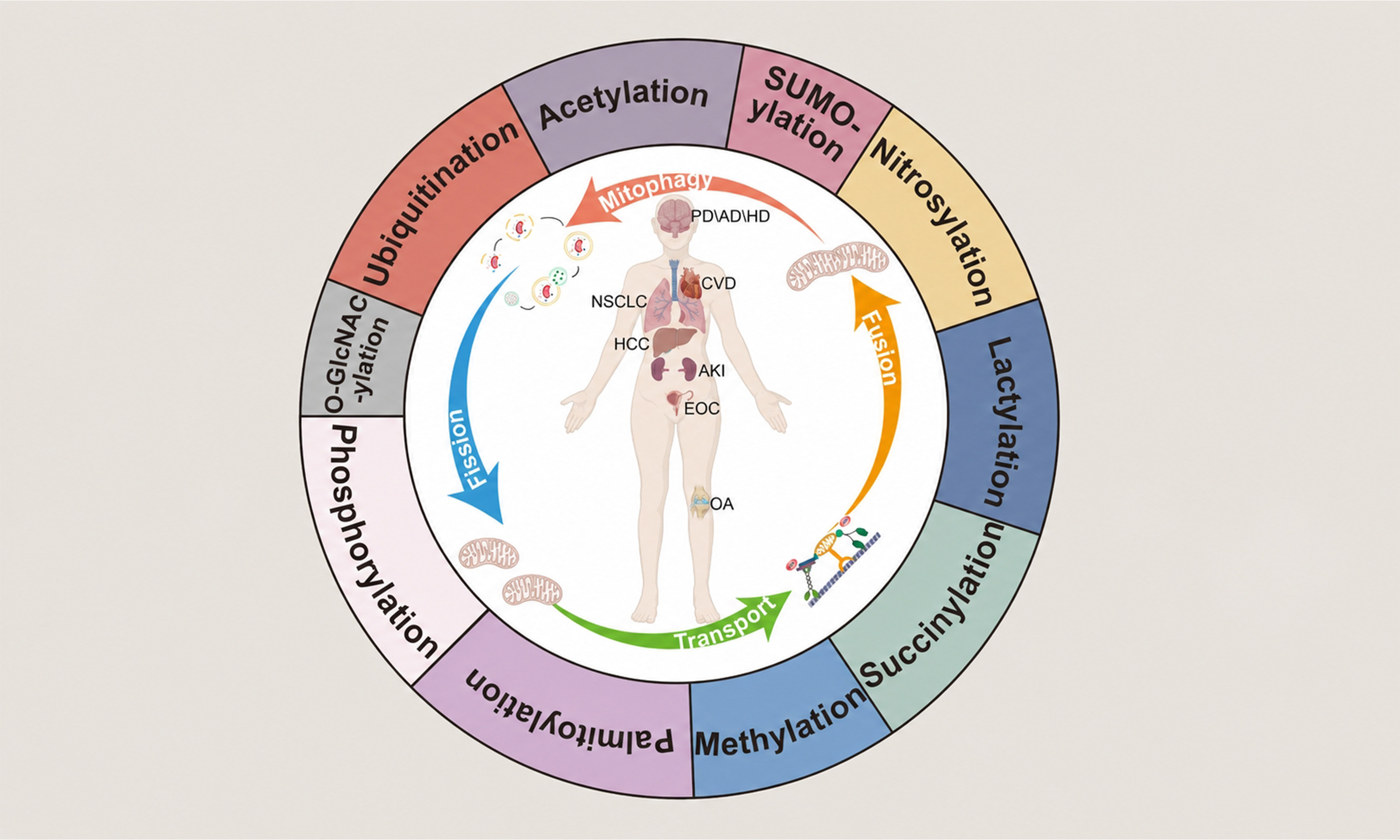
Abstract
Mitochondria are central hubs of metabolic activities in eukaryotic cells and play pivotal roles in various physiological and pathological processes. To accommodate fluctuating metabolic demands and respond to intracellular and extracellular stress, mitochondria undergo highly coordinated fission, fusion, mitophagy, and transport events mediated by dynamics-related proteins. Disrupting this delicate balance impairs mitochondrial bioenergetics and quality control, driving the onset and progression of numerous diseases, including neurodegeneration, heart failure, and cancer. Protein post-translational modifications (PTMs) constitute a critical regulatory layer, conferring remarkable functional diversity upon mitochondrial dynamics proteins beyond their genetic regulation. While extensive studies have characterized canonical PTMs such as phosphorylation and ubiquitination, the intricate roles of a broader spectrum of modifications - particularly those considered “atypical” - in fine-tuning mitochondrial behavior remain underexplored. In this review, we systematically summarize ten key PTMs, including lactylation, succinylation, SUMOylation, and S-nitrosylation, acting on core regulators such as dynamin-related protein 1(DRP1), optic atrophy 1 (OPA1), Parkin, and mitochondrial Rho GTPase 1 (MIRO1). We emphasize how these modifications integrate metabolic cues with mitochondrial dynamics and highlight their potential as diagnostic biomarkers and therapeutic targets. Furthermore, we discuss the sophisticated crosstalk between different PTM types and review emerging protein modification omics and genetic code expansion (GCE) tools designed to dissect the spatiotemporal dynamics of these modifications. By synthesizing current knowledge on these regulatory mechanisms, we aim to provide a comprehensive resource for understanding mitochondrial plasticity in health and disease.
Keywords: Mitochondrial, mitochondrial dynamics, mitochondrial dynamics protein, post-translational modification, physiology, pathology
Key Points
• Ten post-translational modifications converge on core mitochondrial dynamics proteins.
• Post-translational modifications target functional domains with site-specific, context-dependent outcomes.
• Dysregulated post-translational modification sites serve as dual-value candidates for biomarkers and therapeutic targets.
• Post-translational modification crosstalk and emerging tools open new frontiers for dissecting mitochondrial fate.
INTRODUCTION
Mitochondria are essential organelles in eukaryotic cells. Beyond supplying adenosine triphosphate (ATP) to sustain cell survival and proliferation, they support cell-specific demands through diverse metabolic pathways, thereby participating in various physiological and pathological processes. Although research on mitochondria spans over 150 years, it mirrors the evolution of scientific technology. As early as 1857, Swiss embryologist and histologist Rudolph Albert Von Kölliker used an optical microscope to observe a granular compartment with a membrane structure in the cytoplasm in muscle cells, and Retzius called them “Sarcosomes”[1-4]. In 1897, German biologist Carl Benda found abundant granules in muscle cells with either linear or granular morphology, introducing the term “mitochondrion” (derived from the Greek words ‘mitos’ and ‘chondros’, meaning thread and grain)[5]. By 1914, advances in microscopy allowed Lewis and colleagues to demonstrate that mitochondrial morphology was not static. They documented that mitochondria in cells could undergo morphological changes by fission or fusion, giving rise to the concept of mitochondrial dynamics[6,7]. Later in 1931, the dynamic changes of mitochondrial fission and fusion were confirmed during liver development[7-10], solidifying the notion of mitochondria as inherently dynamic organelles [Figure 1].
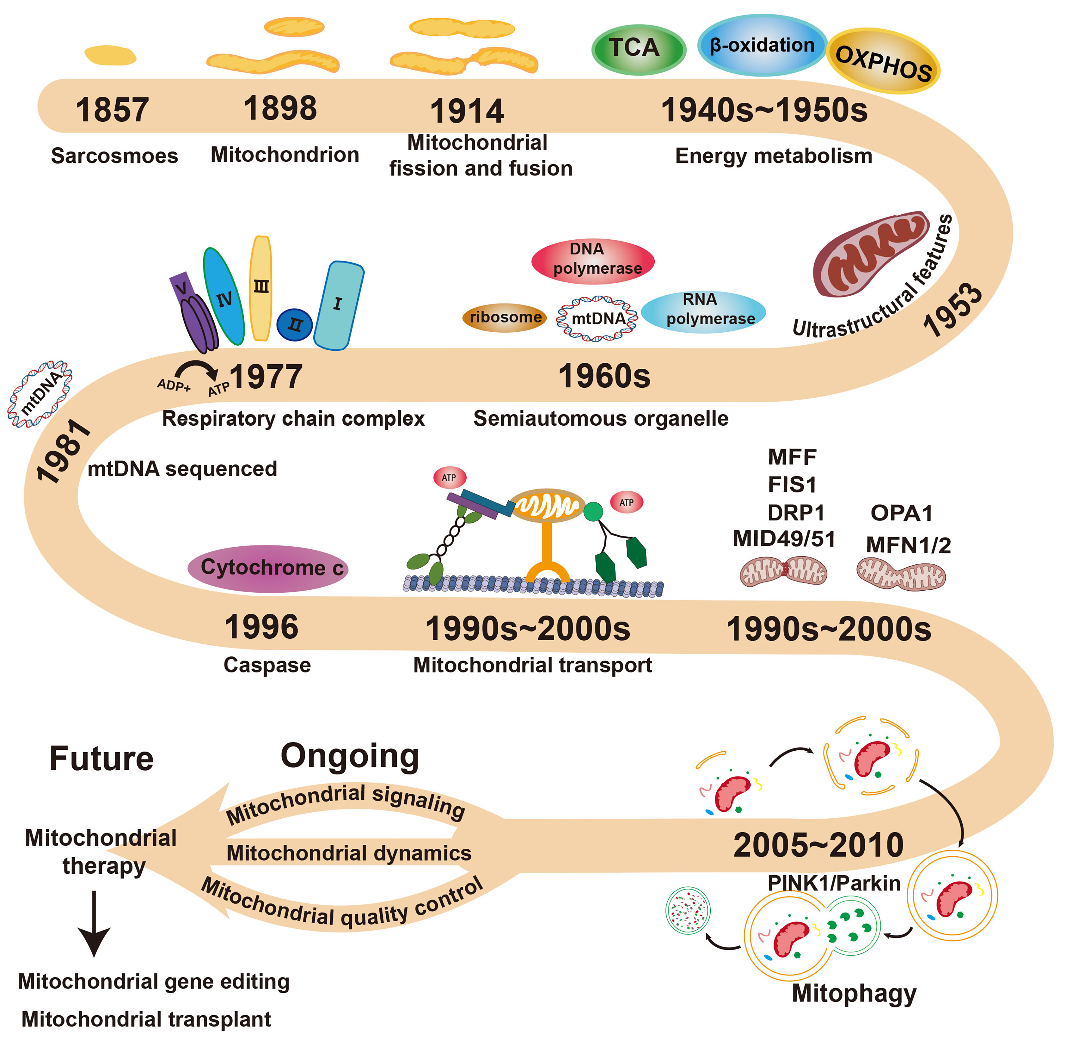
Figure 1. The research history of mitochondria.
Here, we outline key milestones in mitochondrial research history: the initial discovery and naming of sarcosomes; the observation of morphology and coining of the term “mitochondria”; the proposal of mitochondrial dynamics; elucidation of energy metabolism and the respiratory chain; characterization of mitochondrial ultrastructure, DNA, and semi-autonomy; identification of the cytochrome c apoptosis pathway, transport, and mitophagy; and the discovery of dynamics-related proteins, culminating in the analysis of fission/fusion mechanisms and novel mitochondrial functions. The data source for Figure 1 is generated based on references[1,5,6,8,11,13-23,29,35]. The figure was redrawn and designed by the authors based on existing reference images. Some of the mitochondrial icons in this figure were created with BioRender.com. OXPHOS: Oxidative phosphorylation; MFF: mitochondrial fission factor; FIS1: mitochondrial fission 1 protein; DRP1: dynamin-related protein 1; MID49/51: mitochondrial dynamics proteins of 49 and 51 KDa; MFN1/2: mitofusins 1/2; OPA1: optic atrophy 1; TCA: tricarboxylic acid cycle.
Because of the seminal discovery of mitochondria and its potential functions, numerous studies on this organelle were subsequently conducted. For example, the β-oxidation, tricarboxylic acid cycle, and oxidative phosphorylation (OXPHOS) occurring inside mitochondria were elucidated in the 1940s and 1950s, indicating the metabolic function of mitochondria[11,12]. The ultrastructural characteristics of mitochondria as double-membraned organelles with inner cristae were discovered by high-resolution electron microscopy in 1953, confirming their identity as a distinct organelle[13,14]. The genetic material, including DNA/RNA and their polymerases in mitochondria, was found in the 1960s, demonstrating that mitochondria are semi-autonomous organelles[15-17]. The four major complexes of the respiratory chain and the proposal of the chemiosmotic theory were established in the 1960s and 1970s, deepening the understanding of bioenergetics[18,19]. Mitochondrial Rho GTPase (Miro), a member of the Rho GTPase family, and Milton/Trafficking kinesin-binding protein (TRAK) proteins were identified as the core adapter complex linking kinesin/dynein to mitochondria, elucidating the mechanism of mitochondrial transport between the 1970s and 2000s[20-28]. The mitochondrial cytochrome C-mediated apoptosis pathway was uncovered in 1996[29], indicating the important role of mitochondria in apoptosis[30-34]. Mitochondrial fission/fusion proteins including dynamin-related protein 1(DRP1)[35], mitochondrial fission protein 1 (FIS1), mitochondrial dynamics proteins of 49 and 51 KDa (MID49/51), mitochondrial fission factor (MFF), optic atrophy 1 (OPA1), mitofusins (MFN1/2), and their regulatory mechanisms on mitochondrial dynamics were identified in 1990s and 2000s, demonstrating the regulatory role of mitochondrial dynamics in cellular processes[36-40]. Further, the role of phosphatase and tensin homolog (PTEN)-induced putative kinase 1(PINK1) and E3 ubiquitin-protein ligase parkin (Parkin) mediated mitophagy as well as the transmembrane receptor proteins B-cell lymphoma 2 (BCL2)/adenovirus E1B 19 kDa protein-interacting protein 3 (BNIP3) and FUN14 domain-containing protein 1 (FUNDC1) mediated Parkin-independent mitophagy pathways were uncovered in 2005-2010, highlighting the selective degradation of mitochondria in various diseases[41-49]. Currently, research on mitochondria mainly focuses on mitochondrial signaling, mitochondrial quality control, and mitochondrial dynamics in regulating physiological and pathological processes[50-54]. For example, our team demonstrated that superoxide signaling of mitochondrial superoxide bursts during early reprogramming leads to reduced DNA methylation on the Nanog promoter, thereby enhancing Nanog expression and facilitating the reprogramming of somatic cells into induced pluripotent stem cells (iPSCs)[55-57]. Mitochondrial quality control and mitochondrial dynamics were also important for the generation of iPSCs and maintenance of pluripotency in embryonic stem cells (ESCs)[58-60]. Moreover, mitochondrial signaling, quality control, and dynamics have been implicated in many physiological and pathological processes, including early embryonic development, neural activation, aging, cancer development, and immune regulation[61-67]. Among these processes, the regulation of mitochondrial dynamics - encompassing mitochondrial fission/fusion, transport, and mitophagy - plays a central role, linking these mitochondrial processes to cell fate decisions.
Protein post-translational modifications (PTMs) refer to the covalent attachment of functional groups - such as phosphate, ubiquitin, and methyl groups - to proteins, regulating protein activity or stability[68-72]. The activity and function of mitochondrial dynamics-related proteins can also be regulated by different PTMs. Accumulating evidence links specific PTMs on these proteins to disease. For example, phosphorylation, ubiquitination, and acetylation of DRP1 dynamically regulate mitochondrial fission and fusion, impacting neuronal function, metabolic reprogramming, and disease progression[58,73-75]. However, the therapeutic relevance of PTM-mediated regulation of mitochondrial dynamics has been largely overlooked. To better understand the role of PTMs on mitochondrial dynamics-related proteins, we summarize in detail the effects of ten key PTMs on mitochondrial dynamics-related proteins across various physiological and pathological processes in detail. Furthermore, based on the roles and mechanisms of PTMs on mitochondrial dynamics-related proteins, we propose that they can serve as predictive markers for clinical disease prognosis and treatment. Moreover, we discuss the cross-talk of PTMs and highlight novel tools for studying their regulatory functions in mitochondrial dynamics.
PTMS ON MITOCHONDRIAL DYNAMICS-RELATED PROTEINS
Mitochondrial dynamics refers to the process by which mitochondria maintain their morphology, mass, and functional balance through mitochondrial fission/fusion, transport, and mitophagy[51]. Mitochondrial dynamics are necessary for mitochondrial self-renewal and functional stability, serving as a key self-repair mechanism that governs cell energy metabolism, calcium homeostasis, reactive oxygen species (ROS) regulation and cell fate determination[50]. Mitochondrial dynamics related-proteins include fission proteins (DRP1, MID49, MID51, MFF and FIS1)[35,76-78], fusion proteins (OPA1, MFN1 and MFN2), transport proteins (TRAK1, TRAK2, MIRO1 and MIRO2)[20-23], and mitophagy proteins (PINK1 and Parkin)[41-44] [Table 1]. To date, PTMs have been identified in almost all of these mitochondrial dynamics-related proteins. Moreover, PTMs on these proteins exhibit specificity for specific functional domains [Figure 2]. For instance, PTMs on DRP1 predominantly occur in the variable domain (VD) - a region critical for membrane lipid binding, which suggests PTMs may regulate DRP1-membrane interactions[79]. PTMs on FIS1 primarily localize to its N-terminal arm, a domain negatively correlated with DRP1 binding, indicating potential modulation of FIS1-DRP1 complex formation[80]. PTMs on MFF, particularly phosphorylation, are predominantly clustered within its central intrinsically disordered region (IDR), implying a role in modulating MFF-DRP1 interactions[76,81]. PTMs on OPA1 localize to its BSE (bundle signaling element) domain, which is spatially adjacent to the GTPase domain, suggesting that these PTMs may regulate the function of OPA1[82,83]. PTMs on MFN1/2 were concentrated in the GTPase domain, essential for GTP hydrolysis and conformational changes, implying that PTMs may regulate the efficiency of MFN1/2 conformational transitions[84,85]. Phosphorylation of Parkin primarily occurs at its N-terminal region, which constitutes the active site for its ubiquitin ligase activity[86]. This suggests that phosphorylation modulates Parkin’s protein ubiquitination function. Acetylation of Miro1 takes place within its N-terminal GTPase domain, indicating that this modification regulates the protein’s catalytic activity[87]. These findings demonstrate that PTMs in mitochondrial dynamics-related proteins preferentially target functional domains, underscoring their crucial roles in modulating protein function. This domain-specific PTM pattern warrants focused investigation. Below, we discuss the roles of ten key PTMs on mitochondrial dynamics-related proteins in detail [Table 2].
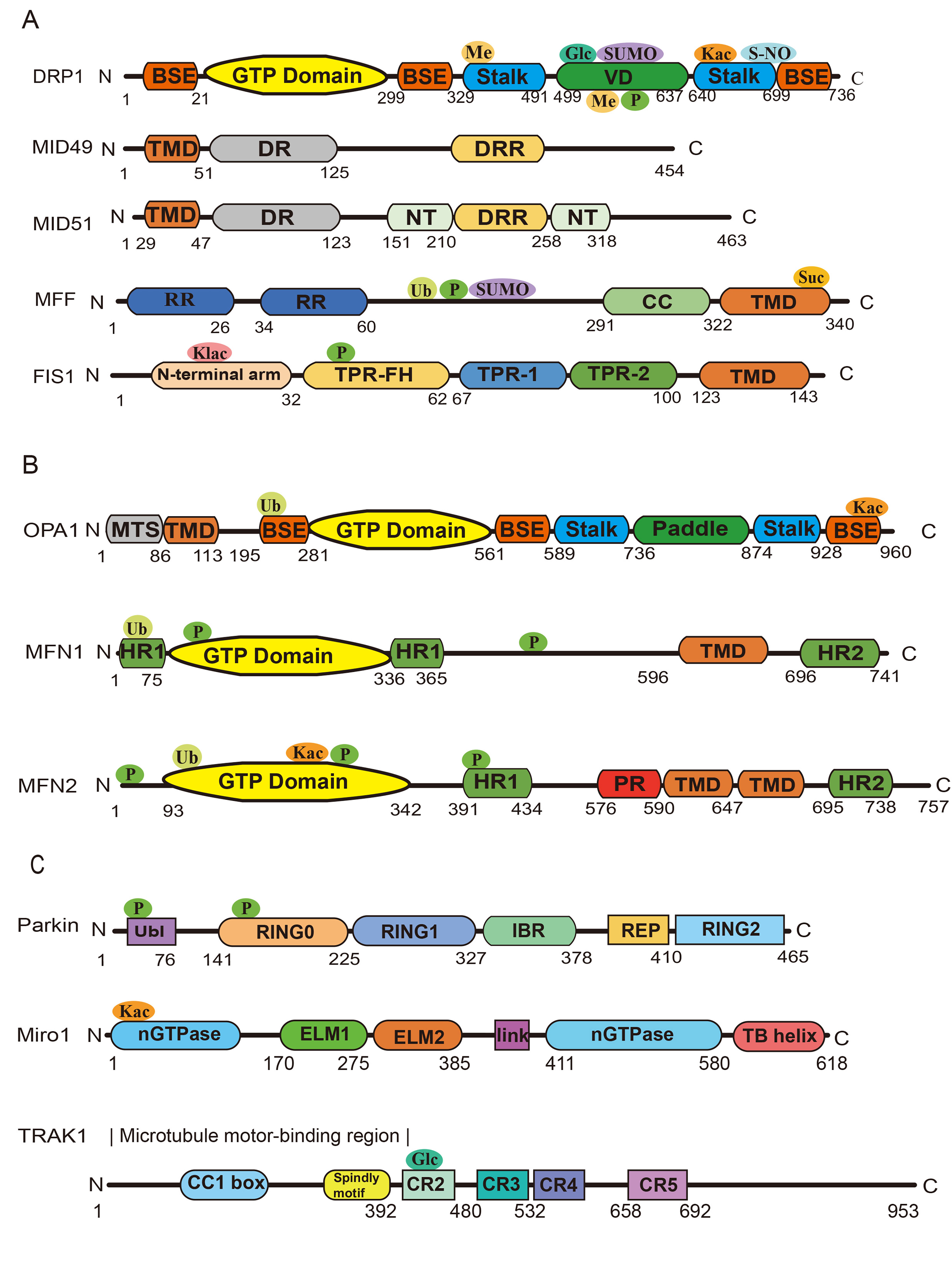
Figure 2. PTMs on mitochondrial dynamics-related proteins.
We mapped all the PTMs on mitochondrial dynamics-related proteins, and annotated their structural locations. The data source for Figure 2 is generated based on references[76,79-87,159]. The figure was redrawn and designed by the authors based on existing reference images. (A) The PTMs on mitochondrial fisson proteins, and annotated their structural locations; (B) The PTMs on mitochondrial fussion proteins, and annotated their structural locations; (C) The PTMs on mitochondrial mitophagy and transoprt proteins, and annotated their structural locations. Ub: Ubiquitination; SUMO: small ubiquitin-like modifier; Kac: lysine acetylation; P: Phosphorylation; Met: Methylation; Glc: GlcNAc; Suc: Succinylation; Klac: lysine lactylation; PTMs: post-translational modifications; DRP1: dynamin-related protein 1; MID49/51: mitochondrial dynamics proteins of 49 and 51 KDa; MFF: mitochondrial fission factor; FIS1: mitochondrial fission 1 protein; OPA1: optic atrophy 1; MFN1: mitofusins 1; Miro1: mitochondrial Rho GTPase 1; TRAK1: trafficking kinesin-binding protein 1.
Mitochondrial dynamics proteins and their function
| Category | Protein | Localization | Core function & Notes |
| Mitochondrial fission | DRP1 | Translocates from the cytoplasm to the OMM upon activation | Core mitochondrial fission executor[35] |
| FIS1 | OMM | Recruits DRP1 to mitochondria[78] | |
| MFF | OMM | Recruit DRP1 to fission sites[76] | |
| MID49/51 | OMM | Recruit DRP1 to fission sites[163] | |
| Mitochondrial fusion | MFN1 | OMM | Mediate OMM fusion[84] |
| MFN2 | OMM | Cooperate with MFN1 for OMM fusion[85] | |
| OPA1 | IMM | Mediate IMM fusion; regulate cristae morphology and respiratory complex assembly[164] | |
| Mitochondrial transport | MIRO1/2 | OMM | Links mitochondria to TRAK proteins[21] |
| TRAK1/2 | OMM/cytoplasm | Connect MIRO to kinesin (anterograde) and dynein (retrograde)[23] | |
| Mitophagy Pathways Ubiquitin-Dependent (PINK1/Parkin) | PINK1 | Stabilized-on-damaged OMM | Accumulate upon loss of membrane potential[155] |
| Parkin (PRKN) | Translocate from cytoplasm to OMM upon activation | Ubiquitinates OMM proteins[44] |
PTMs of mitochondrial dynamics proteins and their sites
| Proteins | PTMs | Sites | Regulatory | Effect on mitochondria |
| OPA1 | Ubiquitination | Lys228 | TRIM22 | Inhibit mitochondrial fusion |
| OPA1 | Acetylation | Lys926/Lys931 | Pathological cardiac stress[165] | Inhibit mitochondrial fusion |
| MFN1 | Phosphorylation | Ser86/Thr562 | βIIPKC[89]/ERK[166] | Inhibit mitochondrial fusion |
| MFN1 | Ubiquitination | Lys48 | MARCH5[116] | Inhibit mitochondrial fusion |
| MFN2 | Phosphorylation | Ser442/Tyr269 | PINK1[91]/c-Abl[90] | Inhibit mitochondrial fusion |
| MFN2 | Ubiquitination | Lys129 | MARCH5[120] | Inhibit mitochondrial fusion |
| MFN2 | Acetylation | Lys243 | Lipid overload[132] | Mitochondrial dysfunction |
| DRP1 | Phosphorylation | Ser616 | PINK1[93]/CDK5[94] | Promote mitochondrial fission |
| DRP1 | Phosphorylation | Ser637 | TBK1[95] | Inhibit mitochondrial fission |
| DRP1 | Acetylation | Lys642 | Lipid overload[133] | Promote mitochondrial fission |
| DRP1 | Methylation | Arg403/Arg634 | CARM1[167] | Promote mitochondrial fission |
| DRP1 | O-GlcNAcylation | Thr585/Thr586 | Diabetic patients[139] | Promote mitochondrial fission |
| DRP1 | S-palmitoylation | Cys | ZDHHC13[142] | Normal mitochondrial fission-fusion dynamics |
| DRP1 | S-Nitrosylation | N/A | Amyloid-β[148] | Promote mitochondrial fission |
| DRP1 | SUMOylation | Multiple sites | IRI[128] | Promote mitochondrial fission |
| DRP1 | SUMOylation | N/A | SENP5L[129] | Inhibit mitochondrial fission |
| DRP1 | Inhibition of SUMOylation | N/A | SENP5S[129] | Promote mitochondrial fission |
| FIS1 | Phosphorylation | Tyr38/Thr34 | Met[96]/DNA-PKcs[97] | Promote mitochondrial fission |
| FIS1 | Lactylation | Lys20 | Nalac[144] | Promote mitochondrial fission |
| MFF | Phosphorylation | Ser131 | AKT[99] | Promote mitochondrial fission |
| MFF | Succinylation | Lys302 | CPT1A[146] | Promote mitochondrial fission |
| MFF | Ubiquitination | N/A | PARKIN[121] | Promote mitophagy |
| MFF | SUMOylation | Lys151 | SUMO1/SUMO2/3[168] | Promote mitochondrial fission |
| MID49 | Ubiquitination | N/A | MARCH5[169] | Inhibit mitochondrial fission |
| PARKIN | phosphorylation | N/A | PINK[109] | Critical for its translocation to mitochondria |
| PINK1 | phosphorylation | Ser228/Ser402 | Autophosphorylation[103] | Promote mitophagy |
| MAP1LC3 | Phosphorylation | Serine 12 | PKA[106] | Inhibit PE binding |
| MIRO1/2 | ubiquitination | N/A | PARKIN[125] | Degradation of hmiro1/2 |
| MIRO1 | deacetylation | lysine 105 | HDAC6[134] | Inhibit mitochondrial transport |
| MID51 | ubiquitination | N/A | N/A | Promote Mid51 degradation[126] |
| TRAK1 | O-GlcNAcylation | N/A | N/A | Reduce mitochondrial motility[170] |
| FIS1 | deSUMOylation | N/A | SENP1 | Maintained mitochondrial integrity[130] |
PTMS DRIVE MITOCHONDRIAL DYNAMICS
Protein phosphorylation
Protein phosphorylation constitutes an enzyme-catalyzed process in which the γ-phosphate of ATP is transferred to amino acid side chains of proteins. This reversible modification is mediated by protein kinases (PKs) for phosphorylation and reversed by protein phosphatases (PPases) for dephosphorylation. As a critical regulatory mechanism for protein activity and function, phosphorylation occurs predominantly on serine (Ser), threonine (Thr), and tyrosine (Tyr) residues[88].
Mitochondrial fusion proteins
During heart failure, beta II protein kinase C (βIIPKC) translocates to the mitochondrial outer membrane and phosphorylates MFN1 at Ser86 within its GTPase domain. Phosphorylation within this domain inhibits GTPase activity and exacerbates mitochondrial fragmentation, leading to cell cytotoxicity. Targeted disruption of the MFN1-βIIPKC interaction using selective antagonists attenuates mitochondrial fragmentation and improves cardiac function[89]. In neurons, extracellular signal-regulated protein kinases (ERK) phosphorylate MFN1 at Thr562, inducing mitochondrial fragmentation via MEK/ERK pathway activation and promoting neuronal apoptosis. In immortalized mouse embryonic fibroblast cells (MEFs) under endoplasmic reticulum (ER) stress, the cellular Abelson gene product(c-Abl) translocates to mitochondria and phosphorylates MFN2 at Tyr269, leading to mitochondrial fragmentation and accelerated neuronal death. c-Abl inhibition reduces fragmentation and delays Amyotrophic Lateral Sclerosis (ALS) progression, suggesting therapeutic potential[90]. In MEFs and human embryonic kidney 293 (HEK293) cells, MFN2 was phosphorylated at Thr111, Ser378 and Ser442 sites by PINK1, leading to mitochondrial fragmentation[91].
Mitochondrial fission proteins
In murine tissues, phosphorylation of DRP1 at Ser600 and Ser579 is mutually correlated and occurs simultaneously upon activation of cyclic adenosine monophosphate (cAMP)-dependent protein kinase A (PKA), leading to mitochondrial fission. Notably, Ser579 phosphorylation serves as a downstream event of Ser600 phosphorylation following PKA activation. Even in the absence of external stimuli, Ser600 phosphorylation on DRP1 alone was sufficient to elevate phosphorylation levels of Ser579 on DRP1, protecting Ser579 from dephosphorylation. However, complete activation of DRP1-mediated mitochondrial fission activity requires simultaneous phosphorylation at both residues[92]. Impeding Ser579 phosphorylation on DRP1 redirects the outcome of Ser600 phosphorylation of DRP1 toward mitochondrial tubulation rather than fission. In Parkinson’s disease (PD) pathogenesis, PINK1-dependent Ser616 phosphorylation of DRP1 facilitates mitophagy by recruiting Parkin to damaged mitochondria, serving as both a potential PD biomarker and therapeutic target. PINK1 knockout models exhibit significantly reduced Ser616 phosphorylation levels[93]. Chronic ethanol exposure in male C57BL/6 mice induces cyclin-dependent kinase 5 (CDK5)-mediated Ser616 phosphorylation on DRP1 in hippocampal neurons, promoting mitochondrial fragmentation, irreversible synaptic damage, and cognitive impairment. Pharmacological modulation of CDK5 activity may mitigate ethanol-induced neurotoxicity[94]. In chondrocytes, Tank-binding kinase 1 (TBK1) overexpression phosphorylates Ser637 on DRP1, inducing mitochondrial elongation, mitophagy, and apoptosis resistance, thereby ameliorating osteoarthritis progression. The TBK1/DRP1 axis represents a promising therapeutic target for osteoarthritis[95]. In hepatocellular carcinoma (HCC) cells, receptor tyrosine kinase Met directly phosphorylated FIS1 at Tyr38, which facilitates DRP1 recruitment to mitochondria, thereby promoting tumor growth and chemoresistance. Pharmacological inhibition of Met effectively suppresses these malignant phenotypes, suggesting this axis as a therapeutic target for metastatic HCC[96]. In renal tubular epithelial cells, DNA-dependent protein kinase catalytic subunit (DNA-PKcs) interacts with FIS1 and phosphorylates it at Thr34, enhancing FIS1-DRP1 binding and inducing pathological mitochondrial fragmentation, thereby contributing to acute kidney injury (AKI). DNA-PKcs inhibitors show protective potential against AKI progression[97]. In ovarian cancer cells (OCCs), knockdown of CRL4CUL4A/DDB1 (E3 ubiquitin ligase) significantly enhances AMP-activated protein kinase (AMPK) activity and induces Ser172/Ser146 phosphorylation of MFF, thereby recruiting DRP1 to mitochondria. Phosphorylation of MFF at Ser172/Ser146 leads to mitochondrial fragmentation and enhanced autophagy in cisplatin-resistant OCCs, indicating the therapeutic significance of CRL4CUL4A/DDB1 knockdown in ovarian cancer treatment[98]. In mice, fasting combined with sensory stimulation (visual/olfactory exposure to inaccessible food or light stimulation of proopiomelanocortin neurons) activates protein kinase B (AKT), which subsequently phosphorylates MFF at Ser131, rapidly inducing hepatic mitochondrial fragmentation. Inhibition of MFF phosphorylation abolishes AKT-driven mitochondrial fragmentation in the liver[99]. In human renal tubular epithelial cells (HK-2), regulator of calcineurin 1 (RCAN1) binds to c-Jun N-terminal kinase (JNK) to increase MFF phosphorylation, promoting DRP1 mitochondrial translocation. MFF phosphorylation induces mitochondrial fragmentation, resulting in mitochondrial dysfunction and apoptosis, and accelerates progression of AKI. RCAN1 deletion suppresses apoptosis and alleviates renal damage[100]. During mammalian cell mitosis (excluding interphase), protein kinase D (PKD) specifically phosphorylates MFF at Ser155, Ser172, and Ser275, driving mitochondrial fission to promote cell survival. PKD/MFF-dependent mitochondrial fission is essential for maintaining genomic integrity during cell division[101]. In human osteosarcoma cells (U2OS) and MEFs, AMPK phosphorylates MFF at Ser155/Ser172, recruiting DRP1 to induce fragmentation. AMPK-dependent MFF phosphorylation signals to the nucleus and initiates mitochondrial biogenesis to replace damaged mitochondria[102].
Beyond mitochondrial fission and fusion, protein phosphorylation also plays pivotal roles in mitophagy. For instance, phosphorylation of PINK1 is essential for recruiting Parkin during mitophagy[103,104]. Furthermore, phosphorylation of Parkin itself is critical for its translocation to mitochondria[105]. In autophagy, phosphorylation of microtubule-associated protein 1 light chain 3A (MAP1LC3A) at serine 12 (S12) inhibits its binding to phosphatidylethanolamine (PE), thereby suppressing autophagy[106]. Phosphorylation of MAP1LC3C at serine 93 (S93) facilitates the formation of a stable lipidated LC3C/GABARAP-L2 coat on early-stage autophagosomes and contributes to maintenance of unidirectional autophagosome-lysosome flux[107]. Concurrently, Parkin is recruited to mitochondria, promoting mitochondrial autophagy and apoptosis[91]. Paradoxically, PINK1-dependent Ser442 phosphorylation in macrophages facilitates Parkin-mediated ubiquitination of mitochondrial outer membrane (MOM) proteins, initiating mitophagy. PINK1 deficiency exacerbates mitochondrial damage during renal fibrosis[108,109]. In doxorubicin-stressed U2OS cells, JNK phosphorylates MFN2 at Ser27, promoting mitochondrial fragmentation and apoptosis[110].
In summary, protein phosphorylation regulates mitochondrial dynamics and quality control through three recurring principles: (i) reversibility via paired kinase/phosphatase modules, allowing rapid toggling; (ii) site-level encoding, whereby a single protein (e.g., DRP1 Ser616 vs. Ser637) can be switched between opposing functional outputs depending on which residue is modified; and (iii) context dependence, wherein the same phosphorylation site may exert pro-pathological or compensatory effects across cancer, neurodegeneration, and metabolic injury. These features collectively make phosphorylation both a mechanistic link and a pharmacologically accessible node in mitochondrial PTM networks.
Protein ubiquitination
Ubiquitination is a multi-component collaborative process requiring ATP, E1 ubiquitin-activating enzyme, E2 ubiquitin-conjugating enzyme, E3 ubiquitin ligase, and substrates. Initially, the E1 activating enzyme utilizes energy to hydrolyze ATP to activate ubiquitin (Ub), forming a thioester bond between the C-terminal glycine (Gly) of Ub and a cysteine residue in E1. The Ub-E1 complex transfers Ub to E2 via transesterification to generate the Ub-E2 complex. Subsequently, the Ub-E2 complex transfers Ub to substrates through two distinct pathways: (1) E3 directly recognizes the substrates and catalyzes the attachment of Ub’s C-terminal Gly to a lysine (Lys) ε-amino group on the target protein; (2) Ub is first transferred to E3 via transesterification, after which E3 specifically recognizes the substrate and links Ub’s C-terminal Gly to the Lys residue’s amino group. Ubiquitin attaches to substrates in a specific manner, with E3 ubiquitin ligases determining the specificity of target protein recognition[111]. Additionally, ubiquitination modifications can occur at the N-terminus of proteins or on other amino acids (cysteine, serine, threonine)[112]. Most mitochondrial dynamics-related proteins undergo ubiquitination modifications, which play critical roles in regulating mitochondrial morphology and function. For instance, in high glucose-induced HK-2 cells and diabetic mouse models, Wilms tumor 1-associating protein (WTAP) upregulates the expression of tripartite motif-containing protein 22 (TRIM22), and promotes TRIM22-OPA1 interaction, inducing ubiquitination at the Lys228 site of OPA1. Ubiquitination decreases OPA1 stability and triggers mitochondrial fragmentation, dysfunction, apoptosis, and renal injury, thereby accelerating diabetic nephropathy (DN) progression. In HK-2 cells, M1 (Mitochondrial fusion promoter) can reverse mitochondrial dysfunction and apoptosis caused by TRIM22 overexpression[113]. In the diabetic mouse model, M1 can alleviate kidney injury and improve pathological features, highlighting that targeting the TRIM22-OPA1 pathway may help improve the progression of DN[113]. In carbonyl cyanide m-chlorophenylhydrazone (CCCP)-treated cells, PINK1/Parkin ubiquitinates MFN1 and MFN2, and inhibition of PINK1/Parkin results in defective mitochondrial accumulation, contributing to PD[114]. Mitochondria-localized apolipoprotein A-I binding protein (AIBP) also modulates MFN1/MFN2 ubiquitination via its N-terminal domain through PINK1/Parkin, thereby inducing mitophagy. MFN1 and MFN2 can also be independently ubiquitinated[115]. In MEFs, membrane-associated RING-CH (MARCH5) could ubiquitinate MFN1 at Lys48 to promote mitochondrial fusion. Treatment with antimycin A (AMA) or CGP 37157 (an inhibitor of mitochondrial calcium efflux) further enhances MARCH5-mediated MFN1 ubiquitination, causing excessive mitochondrial fusion and eventual cell death, highlighting a potential therapeutic target[116]. In melanoma, Parkin directly binds and ubiquitinates MFN2. Inhibition of Parkin decreases MFN2 ubiquitination and suppresses melanoma growth/metastasis, offering a potential therapeutic avenue[117]. In post-subarachnoid hemorrhage (SAH) neuronal cells, downregulation of the deubiquitinating enzyme ubiquitin-specific protease 30 (USP30) enhances Parkin-mediated MFN2 ubiquitination, promoting mitophagy and mediating neuroprotective effects[118]. In HK-2 cells, sirtuin 3 [SIRT3, nicotinamide adenine dinucleotide (NAD+)-dependent histone deacetylase]-mediated MFN2 ubiquitination mitigates mitochondrial damage, reduces fragmentation, and alleviates ischemia reperfusion-induced acute kidney injury (IR-AKI), providing insights for AKI treatment[119]. In neurons, MARCH5 binds and ubiquitinates MFN2 at Lys129, regulating ER-mitochondria tethering to determine mitochondrial fission sites. Then DRP1 is recruited to these sites, facilitating mitochondrial fission and maintaining neuronal functionality[120]. During mitochondrial depolarization in human colorectal human colorectal carcinoma (HCT) 116 cells, Parkin induced MFF ubiquitination at Lys251. CCCP treatment increases MFF ubiquitination, impairing p62 recruitment and clearance of damaged mitochondria[121]. Knockout of MARCH5 suppresses MID49 ubiquitination and proteasomal degradation, causing MID49 accumulation, exacerbated mitochondrial fragmentation, disrupted energy metabolism, and increased cell death[122]. Additionally, Parkin can undergo ubiquitination, which represents a key regulatory mechanism for controlling Parkin protein levels[123,124]. On the other hand, human PINK1 and Parkin coordinately regulate the ubiquitination and degradation of hMIRO1 and hMIRO2. The depletion of hMIRO promotes perinuclear clustering of mitochondria and facilitates mitophagy of damaged mitochondria[125]. In addition, staurosporine (STS) treatment induces the degradation of MIEF1(MID51) via the ubiquitin-proteasome system (UPS), promoting mitophagy[126].
In conclusion, protein ubiquitination regulates mitochondrial dynamics through two features: (i) cascade layering - the E1-E2-E3 architecture delegates substrate choice to the E3 tier, making ubiquitination mechanistically more modular than phosphorylation (kinase/phosphatase pair) and amenable to lineage-specific E3 recruitment; and (ii) bidirectional E3/deubiquitinase (DUB) circuitry, wherein ligases (TRIM22, Parkin, MARCH5) and DUBs (e.g., USP30) impose opposing fluxes on shared substrates, so that the net ubiquitin signal is a balance rather than a binary on/off.
Protein SUMOylation
SUMOylation is a post-translational modification process involving three enzymes: E1 activating enzyme, E2 conjugating enzyme, and E3 ligase. Initially, small ubiquitin-like modifier (SUMO) forms a thioester bond with the E1 activating enzyme (e.g., Aos1/Uba2) and is subsequently transferred to the E2 conjugating enzyme (e.g., Ubc9). With the assistance of E3 ligase, SUMO is conjugated to lysine residues of substrate proteins. This process is dynamically reversible, as SUMO can be removed by SUMO-specific proteases [e.g., sentrin-specific proteases (SENPs)], thereby modulating the SUMOylation status of substrates[127]. For instance, in MEF and 293T cells, treatment with CCCP induces AMPK-mediated phosphorylation of MFF at Ser155 and Ser172, then enhances SUMO1- and SUMO2/3-dependent SUMOylation of MFF at Lys151, accelerating mitochondrial fragmentation and cell apoptosis. AMPK inhibition blocks CCCP-induced MFF SUMOylation and suppresses excessive mitochondrial fission. Thus, targeting SUMOylation represents a viable therapeutic avenue for mitochondrial-related diseases. In hepatocytes from mice with liver ischemia-reperfusion injury (IRI), SUMOylation of DRP1 is markedly increased at multiple sites (K532, K535, K558, K568, K594, K597, K606, K608) within its GTPase domain. SUMOylation on DRP1 promotes mitochondrial fission and impairs energy metabolism, exacerbating hepatocyte apoptosis and liver injury. Augmenter of liver regeneration (ALR) protects against IRI by suppressing DRP1 SUMOylation[128]. In mouse ESCs and mature neurons, the long isoform of SUMO-specific protease 5 (Senp5L) promotes DRP1 deSUMOylation to enhance mitochondrial fusion, while the short isoform (Senp5S) inhibits DRP1 deSUMOylation to drive mitochondrial fission. Balanced expression of Senp5L and Senp5S is essential for neuronal polarization and migration, which is critical for normal cortical development. Mechanistically, Senp5S competitively binds DRP1, blocking Senp5L-mediated deSUMOylation and maintaining DRP1 in a SUMOylated state[129]. Additionally, short-term hypoxia-induced SENP1 translocation to endothelial mitochondria regulates FIS1 deSUMOylation, preserving mitochondrial integrity and ER-mitochondria calcium communication across mitochondrial-associated membranes, subsequently maintaining pulmonary endothelial function and vascular homeostasis[130].
In summary, protein SUMOylation regulates mitochondrial dynamics through two PTM-intrinsic principles: (i) E2 singularity and modification parity - UBC9 serves as the near-unique E2, and SUMO installs predominantly as monomers/short chains, biasing SUMO toward interaction remodeling rather than degradative signaling; (ii) stress-gated reversibility, wherein SENPs and stimulus-coupled E3 recruitment render SUMOylation a rapid, oxidation- and energy-sensitive switch that interfaces closely with phosphorylation and ubiquitination at shared substrates (e.g., DRP1, MFN1/2).
Protein acetylation
Protein acetylation is primarily categorized into two types: N-terminal acetylation of proteins and lysine acetylation. Lysine acetylation occurs when lysine acetyltransferases (KATs) transfer an acetyl group from acetyl-CoA to the ε-amino side chain of lysine residues. This dynamic and highly specific process plays broad and critical roles in both physiological and pathological processes[131]. Under pathological cardiac stress (e.g., chronic angiotensin II infusion or aortic constriction) and diabetic conditions, acetylation modifications at Lys926 and Lys931 of OPA1 reduce its GTPase activity and impair mitochondrial fusion capacity, inducing mitochondrial fragmentation. These alterations ultimately lead to cardiomyocyte dysfunction and disease progression. SIRT3 could directly bind and deacetylate OPA1. SIRT3-mediated deacetylation enhances OPA1 GTPase activity and mitochondrial fusion capacity, protecting cardiomyocytes against stress-induced damage (e.g., doxorubicin-induced cell death) and mitigating myocardial injury. In cardiomyocytes, lipid overload induces acetylation of MFN2 at Lys243, promoting its degradation via the ubiquitin-proteasome pathway. This degradation disrupts mitochondrial-lipid droplet membrane contacts (MLC), hindering fatty acid (FA) transfer from lipid droplets to mitochondria for oxidation. Consequently, intracellular lipid accumulation exacerbates mitochondrial dysfunction and cardiac injury[132]. Additionally, excessive lipid supply reduces NAD+ levels and enhances DRP1 acetylation at Lys642. Acetylation of DRP1 increases its GTPase activity and promotes its binding to mitochondrial voltage-dependent anion channel 1 (VDAC1), triggering excessive mitochondrial fission and cardiomyocyte death[133]. On the other hand, HDAC6 histone deacetylase 6 (HDAC6)-mediated deacetylation at lysine 105 of MIRO1 decreases mitochondrial transport[134].
Overall, protein acetylation regulates mitochondrial dynamics and transport through two PTM-intrinsic principles: (i) charge-based remodeling - ε-acetylation neutralizes lysine positivity, thereby rewiring substrate charge, protein-protein interaction, and stability; and (ii) metabolic linking, wherein KATs or deacetylases activities, particularly NAD+-dependent sirtuins, couple mitochondrial acetylation output to cellular acetyl-CoA, positioning acetylation as a direct metabolic sensor within PTM networks.
Protein methylation
Protein methylation refers to the transfer of methyl groups from methyl donors to protein residues catalyzed by methyltransferases. The primary methyl donor is S-adenosylmethionine (SAM), and common acceptors include the ε-amino group of lysine and the guanidino group of arginine. Methylation can also occur on the imidazole group of histidine, the amide groups of glutamine/asparagine, the sulfhydryl group (-SH) of cysteine, the carboxyl group of cysteine, and the side-chain carboxyl groups of glutamic/aspartic acid[135]. Among mitochondrial dynamics-related proteins, methylation has been specifically identified on arginine residues of DRP1. In MEFs, coactivator-associated arginine methyltransferase 1 (CARM1) methylates DRP1 at Arg403 and Arg634. The methylation promotes its interaction with MFF, its self-assembly and recruitment to mitochondria, triggering mitochondrial fission. Excessive fission reduces oxygen consumption and increases reactive oxygen species (ROS) levels. Elevated ROS further stimulates CARM1 translocation from the nucleus to the cytoplasm, amplifies DRP1 methylation and mitochondrial fission, establishing a self-reinforcing feedback loop that ultimately drives cellular senescence[136].
All in all, protein methylation regulates mitochondrial dynamics through two features: (i) dedicated enzyme dependence - installed by methyltransferases [e.g., protein arginine methyltransferases (PRMTs)] and reversed by demethylases, without the multi-enzyme cascades typical of ubiquitination or SUMOylation; and (ii) narrow substrate targeting, wherein DRP1 Arg methylation is the only confirmed example on fission/fusion/mitophagy machinery and couples fission defects to cellular senescence in aging-related pathologies.
Protein O-GlcNAcylation
Protein glycosylation is an enzymatic process where glycosyltransferases catalyze the attachment of carbohydrate chains to reactive groups on amino acid side chains of proteins via glycosidic bonds. The diversity in carbohydrate composition and linkage patterns contributes to the high structural heterogeneity of glycoproteins. Based on the type of glycosidic bond formed between the carbohydrate chain and the protein, glycosylation is categorized into N-linked glycosylation and O-linked glycosylation[137]. N-linked glycosylation (the most common type) involves the covalent attachment of carbohydrate chains to the free amino group of asparagine (N) residues via an N-glycosidic bond. O-linked glycosylation occurs when carbohydrate chains are attached to the hydroxyl group (-OH) of serine/threonine (S/T) residues via an O-glycosidic bond[137]. Among mitochondrial dynamics-related proteins, O-linked glycosylation has been identified as the predominant modification[138]. In diabetic patients, DRP1 undergoes O-GlcNAcylation at Thr585 and Thr586 residues. O-GlcNAcylation on DRP1 promotes its translocation from the cytoplasm to mitochondria, and accelerates mitochondrial fragmentation, impairing electron transport chain (ETC) activity and OXPHOS. These alterations collectively drive diabetic pathophysiology[139]. In mitochondrial movement, glycosylation of TRAK1 impairs mitochondrial transport, causing motility arrest within cells[140].
In summary, protein O-GlcNAcylation regulates mitochondrial dynamics through two features: (i) nutrient-coupled modification - installed by O-GlcNAc transferase (OGT) and removed by O-GlcNAcase (OGA), with flux dictated by the hexosamine biosynthetic pathway, so that glucose excess directly scales modification output; and (ii) phosphorylation competition, wherein O-GlcNAc and phosphate vie for overlapping Ser/Thr sites (e.g., DRP1 Thr585/586), making glycosylation a context-dependent toggle rather than a standalone switch.
Protein S-palmitoylation
Protein S-palmitoylation refers to a PTM in which a palmitoyl group (C16:0) covalently attaches to the sulfhydryl group of cysteine residues at the C-terminus of proteins via a thioester linkage, a process mediated by palmitoyl acyltransferases (PATs). The modification sites are typically localized on one or more cysteine residues near the C-terminal region[141]. In mice, ZDHHC13 (zinc finger DHHC-type palmitoyltransferase 13) catalyzes the S-palmitoylation of DRP1 at specific cysteine residues. This modification enhances the lipophilicity of DRP1, thereby modulating its subcellular distribution and functional activity. Importantly, S-palmitoylation augments both the enzymatic activity of DRP1 and its binding capacity to mitochondrial membranes, which is essential for maintaining the equilibrium between mitochondrial fission and fusion. ZDHHC13 deficiency leads to reduced S-palmitoylation of DRP1, resulting in mitochondrial dynamic dysregulation characterized by aberrant mitochondrial clustering, impaired OXPHOS, and subsequent cerebral energy metabolism deficits. These pathological changes manifest as neurotransmitter imbalances and behavioral abnormalities related to motor function and anxiety. Pharmacological inhibition of DRP1 S-palmitoylation using 2-bromopalmitate (2-BP) disrupts its mitochondrial targeting and exacerbates mitochondrial dynamic imbalance[142]. Conversely, restoration or enhancement of DRP1 palmitoylation may ameliorate these pathological phenotypes, suggesting that DRP1 palmitoylation represents a potential therapeutic target against neurodegenerative disorders[142].
In conclusion, protein S-palmitoylation regulates mitochondrial dynamics through two features: (i) thioester-based membrane anchoring - PATs (e.g., ZDHHC13) install C16:0 onto substrate cysteines, with reversal by APTs/PPT1 (palmitoyl-protein thioesterase 1); the resulting hydrophobic shift promotes DRP1 translocation to the outer mitochondrial membrane, making palmitoylation a direct switch for fission machinery recruitment; and (ii) substrate narrowness with cell-type nuance, wherein DRP1 is the only confirmed mitochondrial dynamics protein bearing this modification, and ZDHHC13-DRP1 coupling shows neuronal-subtype-specific variation, suggesting brain-region-resolved therapeutic windows.
Protein lactylation
Protein lactylation is a novel PTM characterized by the covalent attachment of a lactyl group to the ε-amino side chain of lysine residues. This modification occurs on both histone and non-histone proteins, playing critical regulatory roles in diverse cellular processes[143]. In renal tubular epithelial cells, downregulation of SIRT3 leads to hyperacetylation and inactivation of the pyruvate dehydrogenase E1 component subunit alpha (PDHA1), leading to excessive lactate production. Subsequently, lactate induces lactylation at Lys20 of FIS1 and promotes pathological mitochondrial hyperfission. This cascade results in ATP depletion, excessive mitochondrial reactive oxygen species (mtROS) generation, and mitochondrial apoptosis[144]. Pharmacological activation of PDHA1 using dichloroacetate (DCA) or SIRT3 overexpression reduces lactate levels and FIS1 lactylation, thereby ameliorating AKI[144].
In a word, protein Lactylation regulates mitochondrial dynamics through two features: (i) lactate-direct supply - the lactyl donor derives from cytosolic glycolysis-derived lactate rather than CoA activation, so that glycolytic flux and hypoxia directly scale modification output without passing through acetyl-CoA pools; and (ii) substrate sparsity with discovery bias, wherein FIS1 is the only confirmed mitochondrial dynamics protein bearing this modification, leaving fusion, mitophagy, and other fission components uncharted and warranting systematic screening.
Protein succinylation
Protein succinylation involves covalent attachment of succinyl groups from succinyl-CoA to lysine residues under the catalysis of succinyltransferases[145]. In OCCs, carnitine palmitoyltransferase 1A (CPT1A) facilitates succinylation at Lys302 on MFF and promotes mitochondrial fission. This process supports the elevated metabolic demands of tumor cells, thereby promoting ovarian carcinogenesis. The succinylation of MFF represents a novel therapeutic target for ovarian cancer[146].
Notably, protein succinylation regulates mitochondrial dynamics through two features: (i) dicarboxylic charge expansion - the succinyl group introduces two negative charges per modified lysine, producing a stronger electrostatic perturbation than acetylation or lactylation and thereby reshaping substrate interaction more markedly; and (ii) TCA-gated substrate sparsity, wherein MFF is the only confirmed mitochondrial dynamics protein bearing this modification, with succinyl-CoA flux coupled to SDH activity and TCA intermediates, suggesting other fission/fusion components may be gated by metabolic-state-dependent succinylation yet to be mapped.
Protein S-nitrosylation
Protein S-nitrosylation is a critical post-translational modification entailing the covalent attachment of nitric oxide (NO) to the sulfhydryl group (-SH) of cysteine residues, forming S-nitrosothiols (-SNO). This modification represents a signaling paradigm analogous to phosphorylation and plays a pivotal role in diverse physiological and pathological processes[147]. In Alzheimer’s disease (AD) patients, β-amyloid (Aβ) oligomers induce S-nitrosylation of DRP1 through a NO-dependent pathway. S-nitrosylation promotes DRP1 dimerization and enhances its GTPase activity, leading to accelerated mitochondrial fission, synaptic damage, and neuronal apoptosis. This finding identifies DRP1 as a critical mediator of Aβ oligomer-induced mitochondrial fragmentation and neurotoxicity. Site-directed mutagenesis of the target cysteine to block DRP1 S-nitrosylation effectively mitigates these effects, positioning DRP1 as a promising pharmacological target for counteracting neurodegenerative progression in AD and related disorders[148]. Conversely, S-nitrosylation of Parkin inhibits mitophagy by inhibiting its E3 ubiquitin ligase activity, thereby promoting the progression of PD[149].
In summary, protein S-nitrosylation regulates mitochondrial dynamics through two features: (i) redox-gated thiol modification - installed by NO· donors and reversed by GSH/Trx systems rather than a dedicated multi-enzyme cascade, making SNO output sensitive to cellular redox and nitrosative tone; and (ii) paired node coverage, wherein established substrates DRP1 (fission) and Parkin (mitophagy) span two arms of dynamics quality control, though whether fission/fusion components beyond DRP1 are broadly S-nitrosylated remains open.
PTMS ON MITOCHONDRIAL DYNAMICS-RELATED PROTEIN AS TARGETS FOR DISEASE PREDICTION AND TREATMENT
In conclusion, the functional homeostasis of the four core mitochondrial processes - fusion, fission, transport, and mitophagy - is governed by site-specific PTMs of key proteins. Dysregulation of PTMs at specific sites constitutes a key driver in the pathogenesis of various diseases [Figure 3]. Therefore, we propose that PTMs on mitochondrial dynamics-related proteins represent dual-value candidates for both molecular biomarkers for disease diagnosis and promising therapeutic targets. As previously discussed, dysregulated PTMs on mitochondrial dynamics-related proteins have already emerged as disease indicators across multiple pathologies. In neurological disorders, elevated S-nitrosylated DRP1 (SNO-DRP1) levels in the brains of AD and Huntington’s disease (HD) patients drive excessive fission and neuronal damage. Furthermore, PINK1-mediated phosphorylation of DRP1 at Ser616 has been linked to PD pathogenesis. Beyond neuroscience, clinical evidence shows that: the level of DRP1 SUMOylation in liver tissue correlates with poor prognosis in patients undergoing liver transplantation; increased phosphorylation of FIS1 at Thr34 in urinary sediment is detected and associated with renal dysfunction in patients with septic AKI; and FIS1 phosphorylation is significantly elevated in HCC tumor tissues compared to adjacent non-tumor tissues. These findings from patient-derived samples highlight the considerable potential of mitochondrial dynamics PTMs as disease biomarkers, a notion strongly supported by extensive evidence from animal and cellular models. Consequently, therapeutically targeting specific PTM sites on mitochondrial dynamics proteins to restore the homeostatic balance represents a precision medicine strategy for treating diseases.
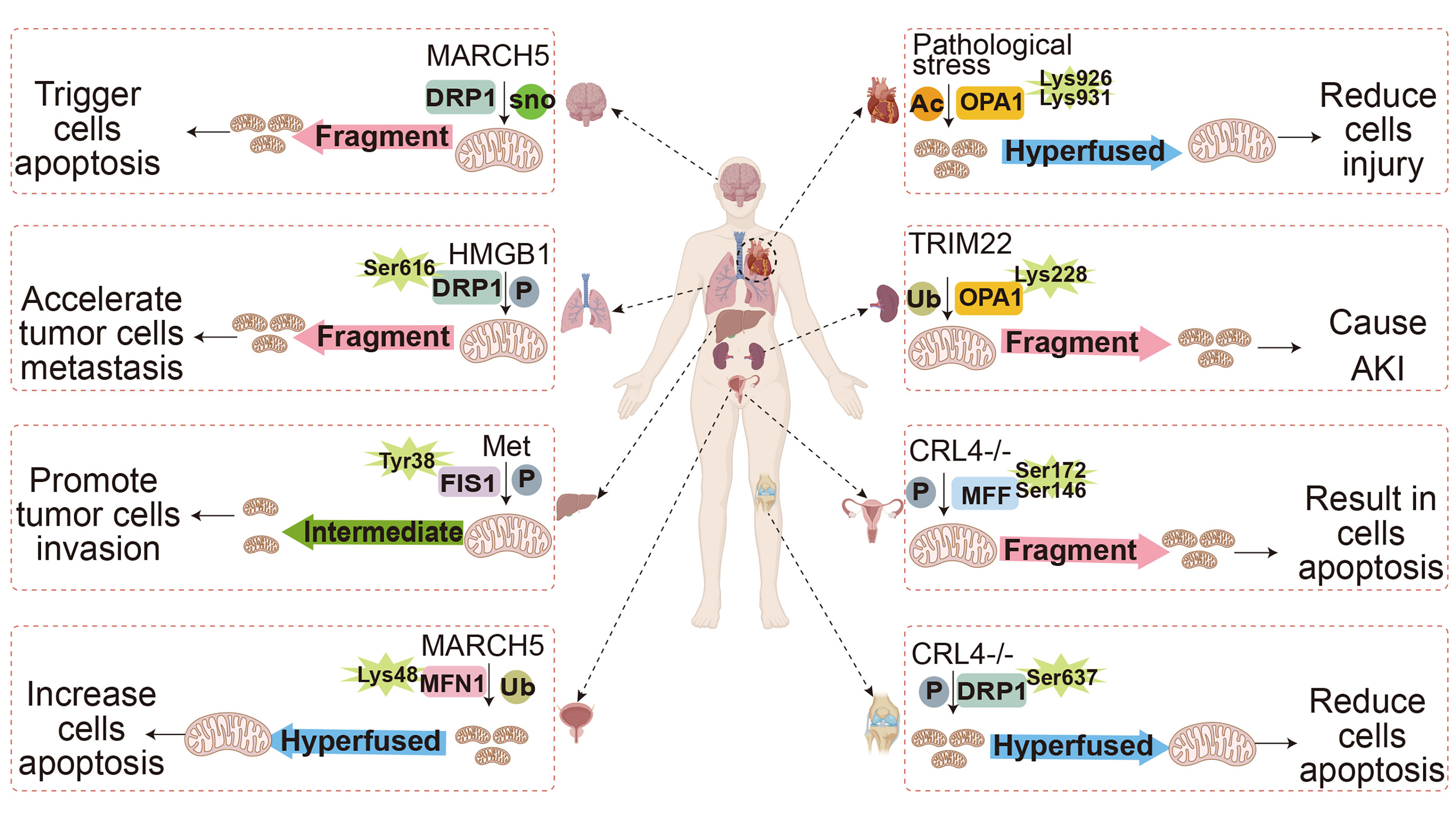
Figure 3. PTMs on mitochondrial dynamics-related proteins regulate many physiological and pathological processes.
Mitochondrial dynamics-related proteins undergo diverse PTMs across tissues, eliciting distinct mitochondrial phenotypes that orchestrate or disrupt physiological and pathological processes. The data source for Figure 3 is generated based on references[96,98,116,148,160-162]. The human organs shown in Figure 3 are created with BioRender.com. P: Phosphorylation; Ub: ubiquitination; Ac: acetylation; sno: nitrosylation; AKI: acute kidney injury; PTMs: post-translational modifications; DRP1: dynamin-related protein 1; FIS1: mitochondrial fission 1 protein; MFN1: mitofusins 1; MFF: mitochondrial fission factor; OPA1: optic atrophy 1.
This novel therapeutic paradigm is defined by three key advantages: high target specificity, reversibility of regulation, and synergy between diagnosis and treatment. First, the extreme site specificity of PTMs allows drugs targeting a particular modified site to avoid disrupting the overall function of the protein, thereby minimizing off-target effects. Second, most PTMs, such as phosphorylation, acetylation, and ubiquitination, are reversible processes. Modulating the activity of the corresponding modifying enzymes via small molecules or gene editing enables dynamic regulation of mitochondrial function, making it suitable for long-term intervention in chronic diseases. However, the writers, erasers, and readers governing these PTMs on mitochondrial dynamics-related proteins remain poorly characterized and warrant further investigation [Table 3]. Third, aberrant levels of specific PTM sites can serve as biomarkers for early disease diagnosis while simultaneously guiding the selection of personalized treatment regimens.
PTM modifying enzymes of mitochondrial dynamics proteins
| PTMs | Proteins | Writer | Eraser |
| Phosphorylation | MFN1 | ERK1/2[166] | PGAM5[171] |
| MFN2 | PKA[172] | Unknown | |
| c-Abl[90] | |||
| PINK1[91] | |||
| DRP1 | CDK5[173] | Calcineurin[174] | |
| ERK1/2[175] | PP2A[176] | ||
| FIS1 | Met[96] | Unknown | |
| DNA-PKcs[97] | |||
| MFF | AKT[177] | Unknown | |
| AMPK[178] | |||
| Ubiquitination | OPA1 | TRIM22[113] | Unknown |
| MFN1 | MARCH5[179] | USP30[180] | |
| MFN2 | RBCK1[181] | Unknown | |
| MARCH5[179] | |||
| RFFL[182] | |||
| DRP1 | TRIM25[183] | Unknown | |
| FIS1 | MARCH5[184] | Unknown | |
| RNF5[185] | |||
| MFF | Parkin[121] | Unknown | |
| MID49 | MARCH5[186] | Unknown | |
| SUMOylation | DRP1 | MAPL[187] | SENP5[129] |
| FIS1 | Unknown | SENP3[188] | |
| MFF | Unknown | SENP3/5[168] | |
| Acetylation | OPA1 | Unknown | SIRT3[165] |
| MFN2 | Unknown | SIRT1[189] | |
| DRP1 | GCN5L1[190] | SIRT1[191] | |
| Unknown | SIRT3[192] | ||
| Miro1 | Unknown | HDAC6[134] | |
| Methylation | DRP1 | CARM1[139] | Unknown |
| O-GlcNAcylation | DRP1 | Unknown | Unknown |
| Palmitoylation | DRP1 | ZDHHC13[142] | Unknown |
| Lactylation | FIS1 | Unknown | SIRT3[144] |
| Succinylation | MFF | CPT1A[146] | Unknown |
| S-Nitrosylation | DRP1 | PDI[193] | GSNOR[194] |
In summary, targeting mitochondrial dynamics protein PTMs holds broad application prospects. Several therapeutics targeting mitochondrial dynamics-related protein PTMs are currently in clinical development or under investigation [Table 4].
Drugs targeting mitochondrial dynamics proteins PTMs
| Name | Target/Mechanism | Development stage | Disease |
| Mdivi-1 | Suppressing the GTPase activity of DRP1, a key regulator of mitochondrial fission, leads to reduced S616 phosphorylation in embryonic tissues[195,196] | Preclinical | Myocardial I/R injury, heart failure, pulmonary hypertension |
| P110 | Blocks DRP1-adaptor protein FIS1 binding; Treatment effectively reduced DRP1 phosphorylation in a mouse model of subarachnoid hemorrhage[197,198] | Preclinical | ALS/motor neuron disease |
| MTX115325 | USP30 (deubiquitinase, removes ubiquitin from OMM)[199] | Phase I | Parkinson’s disease |
CONCLUDING REMARKS
In summary, mitochondrial dynamics are driven by a variety of PTMs. PTMs on fission/fusion, transport, and mitophagy proteins act as triggers that orchestrate mitochondrial dynamics with exquisite spatiotemporal precision - effectively conducting a mitochondrial fate symphony [Figure 4]. Under normal physiological conditions, mitochondria maintain a dynamic equilibrium, balancing fission and fusion to sustain normal function. Disruption of this equilibrium triggers pathological fission or fusion, culminating in mitochondrial dysfunction. Interestingly, emerging evidence extends this regulatory role beyond cellular homeostasis, revealing a vital function for mitochondrial dynamics in sleep pressure control in Drosophila[150]. While this review has focused on ten prevalent PTMs governing mitochondrial dynamics, it is important to recognize that the regulatory landscape extends beyond these modifications. Emerging evidence suggests that other “atypical” PTMs - such as tyrosine ubiquitination[151] and non-enzymatic modifications like carbonyl adducts[152,153] - add further layers of complexity. Although the functional impact of many of these other “atypical” PTM modifications on core dynamics proteins remains largely uncharacterized, future investigations into these less-explored pathways may uncover novel mechanisms linking metabolic flux to mitochondrial architecture, particularly in the context of aging and metabolic disorders.
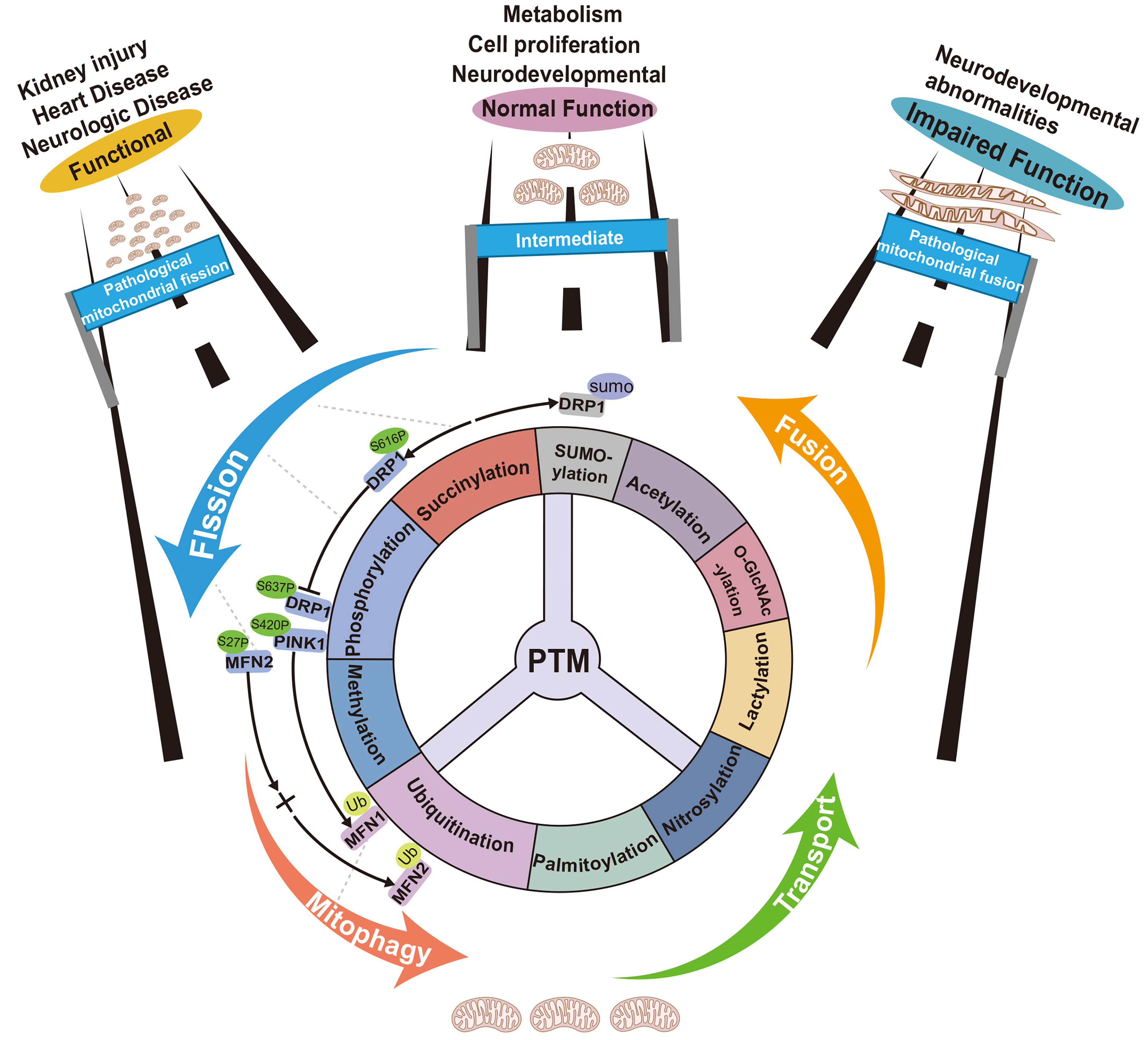
Figure 4. PTMs are the drivers of mitochondrial dynamics balance.
The steering wheel comprising PTMs - including ubiquitination, SUMO, acetylation, phosphorylation, methylation, GlcNAc, palmitoylation, succinylation, lactylation, and S-nitrosylation -coordinates mitochondrial dynamics. PTMs occurring on dynamics-related proteins jointly regulate fission, fusion, transport, and mitophagy, maintaining homeostasis. Disruption of this PTM balance shifts mitochondrial morphology toward excessive fission or fusion, culminating in mitochondrial dysfunction and disease. Graphic reference:⊥: Competitive modification; →: Progressive modification; ←→: Collaborative modification; →+→: Induced modification. The data source for Figure 4 is generated based on references[142,146,148,154-156]. The figure was redrawn and designed by the authors based on existing reference images. DRP1: Dynamin-related protein 1; MFN1: mitofusins; Ub: ubiquitination.
To date, new PTMs on mitochondrial dynamics-related proteins continue to be discovered, reshaping our understanding of mitochondrial biology and disease mechanisms. However, a fundamental question persists: Why have proteins evolved to utilize such a diverse array of PTMs to execute their functions? For example, mitochondrial fission protein DRP1 harbors at least six PTMs. How do these modifications cross-talk on a single protein? Current evidence suggests that multiple PTMs enable a protein to mount differential responses to varying environmental and stress conditions. This multiplicity necessitates precise spatiotemporal regulation, underscoring the protein’s critical role in biological processes. Moreover, our previous perspective on the PTM crosstalk patterns, including “collaborative modification”, ‘‘progressive modification”, ‘‘induced modification”, and ‘‘competitive modification”, which govern pluripotent stem cell fate, offers a conceptual lens through which to dissect the mechanisms of mitochondrial dynamics in disease development[154]. This PTM crosstalk pattern is also readily apparent in the post-translational modifications of mitochondrial dynamics-related proteins. For instance, in human liver transplant patient tissues, elevated DRP1-S616 phosphorylation, paralleled by reduced DRP1-S637 phosphorylation, was identified as a key factor driving hepatic IRI, exemplifying a “competitive modification” pattern. Notably, concurrent upregulation of DRP1 SUMO1 modification in the same pathological context synergizes to enhance DRP1-mediated fission, illustrating the “collaborative modification” mode in mitochondrial dynamics. During mitophagy, PINK1 autophosphorylation and subsequent ubiquitin phosphorylation constitute the core of the PINK1/Parkin mitochondrial clearance pathway, exemplifying a “progressive modification” pattern[155]. In human papillary thyroid carcinoma (PTC) cell lines, LRRK2 (leucine rich repeat kinase 2) overexpression activates the LRRK2-mitogen-activated protein kinase kinase 4 (MKK4)/JNK signaling cascade, triggering phosphorylation of MFN2 at Ser27. This phosphorylation event subsequently induces ubiquitination-mediated degradation of MFN2, following the same “progressive modification” pattern[156] [Figure 4].
OUTSTANDING QUESTIONS
Deciphering the operational logic of the regulatory nexus composed of mitochondrial dynamics proteins represents a new frontier for investigating mitochondrial dynamics. Several questions remain worthy of consideration: (i)What evolutionary advantage drives the diversity of PTMs on proteins like DRP1 - representing redundancy, or a specific combinatorial code for spatial precision? (ii) How do distinct PTM pairs on a single protein (e.g., DRP1 phosphorylation-S616 vs. acetylation-K642) engage in crosstalk to decode specific fission or fusion outputs? (iii) How do fluctuations in mitochondrial metabolites (acetyl-CoA, NAD+) directly alter modification stoichiometry to tip the fusion-fission balance? (iv) Beyond substrate binding, how are PTM-regulating enzymes (writers/erasers) themselves modulated by mitochondrial signals to maintain network homeostasis? (v) What paradigms will enable real-time, multiplexed visualization of PTM dynamics on single proteins within intact mitochondria in living cells? (vi) Can we develop tools targeting specific modification sites (e.g., DRP1 S-nitrosylation) directly, bypassing broad upstream enzyme modulation? Emerging technologies will be instrumental in addressing these questions. For instance, protein modification omics provides a powerful method to study specific modifications under defined cellular states[157]. Prime editing - an accurate and efficient gene editing technology based on CRISPR/Cas system - enables precise site-directed mutagenesis[158]. Furthermore, genetic code expansion (GCE) allows the incorporation of non-canonical amino acids (ncAAs) directly into proteins in vivo[158]. Strategic integration of these tools may efficiently resolve these questions, potentially unlocking new therapeutic avenues for mitochondrial diseases
| Term | Definition |
| Mitochondrial dynamics | A process governing mitochondrial morphology, distribution, and function via antagonistic fusion (MFN1/2, OPA1) and fission (DRP1) cycles. Fusion facilitates content complementation and bioenergetic efficiency; fission enables organellar inheritance and segregation of damaged components for mitophagy. Their equilibrium dictates bioenergetics and apoptosis susceptibility. |
| Mitophagy | A selective autophagy eliminating dysfunctional mitochondria. Loss of ΔΨm stabilizes PINK1 on the outer membrane, which phosphorylates ubiquitin (Ser65) and Parkin. Activated Parkin ubiquitinates outer membrane proteins, recruiting autophagy receptors (p62, OPTN, NDP52) that bridge LC3-phagophores for lysosomal delivery and degradation. |
| Genetic code expansion (GCE) | A synthetic biology method enabling site-specific, co-translational incorporation of noncanonical amino acids (ncAAs) via an engineered orthogonal aaRS/tRNA pair. The orthogonal tRNA decodes a repurposed codon (typically amber UAG), while the engineered aaRS selectively charges it with the desired ncAA. |
| Mitochondrial fission | A conserved GTP-hydrolytic process cleaving a mitochondrial tubule into daughter organelles. Cytosolic DRP1 translocate to the outer membrane, assembling into ring-like helices at constriction sites. GTP hydrolysis drives a conformational twist that mechanistically scissions both outer and inner membranes simultaneously. |
| Mitochondrial transport | Cytoskeleton-based trafficking of mitochondria to sites requiring ATP or Ca2+ buffering. Microtubule motors drive bidirectional transport: kinesin-1 (KIF5B) mediates anterograde (peripheral) movement; cytoplasmic dynein mediates retrograde (perinuclear) movement. Adaptors Miro/Milton couple mitochondria to motors in a Ca2+-sensitive manner. |
| Non-canonical amino acids (ncAAs) | Synthetic amino acid analogs beyond the 20 genetically encoded proteinogenic amino acids. They possess novel side-chain functionalities absent in canonical amino acids, including bioorthogonal handles, photocrosslinkers, spectroscopic probes, and PTM mimics (phosphorylated, acetylated, or ubiquitinated variants). |
| Mitochondrial fusion | A multistep membrane-remodeling process integrating adjacent mitochondria into a continuous network. Outer membrane tethering and fusion are mediated by MFN1/2 via GTP-dependent conformational changes. Inner membrane fusion is subsequently executed by OPA1, a dynamin-like GTPase essential for cristae integrity. |
| PTM crosstalk | Mechanistic interplay between distinct PTMs, comprising four functional patterns: collaborative - synergistic PTMs on same target; progressive - upstream PTM regulates downstream PTM on another protein; induced - one PTM triggers another on same protein; competitive - one PTM antagonizes another at adjacent sites. |
TEXT BOX
Box 1. Translational hurdles of PTM-based biomarkers and proposed strategies
Despite their promise, PTM-based biomarkers face substantial obstacles before clinical adoption.
Key hurdles:
Evidence gap: Current supporting evidence derives predominantly from preclinical models, with limited clinical validation.
Technological barrier: Detecting specific PTMs in clinical samples is technically challenging due to substantial biological variability and a lack of large-scale validation data.
To bridge this gap, we propose the following strategies:
(i) Developing highly sensitive detection technologies (e.g., single-molecule detection or microfluidic enrichment) coupled with site-specific antibodies to overcome the difficulty of detecting low-abundance PTMs, thereby enhancing analytical reliability and reproducibility[200];
(ii) Integrating multi-omics PTM analysis with stable internal references and standardized protocols to establish reference intervals based on large-scale cohorts, addressing current issues of high biological variability;
(iii) Employing multi-omics technologies to systematically screen novel functional PTM sites and clinically validate their associations with disease onset, progression, or prognosis, thereby expanding the therapeutic scope[201].
DECLARATIONS
Acknowledgments
The human organs shown in the graphic abstract were created with BioRender.com.
Authors’ contributions
Conceptualization, writing - original draft, visualization: Ding, H.; E, T.; He, J.; Wu, Y.; Zheng, X.
Conceptualization, writing - review and editing, funding acquisition, supervision: Duan, L.; Liu, X.; Li, L.
Availability of data and materials
Not applicable.
AI and AI-assisted tools statement
Not applicable.
Financial support and sponsorship
This work was financially supported by the National Key Research and Development Program of China (2024YFA0916400), the Strategic Priority Research Program of the Chinese Academy of Sciences (XDB0480000, XDB1410000), the National Key Research and Development Program of China (2022YFA1103800, 2023YFE0210100, 2024YFA1802302, 2025YFA0922001), the National Natural Science Foundation projects of China (32025010, 32488301, 92254301, 92357302, 92157202, 32241002, 32261160376, 32322022, 32370782, 32371007, 32300608, 32300620, 32471358, 32461160288, 32500669, 82505003, 32500691, 32570905, 32422051), NSFC/RGC Joint Grant Scheme 2022/2023 (N_CUHK 428/22, N_CUHK439/24), Major Project of Guangzhou National Laboratory (GZNL2024A03006), the Key Research Program, CAS (ZDBS-ZRKJZ-TLC003), CAS Project for Young Scientists in Basic Research (YSBR-075), the International Partnership Program of Chinese Academy of Sciences(188GJHZ2024048GC), Guangdong Province Science and Technology Program (2023B0303000023, 2023B1212060050, 2023B1212120009, 2024A1515010782, 2024B1515040020, 2024A1515030120, 2023TQ07A024, 2024A1515012839, 2024TQ08A297, 2024TQ08A035), Guangzhou Science and Technology Program (2025A04J2106, 2025A04J7110, 2025A04J5485, 2025B03J0167), Human Cell Lineage Atlas Facility (CLAF) (No. DSS05010101), Shandong Provincial Natural Science Foundation (ZR2025QC1639), the Tianfu Jincheng Laboratory (No. TFJCPI20250043), Health@InnoHK funding support from the Innovation Technology Commission of the Hong Kong SAR, CAS Youth Innovation Promotion Association (to Wu, Y. and Chen, K.), Major Research Project (GIBHMRP25-01), and Basic Research Project of Guangzhou Institutes of Biomedicine and Health, Chinese Academy of Sciences.
Conflicts of interest
All authors declared that there are no conflicts of interest.
Ethical approval and consent to participate
Not applicable.
Consent for publication
Not applicable.
Copyright
© The Author(s) 2026.
REFERENCES
1. Smith, D. S. Reticular organizations within the striated muscle cell: an historical survey of light microscopic studies. J. Biophys. Biochem. Cytol. 10, 61-87 (1961).
3. Atlante, A. ; Valenti, D. Mitochondria have made a long evolutionary path from ancient bacteria immigrants within eukaryotic cells to essential cellular hosts and key players in human health and disease. Curr. Issues Mol. Biol. 45, 4451-79 (2023).
4. Venkatraman, K. ; Lipp, N. F.; Budin, I. Origin and evolution of mitochondrial inner membrane composition. J. Cell Sci. 138, jcs263780 (2025).
5. Cantó, C. Mitochondrial dynamics: shaping metabolic adaptation. Int. Rev. Cell Mol. Biol. (2018) 340, 129-67.
7. El-Hattab, A. W.; Suleiman, J.; Almannai, M.; Scaglia, F. Mitochondrial dynamics: biological roles, molecular machinery, and related diseases. Mol. Genet. Metab. 125, 315-21 (2018).
8. Smith, D. M. The ontogenetic history of the mitochondria of the hepatic cell of the white rat. J. Morphol. 52, 485-511 (1931).
9. Westermann, B. Mitochondrial fusion and fission in cell life and death. Nat. Rev. Mol. Cell Biol. 11, 872-84 (2010).
10. Li, R. ; Toan, S.; Zhou, H. Role of mitochondrial quality control in the pathogenesis of nonalcoholic fatty liver disease. Aging 12, 6467-85 (2020).
11. Bechtel, W. ; Abrahamsen, A. In search of mitochondrial mechanisms: interfield excursions between cell biology and biochemistry. J. Hist. Biol. 40, 1-33 (2007).
12. Pagliarini, D. J.; Rutter, J. Hallmarks of a new era in mitochondrial biochemistry. Genes Dev. 27, 2615-27 (2013).
13. Palade, G. E. An electron microscope study of the mitochondrial structure. J. Histochem. Cytochem. 1, 188-211 (1953).
14. Sjostrand, F. S. Electron microscopy of mitochondria and cytoplasmic double membranes: ultra-structure of rod-shaped mitochondria. Nature 171, 30-2 (1953).
15. Clark-Walker, G. D. Isolation of circular DNA from a mitochondrial fraction from yeast. Proc. Natl. Acad. Sci. U S A. 69, 388-92 (1972).
16. Kummer, E. ; Ban, N. Mechanisms and regulation of protein synthesis in mitochondria. Nat. Rev. Mol. Cell Biol. 22, 307-25 (2021).
17. Nass, M. M.; Nass, S. Intramitochondrial fibers with dna characteristics. I. fixation and electron staining reactions. J Cell Biol. 19, 593-611 (1963).
18. Mitchell, P. Coupling of phosphorylation to electron and hydrogen transfer by a chemi-osmotic type of mechanism. Nature 191, 144-8 (1961).
19. Mitchell, P. Chemiosmotic coupling in oxidative and photosynthetic phosphorylation. Biol. Rev. Camb. Philos. Soc. 41, 445-502 (1966).
20. Glater, E. E.; Megeath, L. J.; Stowers, R. S.; Schwarz, T. L. Axonal transport of mitochondria requires milton to recruit kinesin heavy chain and is light chain independent. J. Cell Biol. 173, 545-57 (2006).
21. Guo, X. et al. The GTPase dMiro is required for axonal transport of mitochondria to Drosophila synapses. Neuron 47, 379-93 (2005).
22. Liu, X. ; Hajnóczky, G. Ca2+-dependent regulation of mitochondrial dynamics by the Miro-Milton complex. Int. J. Biochem. Cell Biol. 41, 1972-6 (2009).
23. Rice, S. E.; Gelfand, V. I. Paradigm lost: milton connects kinesin heavy chain to miro on mitochondria. J. Cell Biol. 173, 459-61 (2006).
24. Kiezun, M. et al. The effect of estrone and estradiol on the expression of the orexin/hypocretin system in the porcine uterus during early pregnancy. Domest. Anim. Endocrinol. 68, 11-24 (2019).
25. Sheng, Z. H. The interplay of axonal energy homeostasis and mitochondrial trafficking and anchoring. Trends Cell Biol. 27, 403-16 (2017).
26. Du, W. et al. The association between constipation and stroke based on the NHANES and Mendelian randomization study. Front. Neurosci. 17, 1276032 (2023).
27. Zaninello, M. ; Bean, C. Highly specialized mechanisms for mitochondrial transport in neurons: from intracellular mobility to intercellular transfer of mitochondria. Biomolecules 13, 938 (2023).
28. Barnhart, E. L. Mechanics of mitochondrial motility in neurons. Curr. Opin. Cell Biol. 38, 90-9 (2016).
29. Liu, X. ; Kim, C. N.; Yang, J.; Jemmerson, R.; Wang, X. Induction of apoptotic program in cell-free extracts: requirement for dATP and cytochrome c. Cell 86, 147-57 (1996).
30. Nguyen, T. T. et al. Mitochondria-associated programmed cell death as a therapeutic target for age-related disease. Exp. Mol. Med. 55, 1595-619 (2023).
31. Vakifahmetoglu-Norberg, H. ; Ouchida, A. T.; Norberg, E. The role of mitochondria in metabolism and cell death. Biochem. Biophys. Res. Commun. 482, 426-31 (2017).
32. Jing, F. ; Zhao, M.; Xiong, H.; Zeng, X.; Jiang, J.; Li, T. Mechanisms underlying targeted mitochondrial therapy for programmed cardiac cell death. Front. Physiol. 16, 1548194 (2025).
33. Zhang, Z. ; Zhang, M.; Jin, H.; Lv, S.; Li, Y.; Li, Y. Mitochondrial quality control and cell death. Int. J. Mol. Sci. 26, 11084 (2025).
34. Zhang, J. et al. Mitochondrial orchestration of PANoptosis: mechanisms, disease pathogenesis, and emerging therapeutic frontiers. Cell Death Discov. 11, 476 (2025).
35. Fonseca, T. B.; Sánchez-Guerrero, Á.; Milosevic, I.; Raimundo, N. Mitochondrial fission requires DRP1 but not dynamins. Nature 570, E34-42 (2019).
36. Chen, H. ; Chan, D. C. Mitochondrial dynamics-fusion, fission, movement, and mitophagy-in neurodegenerative diseases. Hum. Mol. Genet. 18, R169-76 (2009).
37. Wai, T. Is mitochondrial morphology important for cellular physiology? Trends in Endocrinol. Metab. 35, 854-71 (2024).
38. Al Ojaimi, M.; Salah, A.; El-Hattab, A. W. Mitochondrial fission and fusion: molecular mechanisms, biological functions, and related disorders. Membranes 12, 893 (2022).
40. Marsset, M. et al. The Massalia asteroid family as the origin of ordinary L chondrites. Nature 634, 561-5 (2024).
41. Park, J. et al. Mitochondrial dysfunction in Drosophila PINK1 mutants is complemented by parkin. Nature 441, 1157-61 (2006).
42. Narendra, D. ; Tanaka, A.; Suen, D. F.; Youle, R. J. Parkin is recruited selectively to impaired mitochondria and promotes their autophagy. J. Cell Biol. 183, 795-803 (2008).
43. Narendra, D. P. et al. PINK1 Is Selectively Stabilized on Impaired Mitochondria to Activate Parkin. PLoS Biol. 8, e1000298 (2010).
44. Kane, L. A. et al. PINK1 phosphorylates ubiquitin to activate Parkin E3 ubiquitin ligase activity. J. Cell Biol. 205, 143-53 (2014).
45. Wang, S. et al. The mitophagy pathway and its implications in human diseases. Sig. Transduct. Target. Ther. 8, 304 (2023).
46. Picca, A. ; Faitg, J.; Auwerx, J.; Ferrucci, L.; D’Amico, D. Mitophagy in human health, ageing and disease. Nat. Metab. 5, 2047-61 (2023).
47. Tang, M. ; Outissint, I.; Chen, Y.; Gong, X. Comprehensive insights into mitophagy: mechanisms, disease associations, and therapeutic implications. J. Cell. Biochem. 126, e70056 (2025).
48. Shan, W. ; Liu, Y.; Tang, R.; Li, H.; Yang, H.; Lin, L. Targeting mitochondrial autophagy for anti-aging. Cell Death Discov. 12, 78 (2025).
49. Wang, Q. ; Sun, Y.; Li, T. Y.; Auwerx, J. Mitophagy in the pathogenesis and management of disease. Cell Res. 36, 11-37 (2026).
51. Liu, B. H. et al. Mitochondrial quality control in human health and disease. Mil. Med. Res. 11, 32 (2024).
52. Skawratananond, S. et al. Mitophagy in Alzheimer’s disease and other metabolic disorders: a focus on mitochondrial-targeted therapeutics. Ageing Res. Rev. 108, 102732 (2025).
53. Wang, L. ; Zhou, X.; Lu, T. Role of mitochondria in physiological activities, diseases, and therapy. Mol. Biomed. 6, 42 (2025).
54. Ye, L. ; Fu, X.; Li, Q. Mitochondrial quality control in health and disease. MedComm 6, e70319 (2025).
55. Ying, Z. et al. Transient activation of mitoflashes modulates Nanog at the early phase of somatic cell reprogramming. Cell Metab. 23, 220-6 (2016).
56. Ying, Z. et al. Short-term mitochondrial permeability transition pore opening modulates histone lysine methylation at the early phase of somatic cell reprogramming. Cell Metab. 28, 935-45.e5 (2018).
57. Bhutani, N. ; Brady, J. J.; Damian, M.; Sacco, A.; Corbel, S. Y.; Blau, H. M. Reprogramming towards pluripotency requires AID-dependent DNA demethylation. Nature 463, 1042-7 (2009).
58. Prieto, J. et al. Early ERK1/2 activation promotes DRP1-dependent mitochondrial fission necessary for cell reprogramming. Nat. Commun. 7, 11124 (2016).
59. Seo, B. J.; Yoon, S. H.; Do, J. T. Mitochondrial dynamics in stem cells and differentiation. Int. J. Mol. Sci. 19, 3893 (2018).
60. Rasmussen, M. L.; Gama, V. A connection in life and death: the BCL-2 family coordinates mitochondrial network dynamics and stem cell fate. Int. Rev. Cell Mol. Biol. 353, 255-84 (2020).
61. Yildirim, R. M.; Seli, E. The role of mitochondrial dynamics in oocyte and early embryo development. Semin. Cell Dev. Biol. 159-160, 52-61 (2024).
62. Coelho, P. ; Fão, L.; Mota, S.; Rego, A. C. Mitochondrial function and dynamics in neural stem cells and neurogenesis: Implications for neurodegenerative diseases. Ageing Res. Rev. 80, 101667 (2022).
63. Sebastián, D. ; Palacín, M.; Zorzano, A. Mitochondrial dynamics: coupling mitochondrial fitness with healthy aging. Trends Mol. Med. 23, 201-15 (2017).
64. Simula, L. ; Nazio, F.; Campello, S. The mitochondrial dynamics in cancer and immune-surveillance. Semin. Cancer Biol. 47, 29-42 (2017).
65. Kim, S. J.; Ahn, D. G.; Syed, G. H.; Siddiqui, A. The essential role of mitochondrial dynamics in antiviral immunity. Mitochondrion 41, 21-7 (2018).
66. Liao, R. et al. BCL11B and the NuRD complex cooperatively guard T-cell fate and inhibit OPA1-mediated mitochondrial fusion in T cells. EMBO J. 42, e113448 (2023).
67. Wu, Y. et al. Plin2-mediated lipid droplet mobilization accelerates exit from pluripotency by lipidomic remodeling and histone acetylation. Cell Death Differ. 29, 2316-31 (2022).
68. Leutert, M. ; Entwisle, S. W.; Villén, J. Decoding post-translational modification crosstalk with proteomics. Mol. Cell. Proteomics 20, 100129 (2021).
69. Zhong, Q. et al. Protein posttranslational modifications in health and diseases: functions, regulatory mechanisms, and therapeutic implications. MedComm 4, e261 (2023).
70. Guo, H. ; Qian, P. Modulating enzyme activity and specificity by preinstalled posttranslational modifications on substrates: PTM-induced activity stimulation. Biophys. J. 125, 4048-64 (2026).
71. Zhang, S. ; Li, J.; Chen, M.; Huang, H. Post-translational modifications in proteins: prediction methods, biological functions, and diseases. MedComm 7, e70729 (2026).
72. Beltrao, P. ; Bork, P.; Krogan, N. J.; van Noort, V. Evolution and functional cross-talk of protein post-translational modifications. Mol. Syst. Biol. 9, 714 (2013).
73. Xu, D. et al. Blockage of Drp1 phosphorylation at Ser579 protects neurons against Aβ1-42-induced degeneration. Mol. Med. Rep. 24, 657 (2021).
74. Todd, L. R.; Damin, M. N.; Gomathinayagam, R.; Horn, S. R.; Means, A. R.; Sankar, U. Growth factor erv1-like modulates Drp1 to preserve mitochondrial dynamics and function in mouse embryonic stem cells. Mol. Biol. Cell 21, 1225-36 (2010).
75. Wang, X. et al. Impaired balance of mitochondrial fission and fusion in Alzheimer’s disease. J. Neurosci. 29, 9090-103 (2009).
76. Otera, H. et al. Mff is an essential factor for mitochondrial recruitment of Drp1 during mitochondrial fission in mammalian cells. J. Cell Biol. 191, 1141-58 (2010).
77. Yu, R. et al. MIEF1/2 function as adaptors to recruit Drp1 to mitochondria and regulate the association of Drp1 with Mff. Sci. Rep. 7, 880 (2017).
78. Pokhrel, S. et al. A hidden cysteine in Fis1 targeted to prevent excessive mitochondrial fission and dysfunction under oxidative stress. Nat. Commun. 16, 4187 (2025).
79. Rochon, K. et al. Structural basis for regulated assembly of the mitochondrial fission GTPase Drp1. Nat. Commun. 15, 1328 (2024).
80. Egner, J. M. et al. Structural studies of human fission protein FIS1 reveal a dynamic region important for GTPase DRP1 recruitment and mitochondrial fission. J. Biol. Chem. 298, 102620 (2022).
81. Liu, R. ; Chan, D. C. The mitochondrial fission receptor Mff selectively recruits oligomerized Drp1. Mol. Biol. Cell 26, 4466-77 (2015).
82. von der Malsburg, A. et al. Structural mechanism of mitochondrial membrane remodelling by human OPA1. Nature 620, 1101-8 (2023).
83. Nyenhuis, S. B. et al. OPA1 helical structures give perspective to mitochondrial dysfunction. Nature 620, 1109-16 (2023).
84. Cao, Y. L. et al. MFN1 structures reveal nucleotide-triggered dimerization critical for mitochondrial fusion. Nature 542, 372-6 (2017).
85. Li, Y. J. et al. Structural insights of human mitofusin-2 into mitochondrial fusion and CMT2A onset. Nat. Commun. 10, 4914 (2019).
86. Trempe, J. F. et al. Structure of parkin reveals mechanisms for ubiquitin ligase activation. Science 340, 1451-5 (2013).
87. Smith, K. P. et al. Insight into human Miro1/2 domain organization based on the structure of its N-terminal GTPase. J. Struct. Biol. 212, 107656 (2020).
88. Ko, A. R.; Hyun, H. W; Min, S. J.; Kim, J. E. The differential DRP1 phosphorylation and mitochondrial dynamics in the regional specific astroglial death induced by status epilepticus. Front. Cell. Neurosci. 10, 124 (2016).
89. Ferreira, J. C. B. et al. A selective inhibitor of mitofusin 1-βIIPKC association improves heart failure outcome in rats. Nat. Commun. 10, 329 (2019).
90. Martinez, A. et al. c-Abl phosphorylates MFN2 to regulate mitochondrial morphology in cells under endoplasmic reticulum and oxidative stress, impacting cell survival and neurodegeneration. Antioxidants 12, 2007 (2023).
91. Li, J. ; Dang, X.; Franco, A.; Dorn, G. W. Reciprocal regulation of mitofusin 2-mediated mitophagy and mitochondrial fusion by different PINK1 phosphorylation events. Front. Cell Dev. Biol. 10, 868465 (2022).
92. Valera-Alberni, M. et al. Crosstalk between Drp1 phosphorylation sites during mitochondrial remodeling and their impact on metabolic adaptation. Cell Reports 36, 109565 (2021).
93. Han, H. et al. PINK1 phosphorylates Drp1S616 to regulate mitophagy-independent mitochondrial dynamics. EMBO Rep. 21, e48686 (2020).
94. Liu, D. ; Li, J.; Rong, X.; Li, J.; Peng, Y.; Shen, Q. Cdk5 promotes mitochondrial fission via Drp1 phosphorylation at S616 in chronic ethanol exposure-induced cognitive impairment. Mol. Neurobiol. 59, 7075-94 (2022).
95. Hu, S. L. et al. TBK1-medicated DRP1 phosphorylation orchestrates mitochondrial dynamics and autophagy activation in osteoarthritis. Acta Pharmacol. Sin. 44, 610-21 (2023).
96. Yu, Y. et al. Fis1 phosphorylation by Met promotes mitochondrial fission and hepatocellular carcinoma metastasis. Sig. Transduct. Target. Ther. 6, 401 (2021).
97. Wang, S. et al. DNA-PKcs interacts with and phosphorylates Fis1 to induce mitochondrial fragmentation in tubular cells during acute kidney injury. Sci. Signal. 15, eabh1121 (2022).
98. Meng, Y. et al. Targeting CRL4 suppresses chemoresistant ovarian cancer growth by inducing mitophagy. Sig. Transduct. Target. Ther. 7, 388 (2022).
99. Henschke, S. et al. Food perception promotes phosphorylation of MFFS131 and mitochondrial fragmentation in liver. Science 384, 438-46 (2024).
100. Xiao, J. J. et al. Regulator of calcineurin 1 deletion attenuates mitochondrial dysfunction and apoptosis in acute kidney injury through JNK/Mff signaling pathway. Cell Death Dis. 13, 774 (2022).
101. Pangou, E. et al. A PKD-MFF signaling axis couples mitochondrial fission to mitotic progression. Cell Reports 35, 109129 (2021).
102. Toyama, E. Q. et al. AMP-activated protein kinase mediates mitochondrial fission in response to energy stress. Science 351, 275-81 (2016).
103. Kim, Y. et al. PINK1 controls mitochondrial localization of Parkin through direct phosphorylation. Biochem. Biophys. Res. Commun. 377, 975-80 (2008).
104. Okatsu, K. et al. PINK1 autophosphorylation upon membrane potential dissipation is essential for Parkin recruitment to damaged mitochondria. Nat. Commun. 3, 1016 (2012).
105. Shiba-Fukushima, K. ; Arano, T. et al. Phosphorylation of mitochondrial polyubiquitin by PINK1 promotes parkin mitochondrial tethering. PLoS Genet. 10, e1004861 (2014).
106. Cherra, S. J. et al. Regulation of the autophagy protein LC3 by phosphorylation. J. Cell Biol. 190, 533-9 (2010).
107. Herhaus, L. et al. TBK1-mediated phosphorylation of LC3C and GABARAP-L2 controls autophagosome shedding by ATG4 protease. EMBO Rep. 21, e48317 (2020).
108. Chen, Y. ; Dorn, G. W. PINK1-phosphorylated mitofusin 2 is a Parkin receptor for culling damaged mitochondria. Science 340, 471-5 (2013).
109. Bhatia, D. et al. Mitophagy-dependent macrophage reprogramming protects against kidney fibrosis. JCI Insight 4, e132826 (2019).
110. Leboucher, G. P. et al. Stress-induced phosphorylation and proteasomal degradation of mitofusin 2 facilitates mitochondrial fragmentation and apoptosis. Mol. Cell 47, 547-57 (2012).
111. Kim, Y. ; Kim, E. K.; Jang, H. H. Fine-tuning protein fate: mechanisms of E1, E2, and E3 enzymes and deubiquitinases in cell signaling. Int. J. Mol. Sci. 27, 3404 (2026).
112. Huang, B. ; Yang, Y.; Liu, J.; Zhang, B.; Lin, N. Ubiquitination regulation of mitochondrial homeostasis: a new sight for the treatment of gastrointestinal tumors. Front. Immunol. 16, 1533007 (2025).
113. Zhang, Z. et al. WTAP-mediated m6A modification of TRIM22 promotes diabetic nephropathy by inducing mitochondrial dysfunction via ubiquitination of OPA1. Redox Rep. 29, 2404794 (2024).
114. Gegg, M. E.; Cooper, J. M.; Chau, K. Y.; Rojo, M.; Schapira, A. H.; Taanman, J. W. Mitofusin 1 and mitofusin 2 are ubiquitinated in a PINK1/parkin-dependent manner upon induction of mitophagy. Hum. Mol. Genet. 19, 4861-70 (2010).
115. Choi, S. H. et al. Intracellular AIBP (apolipoprotein A-I binding protein) regulates oxidized LDL (low-density lipoprotein)-induced mitophagy in macrophages. Arterioscler. Thromb. Vasc. Biol. 41, e82-96 (2021).
116. Park, Y. Y.; Nguyen, O. T. K.; Kang, H.; Cho, H. MARCH5-mediated quality control on acetylated Mfn1 facilitates mitochondrial homeostasis and cell survival. Cell Death Dis. 5, e1172 (2014).
117. Lee, Y. S. et al. Deficiency of parkin suppresses melanoma tumor development and metastasis through inhibition of MFN2 ubiquitination. Cancer Lett. 433, 156-64 (2018).
118. Liu, Y. et al. Inhibition of USP30 promotes mitophagy by regulating ubiquitination of MFN2 by parkin to attenuate early brain injury after SAH. Transl. Stroke Res. 16, 448-66 (2025).
119. Shen, L. ; Zhang, Q.; Tu, S.; Qin, W. SIRT3 mediates mitofusin 2 ubiquitination and degradation to suppress ischemia reperfusion-induced acute kidney injury. Exp. Cell Res. 408, 112861 (2021).
120. Sugiura, A. et al. MITOL regulates endoplasmic reticulum-mitochondria contacts via Mitofusin2. Mol. Cell 51, 20-34 (2013).
121. Lee, L. ; Seager, R.; Nakamura, Y.; Wilkinson, K. A.; Henley, J. M. Parkin-mediated ubiquitination contributes to the constitutive turnover of mitochondrial fission factor (Mff). PLoS One 14, e0213116 (2019).
122. Cherok, E. et al. Novel regulatory roles of Mff and Drp1 in E3 ubiquitin ligase MARCH5-dependent degradation of MiD49 and Mcl1 and control of mitochondrial dynamics. Mol. Biol. Cell 28, 396-410 (2017).
123. Xiong, H. et al. Parkin, PINK1, and DJ-1 form a ubiquitin E3 ligase complex promoting unfolded protein degradation. J. Clin. Invest. 119, 650-60 (2009).
124. Spratt, D. E. et al. A molecular explanation for the recessive nature of parkin-linked Parkinson’s disease. Nat. Commun. 4, 1983 (2013).
125. Liu, S. et al. Parkinson’s disease-associated kinase PINK1 regulates Miro protein level and axonal transport of mitochondria. PLoS Genet. 8, e1002537 (2012).
126. Xian, H. ; Liou, Y. C. Loss of MIEF1/MiD51 confers susceptibility to BAX-mediated cell death and PINK1-PRKN-dependent mitophagy. Autophagy 15, 2107-25 (2019).
127. Princz, A. ; Pelisch, F.; Tavernarakis, N. SUMO promotes longevity and maintains mitochondrial homeostasis during ageing in Caenorhabditis elegans. Sci. Rep. 10, 15513 (2020).
128. Huang, J. ; Xie, P.; Dong, Y.; An, W. Inhibition of Drp1 SUMOylation by ALR protects the liver from ischemia-reperfusion injury. Cell Death Differ. 28, 1174-92 (2021).
129. Yamada, S. ; Sato, A.; Ishihara, N.; Akiyama, H.; Sakakibara, S. I. Drp1 SUMO/deSUMOylation by Senp5 isoforms influences ER tubulation and mitochondrial dynamics to regulate brain development. iScience 24, 103484 (2021).
130. Zhou, X. et al. Endothelial FIS1 DeSUMOylation protects against hypoxic pulmonary hypertension. Circ. Res. 133, 508-31 (2023).
131. Keller, M. A.; Ivessa, A.; Liu, T.; Li, H.; Romanienko, P. J.; Nakamura, M. KAT6A acetylation drives metabolic adaptation to mediate cellular growth and mitochondrial metabolism through AMPK interaction. bioRxiv (2025) Epub ahead of print.
132. Hu, L. et al. Mfn2/Hsc70 complex mediates the formation of Mitochondria-lipid droplets membrane contact and regulates myocardial lipid metabolism. Adv. Sci. 11, e2307749 (2024).
133. Jin, J. Y.; Wei, X. X.; Zhi, X. L.; Wang, X. H.; Meng, D. Drp1-dependent mitochondrial fission in cardiovascular disease. Acta Pharmacol. Sin. 42, 655-64 (2021).
134. Kalinski, A. L. et al. Deacetylation of Miro1 by HDAC6 blocks mitochondrial transport and mediates axon growth inhibition. J. Cell Biol. 218, 1871-90 (2019).
135. Paik, W. K.; Paik, D. C.; Kim, S. Historical review: the field of protein methylation. Trends Biochem. Sci. 32, 146-52 (2007).
136. Cho, Y. ; Kim, Y. K. ROS-mediated cytoplasmic localization of CARM1 induces mitochondrial fission through DRP1 methylation. Redox Biol. 73, 103212 (2024).
137. Onigbinde, S. et al. Glycomics in human diseases and its emerging role in biomarker discovery. Biomedicines 13, 2034 (2025).
138. Jóźwiak, P. et al. Increased O-GlcNAcylation by upregulation of Mitochondrial O-GlcNAc transferase (mOGT) inhibits the activity of respiratory chain complexes and controls cellular bioenergetics. Cancers 16, 1048 (2024).
139. Akinbiyi, E. O. et al. Blocked O-GlcNAc cycling alters mitochondrial morphology, function, and mass. Sci. Rep. 11, 22106 (2021).
140. Iyer, S. P. N.; Akimoto, Y.; Hart, G. W. Identification and cloning of a novel family of coiled-coil domain proteins that interact with O-GlcNAc transferase. J. Biol. Chem. 278, 5399-409 (2003).
141. Liu, Y. et al. MAVS Cys508 palmitoylation promotes its aggregation on the mitochondrial outer membrane and antiviral innate immunity. Proc. Natl. Acad. Sci. U S A. 121, e2403392121 (2024).
142. Napoli, E. et al. Zdhhc13-dependent Drp1 S-palmitoylation impacts brain bioenergetics, anxiety, coordination and motor skills. Sci. Rep. 7, 12796 (2017).
143. Dong, W. ; Huang, S. X.; Qin, M. L.; Pan, Z. Mitochondrial alanyl-tRNA synthetase 2 mediates histone lactylation to promote ferroptosis in intestinal ischemia-reperfusion injury. World J. Gastrointest. Surg. 17, 106777 (2025).
144. An, S. et al. PDHA1 hyperacetylation-mediated lactate overproduction promotes sepsis-induced acute kidney injury via Fis1 lactylation. Cell Death Dis. 14, 457 (2023).
145. Wang, L. et al. Identification of mitochondrial and succinylation modification-related gene signature in ischemic stroke. Mol. Neurobiol. 62, 11130-44 (2025).
146. Zhu, Y. et al. Carnitine palmitoyltransferase 1A promotes mitochondrial fission by enhancing MFF succinylation in ovarian cancer. Commun. Biol. 6, 618 (2023).
147. Han, F. et al. S-nitrosylation of peroxiredoxin 2 exacerbates hyperuricemia-induced renal injury through regulation of mitochondrial homeostasis. Free Radic. Biol. Med. 230, 66-78 (2025).
148. Cho, D. H. et al. S-Nitrosylation of Drp1 mediates β-amyloid-related mitochondrial fission and neuronal injury. Science 324, 102-5 (2009).
149. Chung, K. K. K. et al. S-nitrosylation of parkin regulates ubiquitination and compromises parkin’s protective function. Science 304, 1328-31 (2004).
150. Sarnataro, R. ; Velasco, C. D.; Monaco, N.; Kempf, A.; Miesenböck, G. Mitochondrial origins of the pressure to sleep. Nature 645, 722-8 (2025).
151. Shen, S. ; Yin, H. E2-Ub-R74G strategy reveals E2-specific ubiquitin conjugation profiles in live cells. Nat. Chem. Biol. 21, 1089-99 (2025).
152. Estévez, M. ; Díaz-Velasco, S.; Martínez, R. Protein carbonylation in food and nutrition: a concise update. Amino Acids 54, 559-73 (2022).
153. Akagawa, M. Protein carbonylation: molecular mechanisms, biological implications, and analytical approaches. Free Radic. Res. 55, 307-20 (2021).
154. Fan, W. et al. The role of protein post-translational modifications and their crosstalk in determining pluripotent stem cells fate. Science Bulletin 70, 3453-6 (2025).
155. Koyano, F. et al. Ubiquitin is phosphorylated by PINK1 to activate parkin. Nature 510, 162-6 (2014).
156. Zhang, S. et al. LRRK2 aggravates kidney injury through promoting MFN2 degradation and abnormal mitochondrial integrity. Redox Biol. 66, 102860 (2023).
157. Guo, T. ; Steen, J. A.; Mann, M. Mass-spectrometry-based proteomics: from single cells to clinical applications. Nature 638, 901-11 (2025).
158. Nelson, J. W. et al. Engineered pegRNAs improve prime editing efficiency. Nat. Biotechnol. 40, 402-10 (2022).
159. Ravitch, E. E. et al. Structural-functional characterization of the MIRO1-TRAK1 complex. Nat. Commun. 16, 6173 (2025).
160. Feng, W. et al. ERK/Drp1-dependent mitochondrial fission contributes to HMGB1-induced autophagy in pulmonary arterial hypertension. Cell Prolif. 54, e13048 (2021).
161. Ko, H. J. et al. The phosphorylation status of Drp1-Ser637 by PKA in mitochondrial fission modulates mitophagy via PINK1/parkin to exert multipolar spindles assembly during mitosis. Biomolecules 11, 424 (2021).
162. Wang, R. et al. SIRT3 promotes metabolic maturation of human iPSC-derived cardiomyocytes via OPA1-controlled mitochondrial dynamics. Free Radic. Biol. Med. 195, 270-82 (2023).
163. Palmer, C. S.; Osellame, L. D.; Laine, D.; Koutsopoulos, O. S.; Frazier, A. E.; Ryan, M. T. MiD49 and MiD51, new components of the mitochondrial fission machinery. EMBO Rep. 12, 565-73 (2011).
164. Ban, T. et al. Molecular basis of selective mitochondrial fusion by heterotypic action between OPA1 and cardiolipin. Nat. Cell Biol. 19, 856-63 (2017).
165. Samant, S. A. et al. SIRT3 deacetylates and activates OPA1 to regulate mitochondrial dynamics during stress. Mol. Cell. Biol. 34, 807-19 (2023).
166. Pyakurel, A. ; Savoia, C.; Hess, D.; Scorrano, L. Extracellular regulated kinase phosphorylates mitofusin 1 to control mitochondrial morphology and apoptosis. Mol. Cell 58, 244-54 (2015).
167. Zhou, J. et al. N6-methyladenosine facilitates mitochondrial fusion of colorectal cancer cells via induction of GSH synthesis and stabilization of OPA1 mRNA. Natl. Sci. Rev. 11, nwae039 (2024).
168. Seager, R. ; Ramesh, N. S.; Cross, S.; Guo, C.; Wilkinson, K. A.; Henley, J. M. SUMOylation of MFF coordinates fission complexes to promote stress-induced mitochondrial fragmentation. Sci. Adv. 10, eadq6223 (2024).
169. Xu, S. et al. Mitochondrial E3 ubiquitin ligase MARCH5 controls mitochondrial fission and cell sensitivity to stress-induced apoptosis through regulation of MiD49 protein. Mol. Biol. Cell 27, 349-59 (2016).
170. Pekkurnaz, G. ; Trinidad, J. C.; Wang, X.; Kong, D.; Schwarz, T. L. Glucose regulates mitochondrial motility via milton modification by O-GlcNAc transferase. Cell 158, 54-68 (2014).
171. Nag, S. ; Szederkenyi, K.; Gorbenko, O.; Tyrrell, H.; Yip, C. M.; Mcquibban, G. A. PGAM5 is an MFN2 phosphatase that plays an essential role in the regulation of mitochondrial dynamics. Cell Rep. 42, 112895 (2023).
172. Zhou, W. et al. Mutation of the protein kinase A phosphorylation site influences the anti-proliferative activity of mitofusin 2. Atherosclerosis 211, 216-23 (2010).
173. Cho, B. et al. CDK5-dependent inhibitory phosphorylation of Drp1 during neuronal maturation. Exp. Mol. Med. 46, e105 (2014).
174. Cereghetti, G. M. et al. Dephosphorylation by calcineurin regulates translocation of Drp1 to mitochondria. Proc. Natl. Acad. Sci. U S A. 105, 15803-8 (2008).
175. Serasinghe, M. N. et al. Mitochondrial division is requisite to RAS-induced transformation and targeted by oncogenic MAPK pathway inhibitors. Mol. Cell 57, 521-36 (2015).
176. Xia, W. et al. Obesity causes mitochondrial fragmentation and dysfunction in white adipocytes due to RalA activation. Nat. Metab. 6, 273-89 (2024).
177. Zhi, M. ; Cao, R.; Fu, X.; Li, L. Mitochondrial fission factor: a living way for mitochondria sensing food. MedComm 5, e770 (2024).
178. Hanada, Y. et al. Alternative splicing of Mff regulates AMPK-mediated phosphorylation, mitochondrial fission and antiviral response. Pharmacol. Res. 209, 107414 (2024).
179. Wang, Z. et al. Pathological roles of ubiquitination and deubiquitination systems in sepsis-induced myocardial dysfunction. Biomol. Biomed. 25, 1444-58 (2025).
180. Yue, W. et al. A small natural molecule promotes mitochondrial fusion through inhibition of the deubiquitinase USP30. Cell Res. 24, 482-96 (2014).
181. Su, D. et al. E3 ubiquitin ligase RBCK1 confers ferroptosis resistance in pancreatic cancer by facilitating MFN2 degradation. Free Radic. Biol. Med. 221, 136-54 (2024).
182. Narendradev, N. D. et al. Endosomal RFFL ubiquitin ligase regulates mitochondrial morphology by targeting mitofusin 2. J. Cell Sci. 138, jcs263830 (2025).
183. Das, P. ; Chakrabarti, O. ISGylation of DRP1 closely balances other post-translational modifications to mediate mitochondrial fission. Cell Death Dis. 15, 184 (2024).
184. Yonashiro, R. et al. A novel mitochondrial ubiquitin ligase plays a critical role in mitochondrial dynamics. EMBO J. 25, 3618-26 (2006).
185. Zhang, Q. et al. DJ-1 promotes the proteasomal degradation of Fis1: implications of DJ-1 in neuronal protection. Biochem. J. 447, 261-9 (2012).
186. Tilokani, L. et al. Mitochondrial dynamics: overview of molecular mechanisms. Essays Biochem. 62, 341-60 (2018).
187. Lin, Z. et al. The mitochondrial E3 ligase MAPL SUMOylates Drp1 to facilitate mitochondrial fission in intervertebral disc degeneration. Bone Res. 13, 72 (2025).
188. Waters, E. et al. The SUMO protease SENP3 regulates mitochondrial autophagy mediated by Fis1. EMBO Rep. 23, e48754 (2022).
189. Sooyeon, L. ; Go, K. L.; Kim, J. S. Deacetylation of mitofusin-2 by sirtuin-1: a critical event in cell survival after ischemia. Mol. Cell. Oncol. 3, e1087452 (2015).
190. Zhang, J. et al. Drp1 acetylation mediated by CDK5-AMPK-GCN5L1 axis promotes cerebral ischemic injury via facilitating mitochondrial fission. Mol. Med. 30, 173 (2024).
191. Yin, S. ; Dai, D.; Wang, G.; Zhou, S. SIRT1-mediated deacetylation of DRP1 suppresses excessive mitophagy and ameliorates pediatric functional dyspepsia. BMC Immunol. 27, 58 (2026).
192. Xi, Y. et al. SIRT3-mediated deacetylation of DRP1K711 prevents mitochondrial dysfunction in parkinson’s disease. Adv. Sci. 12, 2411235 (2025).
193. Lee, D. S.; Kim, J. E. PDI-mediated S-nitrosylation of DRP1 facilitates DRP1-S616 phosphorylation and mitochondrial fission in CA1 neurons. Cell Death Dis. 9, 869 (2018).
194. Benhar, M. ; Forrester, M. T.; Stamler, J. S. Protein denitrosylation: enzymatic mechanisms and cellular functions. Nat. Rev. Mol. Cell Biol. 10, 721-32 (2009).
195. Aishwarya, R. et al. Pleiotropic effects of mdivi-1 in altering mitochondrial dynamics, respiration, and autophagy in cardiomyocytes. Redox Biol. 36, 101660 (2020).
196. Li, Y. ; Mei, N. H.; Cheng, G. P.; Yang, J.; Zhou, L. Q. Inhibition of DRP1 impedes zygotic genome activation and preimplantation development in mice. Front. Cell Dev. Biol. 9, 788512 (2021).
197. Hao, S. et al. The DRP1 inhibitory peptide P110 provides neuroprotection after subarachnoid hemorrhage by suppressing neuronal apoptosis and stabilizing the blood-brain barrier. Free Radic. Biol. Med. 240, 1-14 (2025).
198. Joshi, A. U.; Saw, N. L.; Vogel, H.; Cunnigham, A. D.; Shamloo, M.; Mochly-Rosen, D. Inhibition of Drp1/Fis1 interaction slows progression of amyotrophic lateral sclerosis. EMBO Mol. Med. 10, e08166 (2018).
199. Fang, T. Z. et al. Knockout or inhibition of USP30 protects dopaminergic neurons in a Parkinson’s disease mouse model. Nat. Commun. 14, 7295 (2023).
200. Cao, C. et al. Deep learning-assisted single-molecule detection of protein post-translational modifications with a biological nanopore. ACS Nano 18, 1504-15 (2024).
Cite This Article
 Open Access
How to Cite
Download Citation
Export Citation File:
Type of Import
Tips on Downloading Citation
Citation Manager File Format
Type of Import
Direct Import: When the Direct Import option is selected (the default state), a dialogue box will give you the option to Save or Open the downloaded citation data. Choosing Open will either launch your citation manager or give you a choice of applications with which to use the metadata. The Save option saves the file locally for later use.
Indirect Import: When the Indirect Import option is selected, the metadata is displayed and may be copied and pasted as needed.
Article Notes
About This Article
Copyright






Data & Comments
Data
 0
0





Comments
Comments must be written in English. Spam, offensive content, impersonation, and private information will not be permitted. If any comment is reported and identified as inappropriate content by OAE staff, the comment will be removed without notice. If you have any queries or need any help, please contact us at [email protected].