





Mechanistic roles and clinical applications of tertiary lymphoid structures in colorectal cancer drug resistance
 0
0 Abstract
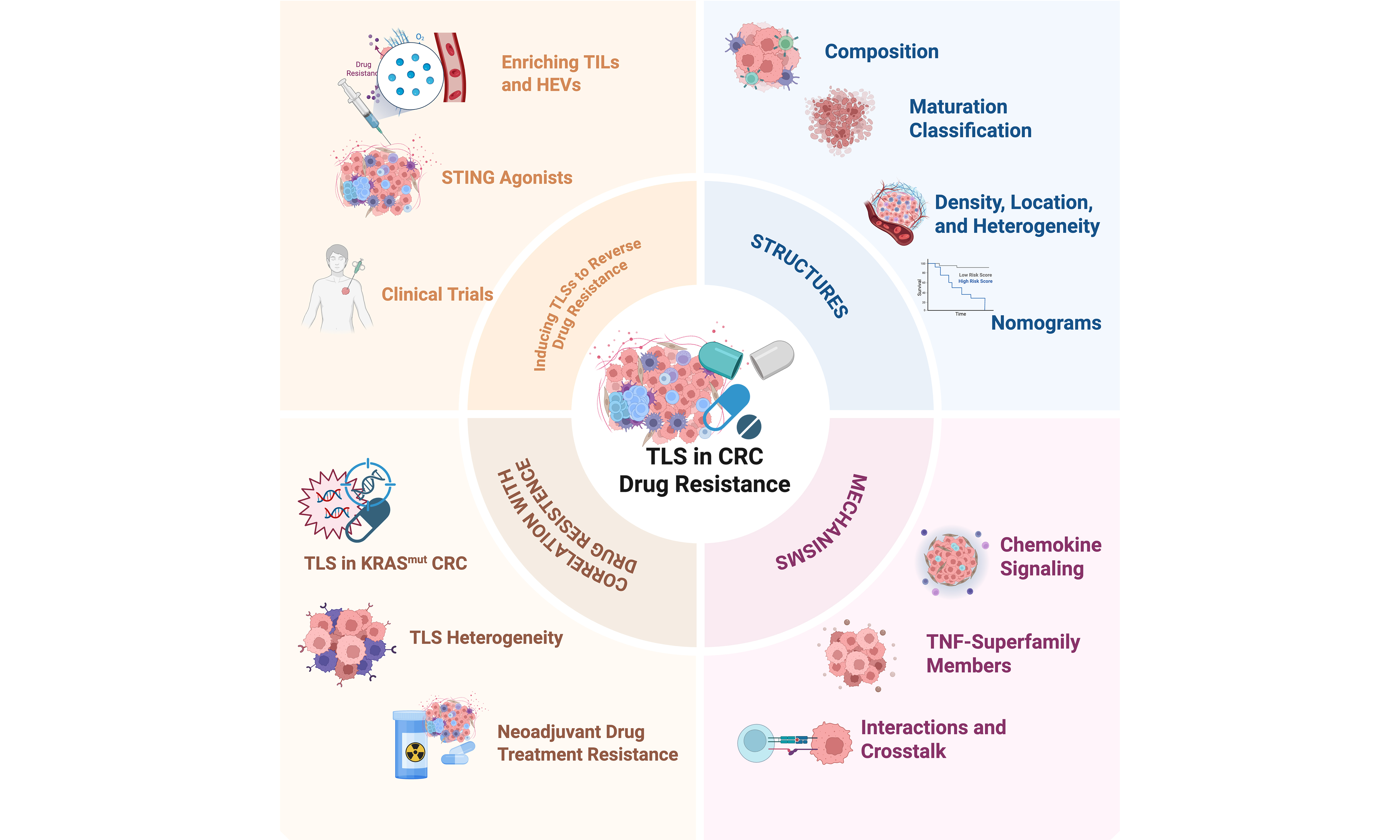
Therapeutic resistance represents a formidable bottleneck in colorectal cancer (CRC) management, severely limiting the clinical efficacy of both conventional regimens and novel immunotherapies. Emerging evidence underscores tertiary lymphoid structure (TLS) - ectopic lymphoid aggregates newly sculpted within the tumor microenvironment (TME) - as pivotal orchestrators of localized antitumor immunity and prime therapeutic targets to circumvent resistance. Distinct from secondary lymphoid organs, TLS facilitate in situ immune cell priming, clonal expansion, and functional differentiation, thereby sustaining a robust adaptive immune response. This review systematically dissects the cellular architecture, maturation dynamics, and spatial heterogeneity of TLS in primary and metastatic CRC, clarifying how these attributes dictate clinical outcomes and treatment responsiveness. Mechanistically, we delineate the molecular networks driving TLS neogenesis, encompassing chemokine cascades, tumor necrosis factor (TNF) superfamily signaling, and interactive crosstalk with the gut microbiota. Furthermore, we summarize cutting-edge preclinical strategies - including stimulator of interferon genes agonists, bio-nanovaccines, and hypofractionated radiotherapy combinations - engineered to trigger TLS neogenesis, promote germinal center (GC) maturation, and enrich stem-like effector tumor-infiltrating lymphocytes (TILs) to convert immunologically “cold” tumors into “hot” niches. By integrating mechanistic paradigms with clinical applications, this review provides a definitive framework for leveraging TLS targeting as a transformative modality to modulate drug resistance and optimize therapeutic trajectories in CRC; the graphical abstract is shown below.
Keywords
INTRODUCTION
Colorectal cancer (CRC) remains the third most common malignancy and the second-leading cause of cancer-related mortality worldwide, with over 1.9 million new CRC cases and 930,000 deaths annually[1]. Despite decades of clinical exploration and therapeutic advancements, including surgery, chemotherapy, and radiotherapy, the overall survival (OS) of CRC patients still necessitates further improvement through personalized and targeted anticancer strategies[2,3]. In the emerging era of immunotherapy, immune checkpoint inhibitors (ICIs) targeting CTLA-4 or the PD-1/PD-L1 pathway have demonstrated promising efficacy and favorable tolerability[4-6]. However, the clinical benefit of ICIs is predominantly observed in the 15%-20% of CRC patients with microsatellite instability-high (MSI-H) status, a consequence of DNA mismatch repair deficiency (dMMR), which is associated with an immunologically active tumor microenvironment (TME) and high tumor mutational burden (TMB) that collectively foster robust anti-tumor immunity compared with proficient mismatch repair-proficient (pMMR) tumors[7,8]. Consequently, there is an urgent need to identify predictive biomarkers and develop effective strategies to overcome therapeutic resistance, particularly among the majority of patients with microsatellite-stable (MSS) tumors who derive limited benefit from current immunotherapies.
Consequently, tertiary lymphoid structures (TLSs) have attracted increasing attention as important regulators of the TME. TLS are ectopic lymphoid aggregates that arise in non-lymphoid tissues under conditions of chronic inflammation, including persistent infections, autoimmune diseases, transplant rejection, and malignancies[9]. Structurally analogous to secondary lymphoid organs (SLOs), TLS is composed of a diverse array of immune cells, including T cells, B cells, dendritic cells (DCs), follicular dendritic cells (FDCs), myeloid cells, high endothelial venules (HEVs), and fibroblasts, all of which contribute to their formation and function[10,11]. Functionally, TLS serves as a privileged site for local tumor antigen presentation by DCs and B cells, enabling the rapid initiation of innate and adaptive anti-tumor immune responses while providing a supportive niche for T- and B-cell proliferation and differentiation. The presence of TLS has been associated with reduced recurrence rates, improved prognosis, and enhanced responsiveness to immunotherapy across multiple cancer types, including melanoma, soft tissue sarcoma, renal cell carcinoma, urothelial carcinoma, and CRC[12-16]. Although the classification of TLS maturation remains a subject of ongoing discussion, mature TLS - characterized by the presence of germinal centers (GCs) and organized B-cell follicles with associated T-cell zones - is generally considered to confer superior prognostic value compared with immature or single-zone TLS[17,18]. Furthermore, TLS exhibits considerable heterogeneity in spatial distribution, occurring within intratumoral regions, at the invasive margin, or in peritumoral areas, with varying densities across different CRC tumor sites. This heterogeneity in maturation status, localization, and density contributes to diverse immunological functions and distinct clinical outcomes in CRC patients[19-21].
Given the established association between TLS and improved immune responses, emerging evidence suggests that TLS may play a pivotal role in modulating drug resistance in CRC. In this review, we aim to provide a comprehensive framework for understanding the functional and clinical relevance of TLS, with a particular focus on their relationship with therapeutic resistance. We systematically summarize the composition, formation, and maturation of TLS in CRC, and elaborate on their clinical implications in modulating responses to chemotherapy, immunotherapy, and other cutting-edge treatment modalities, thereby offering insights into how TLS-based strategies may help overcome drug resistance and improve patient outcomes.
Literature for this review was retrieved from databases including PubMed and Web of Science (2010 - March 2026) using relevant keywords, and the inclusion/exclusion criteria (original research, reviews, and clinical trials in English; studies focusing on TLS in solid tumors, with priority given to CRC). The aim of this review is to synthesize current knowledge and provide a forward-looking perspective, rather than to perform a quantitative meta-analysis or systematic review.
TLSS AND RELATIONSHIPS TO DRUG RESISTANCE IN CRC
Composition of TLS and implications for drug resistance
Structurally, TLSs are organized similarly to SLOs, featuring a central B cell zone (predominantly CD20+ B cells) surrounded by a T cell zone containing CD4+ helper T cells, CD8+ cytotoxic T cells, and DCs[10,22]. Unlike SLOs, TLS lack a surrounding capsule, enabling rapid migration of T cells and high-affinity antibodies into tumor tissues to exert anti-tumor effects. Within CRC, specific B cell subsets, including IGHD+ naïve B cells and CD27+ memory B cells, are predominantly localized within TLS[23].
The maturation status of TLS, classified by two- or three-grade systems, critically influences their immunological function. Histologically, TLS range from simple T- and B-cell aggregates to organized structures with primary follicles or secondary follicles containing GCs[24]. The presence of GCs - comprising dark zones of Ki67+ proliferating B cells and light zones where B cells interact with CD21+CD23+ FDCs and T follicular helper (Tfh) cells - drives enhanced B cell maturation, memory B cell formation, plasma cell differentiation, and production of high-affinity antibodies[25-29]. Consequently, mature TLS with GCs correlate with better clinical prognosis and increased response to immune ICIs and CTLA-4 blockade[30]. The T cell zone contains CD4+ and CD8+ T cells, along with smaller populations of macrophages, DCs, and regulatory T cells (Tregs)[31-34].
HEVs, characterized by MECA-79 and PNAd expression, are present within TLS in most primary CRC tumors. HEVs facilitate lymphocyte trafficking by supporting the entry of naïve T cells (CCR7+, CD44-) and central memory T cells (CD45RA-, CD62L+) into TLS[35-37]. Tumors with high HEV density exhibit increased infiltration of both CD3+ T cells and CD20+ B cells, promoting adaptive immune responses.
From a drug-resistance perspective, the structural and cellular composition of TLS may influence therapeutic outcomes. Mature TLS with fully formed GCs and functional HEVs support sustained anti-tumor immunity by generating high-affinity antibodies and facilitating effective T cell recruitment - mechanisms that are often compromised in drug-resistant tumors. Conversely, TLS that lack GCs or exhibit an enriched Treg compartment may contribute to an immunosuppressive microenvironment that promotes immune evasion and resistance to immunotherapy. The absence or immaturity of HEVs can impair lymphocyte trafficking, limiting the infiltration of effector T cells into the tumor and reducing sensitivity to ICIs. Thus, the compositional features of TLS - particularly the presence of GCs, functional HEVs, and balanced immune cell subsets - serve not only as prognostic indicators but also as contributors of therapeutic susceptibility, highlighting TLS as potential targets for overcoming drug resistance in CRC. Notably, the cellular and structural integrity of these structures is closely associated with therapeutic sensitivity. Tumors lacking these organized components often display immune exclusion, T-cell exhaustion, and impaired TLS maturation, which are clinically correlated with poor responses to conventional chemotherapy and ICIs. Detailed cellular composition and mature construction of TLS in CRC are presented in Figure 1.
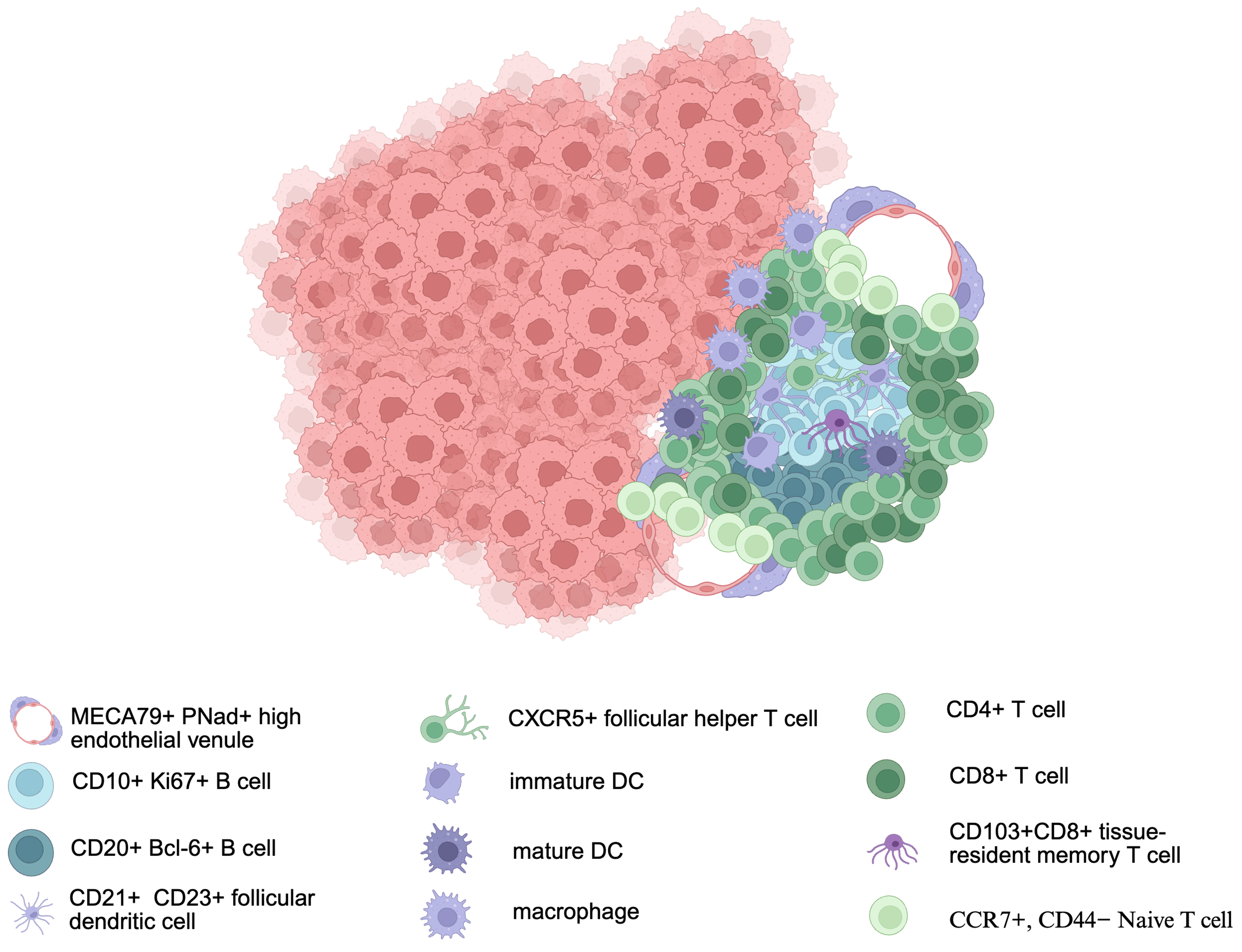
Figure 1. Detailed cellular composition and mature construction of TLS in CRC. This schematic delineates the architectural arrangement and key immune cell subsets within a mature TLS in CRC. Copyright permission link: https://BioRender.com/h060tsk. TLS: Tertiary lymphoid structure; CRC: colorectal cancer; MECA79: MECA-79 antigen; PNad: peripheral node addressin; CD: cluster of differentiation; CXCR: CXC chemokine receptor; DC: dendritic cell; CCR: CC chemokine receptor.
Maturation classification and implications for drug resistance of TLS in CRC
Based on a synthesis of previously published literature, the maturation classification of TLS can be broadly categorized into two-grade or three-grade systems, which reflect different understanding in organizational architecture, cellular composition, and genetic heterogeneity. The two-grade classification, primarily reported in esophageal squamous cell carcinoma (ESCC) and oral squamous cell carcinoma (OSCC), distinguishes between Agg-TLS (lymphocytic aggregates without FDCs or lymphoid follicles) and Fl-TLS (presence of at least one lymphoid follicle)[38,39]. In contrast, the three-grade classification is more commonly applied in CRC. In non-metastatic CRC (nmCRC, stage II-III), the three maturation stages are defined by progressive changes in cellular composition, particularly increasing frequencies of FDCs and mature B cells[40]. In CRC liver metastases (CRCLMs), TLS maturation is characterized primarily by organizational structure[41]. By integrating these two classification frameworks with histological and immunofluorescence features, a unified three-tier classification emerges: (1) early TLS (Agg), consisting of dense lymphocytic aggregates without differentiated FDCs or vascularization, negative for CD21 and CD23 expression; (2) primary follicle-like TLS (PFL-TLS), featuring closely packed clusters of predominantly naïve B cells surrounding a central FDC network (CD21+) but lacking GCs (CD23-); and (3) c TLS (SFL-TLS), characterized by the presence of GCs with HEVs, a T-cell zone containing mature DCs, and a GC comprising FDCs and B cells (CD21+CD23+). Notably, the gene expression profile associated with mature TLS includes not only chemokines with established roles in TLS formation and function (e.g., CXCL13, CXCL11, CXCL10) but also genes such as TGFB2 and VEGFB, which are linked to an immunosuppressive TME and may paradoxically inhibit TLS development. Collectively, the presence of GC is considered a prerequisite for mature TLS[42-44]. Detailed classification criteria are summarized in Tables 1 and 2.
| TLS maturation stage | Histological definition | Key immunohistochemical markers |
| Early TLS[40,42] | Dense lymphocytic aggregates, without FDCs or vascularization | CD21-, CD23- |
| PFL-TLS[41-44] | Central FDC network, surrounded by naïve B cells | CD21+, CD23- |
| SFL-TLS[41-44] | Presence of GCs (FDCs and B cells), with HEVs, T-cell zone | CD21+, CD23+, chemokines (CXCL13, CXCL11, CXCL10, etc.) |
| TME | Classification standard | Prognostic factor | Prognostic value |
| nmCRC, stage II-III[40] | [1] Early TLS (E-TLS, dense lymphocytic aggregates without differentiated FDCs) | TLS maturation | Recurrence; sensitivity to ICI |
| [2] PFL-TLS (B cell clusters with FDC network but without GCs) | |||
| [3] SFL-TLS (with GCs) | |||
| CRCLM[41,43] | [1] Aggregates (Agg): vague clusters of lymphocytes | Intratumor region of TLS | Clinical outcome |
| [2] Lymphoid follicles I (Fol-I): lymphoid follicles without GCs formation | |||
| [3] Lymphoid follicles II (Fol-II): lymphoid follicles with GCs formation | |||
| Six common gastrointestinal cancers[45,48] | [1] Lymphoid aggregates | TLS with GC | Independent prognostic factor |
| [2] Primary follicles | |||
| [3] Secondary follicles with a GC | |||
| ESCC, OSCC[46,47] | [1] Agg-TLS: tumors with only Agg and no Fl | Not directly transferable to CRC without validation | |
| [2] Fl-TLS: tumors with at least one Fl structure | |||
Furthermore, the organizational architecture of TLS and its corresponding immunological functionality can be categorized into distinct maturational classes with differential implications for drug resistance. Immature TLS, characterized by loose aggregates of T and B cells with sparse DCs, fail to support efficient anti-tumor immune responses and are instead associated with a T-cell-exhausted, inflamed, and immunosuppressive TME - features that collectively contribute to therapeutic resistance. In contrast, mature TLS (mTLS) exhibit well-organized structures in which mature DCs engage in active interactions with T cells, and immune hubs - such as Tfh cells expressing CD4, PD-1, and CXCR5 - establish productive contact with B cells, positioning mTLS as central anti-tumor immune niches within the TME. Accumulating evidence indicates that mTLS serve as important factors in the tumor immune contexture and carry significant prognostic and potential predictive value for treatment response. Notably, in non-metastatic stage II and III CRC, the presence of immature TLS (including early TLS and PFL-TLS) exhibits a strong association with increased recurrence risk, whereas patients with low densities of mature TLS (SFL-TLS) face a significantly higher cumulative risk of recurrence[40,49-51]. These observations underscore the functional divergence between immature and mature TLS in shaping immune competence and suggest that promoting TLS maturation may represent a viable strategy to overcome immune resistance and improve clinical outcomes in CRC.
Density, location, and heterogeneity of TLS as prognostic and drug resistance-related factors in CRC
In nmCRC, TLS are predominantly located in the interstitial or peritumoral regions and are composed mainly of B and T cells. TLS density is notably higher in right-sided colon cancer (RCC) and serves as an independent prognostic factor for 5-year OS, comparable to vascular invasion and AJCC stage[52,53]. Spatially, TLS can be classified as intratumoral (In-TLS), peritumoral (P-TLS), or distant-tumoral (D-TLS), with ongoing debate regarding their functional roles. In nmCRC, low P-TLS density - particularly when associated with immature TLS - correlates with reduced recurrence-free survival (RFS) and OS, independent of TNM stage, whereas In-TLS shows no significant prognostic value, possibly due to a higher proportion of Tregs within In-TLS[54,55]. This spatial heterogeneity extends to other cancer types and metastatic settings, underscoring the need for context-specific evaluation.
The prognostic and resistance-related implications of TLS are further complicated by their structural heterogeneity. Highly vascularized TLS containing HEVs and organized TLS with GCs are associated with increased infiltration of CD3+ T cells and enhanced anti-tumor immunity, predicting better outcomes in stage II CRC and other solid tumors[56-59]. Conversely, TLS with immature features or immunosuppressive cellular compositions may contribute to therapeutic resistance.
In CRCLMs, the relationship between TLS location and prognosis reverses compared to nmCRC: In-TLS correlates with superior outcomes, while P-TLS is associated with poor survival. Multiplex immunohistochemistry reveals that intratumoral TLS harbor higher densities of Tregs, M2 macrophages, and Tfh cells, all of which significantly influence RFS and OS[60]. Notably, recent evidence indicates that both mature In-TLS and P-TLS enriched with IgG+ plasma cells are linked to improved clinical outcomes, whereas TLS- areas are characterized by IgA+ plasma cells. The chemokines CCL19 and CXCL13 are enriched in TLS+ regions, with CCL19-producing cancer-associated fibroblasts (CAFs) promoting B cell recruitment via the CCL19-CCR7 axis, thereby facilitating TLS formation and the generation of therapeutic antibodies that may help overcome drug resistance[61]. Additionally, TLS presence in CRCLM is positively associated with specific KRAS mutation subtypes, suggesting a molecular link between TLS formation and resistance mechanisms[62].
In CRC pulmonary metastases (CRCPM), TLS correlate with increased immune cell infiltration, though their prognostic value remains controversial. Notably, the density and distribution of tumor-infiltrating lymphocytes (TILs) and TLS are consistent between pulmonary metastases and primary tumors, with CD3+, CD8+, CD45RO+, and FoxP3+ TILs evident in both sites[63-65]. This consistency suggests that TLS-associated immune landscapes may be preserved during metastasis and could represent stable determinants of treatment response.
Overall, the density, location, and structural maturity of TLS collectively shape their prognostic significance and influence drug resistance. Understanding these heterogeneous features is essential for leveraging TLS as both a predictive biomarker and a therapeutic target to overcome resistance in CRC.
Nomograms integrating TLS for predicting clinical outcomes and drug resistance level in CRC
Accumulating evidence supports that TLS density, location, and maturation status serve as independent prognostic factors in CRC, alongside traditional parameters such as TNM stage and tumor budding. Based on multivariate Cox regression analyses, several nomograms incorporating TLS features have been developed to enhance prognostic accuracy and, importantly, to reflect underlying mechanisms of therapeutic resistance.
One nomogram integrating TLS density, AJCC stage, and venous invasion accurately predicts 3- and 5-year OS in RCC, demonstrating superior predictive performance compared with models that use AJCC stage or TLS density alone[53]. Another nomogram incorporating P-TLS density, tumor stroma percentage, and TNM stage effectively predicts RFS and OS in nm CRC, with improved accuracy over TNM staging alone[66]. Notably, the inclusion of P-TLS density - which reflects immune competence at the tumor margin - may capture immune-mediated resistance mechanisms that are not accounted for by anatomical staging.
At the structural level, a nomogram incorporating the proportion of HEVs within TLS has been proposed. A high HEV/TLS ratio correlates with favorable prognosis, enhanced recruitment of CD3+ and CD8+ T cells and M1 macrophages, and reduced angiogenesis, suggesting that HEV-rich TLS facilitate effective anti-tumor immune infiltration and may overcome immune exclusion - a key mechanism of resistance to immunotherapy[67]. Conversely, low HEV expression alone does not confer prognostic benefit, underscoring the importance of TLS integrity in mediating immune function.
In the context of curative resection, TLS combined with low fibroblast activation protein (FAP) expression in the central tumor region is associated with significantly improved RFS, whereas high FAP expression in TLS-absent tumors predicts poor outcomes. This interaction suggests that TLS may counteract the immunosuppressive effects of CAFs, thereby mitigating resistance to adjuvant therapies[68].
At the molecular level, a 12-chemokine signature (including CCL2, CCL3, CCL4, CCL5, CCL8, CCL18, CCL19, CCL21, CXCL9, CXCL10, CXCL11, and CXCL13) has been linked to TLS expression and immune infiltration. High expression of this signature is associated with RCC, high TLS density, BRAF mutation, CIMP-high status, MSI-H status, increased tumor-infiltrating cytotoxic T lymphocytes and myeloid DCs, and reduced recurrence rates[69-72]. These chemokines are directly involved in lymphocyte recruitment and TLS organization; their coordinated expression may therefore serve as a surrogate for a functionally competent TLS that can sustain anti-tumor immunity and prevent resistance. Conversely, loss or downregulation of this chemokine network may contribute to immune evasion and poor therapeutic response.
Collectively, these TLS-integrated nomograms - spanning clinical, structural, and molecular dimensions - offer refined prognostic stratification and provide insights into immune-mediated resistance mechanisms. By capturing the functional status of TLS, these models may help identify patients who are more likely to benefit from immunotherapy and other treatments, thereby guiding strategies to overcome drug resistance in CRC. Although TLS-integrated nomograms show promising stratification performance, the level of evidence varies. For example, the TLS density and venous invasion model achieved multi-center validation via propensity score matching, enhancing its reproducibility. Conversely, other models (e.g., HEV/TLS ratio) remain constrained by retrospective, single-center designs. Consequently, extensive cross-cohort validation in diverse populations and integration into prospective trials (e.g., EMPIRE or TAYLOR) are essential before clinical guideline adoption; the nomogram is shown in Table 3.
Nomograms integrating TLS features for predicting clinical outcomes and patient recurrence in CRC[52,53,67-69]
| Detailed nomogram model | Prognostic indicator | Cancer type | Validation status/level of evidence |
| Density of TLS, AJCC stage and venous invasion[53] | 3-year OS, 5-year OS | RCC | Retrospective, multi-center validated (propensity score-matched) |
| P-TLS density, TSP and TNM stage[52] | 2-year OS, 5-year OS | nmCRC | Retrospective, single-center; requires multi-center verification |
| HEV/TLS ratio, TLS[67] | OS and DFS | CRC | Retrospective, single-center cohort; requires prospective validation |
| FAP expression in central tumor, TLS[68,69] | RFS | CRC and OSCC | Retrospective cohort; lacks prospective clinical adoption trial |
Association of TLS with MSI-H or high-TMB CRC and implications for drug resistance
In CRC, the formation and maturation of TLS - historically described as Crohn’s-like lymphoid reactions - are significantly more prevalent in MSI-H tumors and those harboring BRAF, KRAS, CIMP-high, or HER2-amplified mutations compared with MSS or low-mutant tumors[73]. This enrichment is largely attributable to the high TMB characteristic of MSI-H CRC, which generates abundant neoepitopes that drive sustained immune activation. Consequently, the TME of MSI-H CRC exhibits heightened infiltration of activated natural killer (NK) cells, CD8+ T cells, and T helper cells[74].
B cell-rich TLS, characterized by robust antigen presentation, accumulation of IgG+ plasma cells, and perforin-expressing B cells, are prominently observed in MSI-H and high-TMB CRC patients[23,75]. These TLS are further supported by CXCL13+ TH1-like cells, which recruit CCR5+ cells and promote TLS formation[76]. Additionally, TIM4 and FOLR2 double-positive macrophages localized within the T-cell zones of MSI-H-associated TLS correlate with high infiltration of B cells and effector CD8+ T cells, underscoring their role in sustaining productive anti-tumor immunity[77]. High-TMB tumors also exhibit elevated expression of lymphotactin 1 and 2 (XCL1 and XCL2), which facilitate cDC1 recruitment and T cell proliferation[78].
Recent single-cell analyses have identified a distinct gene signature in MSI-H TLS, including high expression of class I MHC genes, chemokines that attract memory T cells (CXCL9, CXCL10), T cell-activating cytokines (IFNG), and factors supporting T and NK cell proliferation (IL15, CXCL16). These molecules serve as important drivers of TLS formation and immune activation[79-87].
From the perspective of drug resistance, the robust TLS formation observed in MSI-H CRC represents a key factor of enhanced sensitivity to ICIs. In contrast, MSS CRC - characterized by lower TLS prevalence, reduced immune infiltration, and an immunosuppressive TME - exhibits intrinsic resistance to immunotherapy. Notably, the molecular features that drive TLS formation in MSI-H tumors, including high chemokine expression and antigen presentation capacity, are frequently absent or suppressed in MSS tumors, contributing to immune evasion and poor treatment response. Thus, TLS not only serve as a distinguishing immunological hallmark between MSI-H and MSS CRC but also represent a potential contributor of ICI sensitivity. Strategies aimed at inducing TLS formation in MSS tumors may therefore offer a promising approach to overcome intrinsic drug resistance and expand the therapeutic benefits of immunotherapy to a broader CRC patient population. Here, the gene signatures of MSI-H TL in CRC are summarized in Table 4, and specific cells in CRC are described in Table 5, respectively.
| Researchers | Gene names | Functional roles of upregulated genes |
| Becht et al.[79] | CXCL9, CXCL10, CXCL16, IFNG, IL15 | Production of IFN-γ, activation of NK cells |
| Kim et al.[80] | CTSC, CCL5, CCL18, CXCL9 | Recruitment of immune cells, naïve T cells |
| Wang et al.[81] | GBP2, CXCL10, CXCL11 | Simulators of IFN, infiltration of TILs |
| Masuda et al.[82] | CXCL13, CD4, HAVCR2 | Formation of TLS, negative regulator of anti-tumor immunity |
| Mlecnik et al.[83] | IL15, IFNG, GNLY, CCL3, CXCL16 | Production of IFN-γ, enrichment of NK cells and CD8+ effector T cell |
| Xu et al.[84] | AEN, CD3E, TRGC2, ULBP3 | Enrollment of ILCs, response to ICI |
| Yang et al.[85] | CCL19, CCL21, CXCL13, CCR7 | Formation of TLS, activation and recruitment of Lti cells |
| Jia et al.[86] | KRAS, B2M, CTSC, IKZF3, CCL5 | Recruitment of immune cells, differentiation, proliferation of B cells |
| Cell in CRC TLS | Main cell markers | Representative markers / functional annotation |
| B cells[26,32,89-91] | ||
| CD20+ naïve B cells | IGHD, FCER2, TCL1A, IL4R | |
| CD27+ memory B cells | CD27, AIM2, TNFRSF13B | |
| CD38+ GC B cells | BANK1, TNFRSF13C, MKI67, AICDA | Proliferation and somatic hypermutation (MKI67, AICDA) |
| CD138+ plasma B cells | MZB1, DERL3, TNFRSF17, JCHAIN | |
| T cells[44,45,87,88] | ||
| Naïve T cells (Tn) | CCR7, LEF1, SELL, CD27, CD28 | |
| Central memory T cells (T-CM) | CCR7, SELL, IL7R, PTGER2 | Memory maintenance and lymphocyte trafficking |
| Effector memory T cells (T-EM) | GZMK, CXCR4, CXCR3, CD44 | |
| Effector T cells | GNLY, NKG7, TBX21, PRF1, CX3CR1 | |
| Recently activated effector memory or effector T cells (T-EMRA) | KLRG1, CX3CR1, FCGR3A, NKG7, PRF1, GNLY | S1PR1, S1PR5 and ITGB7: capability to circulate in the periphery and home to normal mucosa and tumors |
| CD4+ T cells | ||
| Th1 cells | IFNG, TIGIT, ZEB2, ICOS | |
| - GZMK+ Th1 cells | GZMK, IFNG, CXCR3 | |
| - CXCL13+ Th1 cells | CXCL13, BHLHE40, PDCD1, HAVCR2 | |
| Th2 cells | IL13, CD69, GZMK, NKG7 | |
| TH17 cells | IL23R, RORC, IL17A, FURIN | |
| Follicular T helper cells | CXCR5, BCL6, ICA1, TOX, TOX2 | |
| Follicular T regulatory cells | FOXP3, IL10, IL2RA, CXCR5, PDCD1, IL10 | |
| Treg cells | FOXP3, IL2RA, IKZF2 | |
| - Tumor-Treg | FOXP3, CCR8, TNFRSF18, LAYN | |
| - Blood-Treg | FOXP3, IL2RA, CXCR5, PDCD1, IL10 | |
| CD8+ T cells | ||
| Mucosal-associated invariant T (MAIT) cells | SLC4A10, KLRB1, ZBTB16, NCR3, RORC, RORA | |
| Cytotoxic T cells | GZMK, PRF1, HOPX, LAG3, GZMA, GZMB | |
| Naïve CD8+ T cells | TCF7, SELL, ITGAE, IL2RA | |
| Intraepithelial lymphocytes | ||
| Tissue-resident memory T cells (TRM) | CD69, ITGAE, CXCR6 | |
| - Lamina propria TRM | CD6, CXCL1, MYADM, CAPG | |
| - Intraepithelial lymphocytes (IELs) | CD160, KIR2DL4, TMIGD2, KLRC1/2/3 | Highly expressed NK cell markers |
| CTLA4+ Treg | IL32, KLF6, HLA-DRB1, ETS1, IL7R, KMT2E | |
| CTLA4- Tregs | TOX2, ANKRD17 | |
| Exhausted CD8+ T cells (T-EX) | HAVCR2, CXCL13, PDCD1, LAYN, TOX | tumor exclusivity |
| Myeloid cells[88-91] | ||
| Monocyte | C1QA, C5AR1, CCL19, CCL23 | |
| - CD16-monocytes | CD16, FCN1, and VCAN | |
| - CD14-monocytes | CD14, IFITM2, LST1 | |
| TAM macrophage | ||
| - SPP1-macrophages | SPP1, VEGFA, FCN1 | |
| - FOLR2-macrophages | FOLR2, MARCO, C1QA | |
| - CXCL5-macrophage | MMP1, CXCL2, S100A8 | |
| DCs | XCR1, CLEC9A, CD11c | Antigen processing- and presentation-related pathway |
| NK cells | NKG7, PTPRC, CST7, DNAM-1 | |
Conclusion: TLSs as biomarks in drug resistance of CRC
Predictive biomarkers
TLS serves as an indispensable predictive biomarker for therapeutic responsiveness, particularly in partitioning responses to ICIs. In MSI-H or high-TMB CRC, sustained chemokine networks (e.g., CXCL13, CCL19) drive robust TLS neogenesis and GC formation, rendering these tumors highly sensitive to immunotherapy. In stark contrast, MSS tumors typically exhibit low TLS prevalence and defective maturation, representing a hallmark of intrinsic immunotherapy resistance[40,53,56].
Functional mediators of resistance
In addition, TLS functions as active functional mediators of resistance in CRC. Structurally mature TLSs equipped with functional HEVs and active GCs serve as productive local immune hubs. They facilitate high-affinity antibody production and the entry of tumor-reactive stem-like CD8+ T cells, effectively counteracting immune exclusion and mitigating drug resistance. However, when tumors actively disrupt these structures - resulting in HEV deficiency, a lack of GCs, or an enrichment of immunosuppressive cells like Tregs and LAMP3+ DCs. Notably, LAMP3+ DCs exhibit a context-dependent dual role throughout the TLS life cycle. Structurally, mature LAMP3+ DCs secrete homing chemokines (CCL19/CCL21) to guide naïve T cells and support early lymphoid aggregation. However, within the heavily suppressed microenvironment of conventionally resistant MSS rectal cancers, these cells shift toward a regulatory phenotype. In this therapy-refractory context, they upregulate immune checkpoint ligands and secrete anti-inflammatory cytokines (IL-10 and TGF-β). These factors may interfere with the CXCL13-mediated B-cell homing pathway and impair local T-cell activation, acting as potential negative regulators that may contribute to maintaining TLS in an immature state. Consequently, these observations should be interpreted with caution, and future studies are required to clarify the precise molecular mechanisms underlying these context-dependent effects[40,67,88].
FORMATION AND MATURATION OF TLS: MECHANISMS AND IMPLICATIONS FOR DRUG RESISTANCE
Despite ongoing investigations into the precise mechanisms underlying TLS generation and development, the fundamental prerequisites for TLS formation in tumors are increasingly well-defined. TLS arise in settings of chronic inflammation, sustained by persistent expression of lymphoid chemokines - particularly CXCL13, CCL19, and CCL21 - and are closely associated with high TMB. Tumor cells contribute by presenting antigens derived from aberrant gene expression, mutated oncogenes (e.g., KRAS, BRAF), MSI-H status, epigenetically modified self-molecules, cancer-testis antigens, and virally encoded proteins[92-94]. Given their structural and functional similarity to SLOs, TLS are thought to share core developmental mechanisms with SLOs, with both tumor cells and the tumor stroma - particularly CAFs - playing critical roles in inducing and localizing TLS formation[92-94].
From the perspective of drug resistance, the presence and maturity of TLS serve as key contributors to therapeutic sensitivity. Tumors with high TMB and sustained chemokine expression - such as MSI-H CRC - exhibit robust TLS formation, which in turn supports durable anti-tumor immunity and favorable responses to ICIs. Conversely, tumors lacking these TLS-promoting features, including the majority of MSS CRC, often exhibit impaired TLS development, immune exclusion, and intrinsic resistance to immunotherapy. Moreover, the tumor stroma can exert dual effects: while certain stromal components facilitate TLS organization, others may suppress TLS formation through immunosuppressive signals. Understanding the mechanisms relevant to TLS formation - from chemokine signaling to stromal interactions - is therefore essential for developing strategies to induce TLS neogenesis in resistant tumors and expand the clinical benefits of immunotherapy to a broader patient population.
Chemokine signaling pathways in TLS formation and their implications for drug resistance
CXCL13-CXCR5 axis
CXCL13 is an important chemokine driving TLS formation and localization. Induced by TGF-β and IL-7/21, CXCL13 is expressed by various cell types, including CD4+ TILs (Tfh, Tph, Th1), tumor-reactive CD8+ T cells, FDCs, HEVs, type 3 innate lymphoid cells (ILC3s), and even CAFs and tumor cells[53,95-97]. In CRC, CXCL13-expressing CD4+ and CD8+ T cells are frequently observed near carcinoma cells, particularly in dMMR tumors[98,99]. CXCL13 acts through its receptor, CXCR5, to recruit B cells and lymphoid tissue inducer (LTi) precursors, initiating TLS formation[23,100-104]. Neutralization of CXCL13 or its loss results in reduced infiltration of B cells and Tfh cells, impairing TLS development[23,100].
Lymphotoxin-mediated positive feedback
CXCL13 recruits CXCR5+ LTi cells expressing lymphotoxin-α1β2 (LT-α1β2), which interact with LTβR on mesenchymal lymphoid-tissue organizer (LTo) cells. This interaction activates canonical and non-canonical NF-κB signaling, promoting LTo cells to upregulate chemokines (CXCL13, CCL19, CCL21) and adhesion molecules (ICAM-1, VCAM-1, MAdCAM-1), thereby facilitating further lymphocyte recruitment via HEVs[105-112]. Subsequently, LTi cells express TRANCE, activating TRANCER signaling that induces LT-α1β2 and IL-7 expression, establishing a positive feedback loop that expands lymphatic vessels, enhances FDC networks, and amplifies chemokine expression, ultimately supporting TLS organization and maturation[113,114].
CCL19/21-CCR7 axis
CCL19 and CCL21, secreted by LAMP3+ DCs, HEVs, cancer cells, and fibroblastic reticular cells (FRCs), recruit naïve and central memory T cells expressing CCR7 and CD62L[115-117]. These T cells are guided to T-cell zones, while CXCR5+ B cells are recruited to B-cell zones in response to CXCL13, enabling GC formation and T cell–B cell cooperation essential for productive anti-tumor immunity[118-121].
CCL28-CCR10 axis
The CCL28-CCR10 axis, primarily involving epithelial cell-derived CCL28, mediates the migration of IgG+ and IgA+ CCR10+ plasma cells from TLS peripheries into the tumor stroma, supporting humoral anti-tumor responses[23,122]. Additionally, CCR10+ CD4+ T cells enriched in IL-17- and IL-22-producing subsets (Th22 cells) suggest a role in T cell homing within the CRC TME[123,124].
Implications for drug resistance
These chemokine networks are central to TLS formation and function and are directly relevant to drug resistance. Tumors with high expression of TLS-associated chemokines (e.g., CXCL13, CCL19, CCL21) exhibit enhanced lymphocyte infiltration, organized TLS, and improved sensitivity to ICIs. Conversely, loss or downregulation of these chemokine axes - often observed in MSS CRC or following immune evasion - impairs TLS formation, leading to diminished T and B cell recruitment, reduced GC activity, and compromised anti-tumor immunity. This chemokine-deficient state contributes to an immune-excluded or immune-desert phenotype that underlies resistance to immunotherapy and other treatments. Therefore, strategies aimed at restoring or enhancing the chemokine signaling pathways may represent a promising approach to promote TLS neogenesis and overcome drug resistance in CRC.
Functional roles of tumor necrosis factor superfamily members in TLS formation and drug resistance
The tumor necrosis factor (TNF) superfamily, including TNF-α, TNF-β, CD40L, and LIGHT, plays key roles in TLS formation through complex interactions involving LTα1β2/LTβR signaling.
LTα1β2/LTβR axis
LTαβ expression on activated CD4+ T cells is essential for DC function, while LTβR signaling in DCs provides maturation signals downstream of CD40 activation[125-127]. For CD8+ T cells, the LT pathway supports clonal expansion but not effector function, whereas the CD40 pathway is required for CD8+ T cell effector function and induces CD86 expression on DCs[125-127]. LTα1β2/LTβR interactions between B cells and follicular DCs induce CXCL13 and CCL19 production, promoting DC and CD4+ T cell recruitment that supports helper T cell and Tfh responses[128-130].
TNF-α, LIGHT, and cellular contributors
TNF-α secreted by tumor-associated neutrophils (TANs) recruits B cells to the TME and promotes their differentiation into plasma cells, a process mediated in part by membrane-bound BAFF on TANs[131,132]. LIGHT (TNFSF14), produced by Tfh cells, induces CCL21 production by tumor endothelial cells and fibroblasts; upon binding LTβR, LIGHT promotes blood vessel normalization and triggers intratumoral TLS formation[74,133,134]. Emerging evidence suggests that CD8+ T cells, NK cells, and M1 macrophages may function as alternative LTi cells in TLS via LTα3-TNFR signaling or TNF-α expression[135-137].
TGF-β in T cell recruitment and retention
TGF-β1, abundant in epithelial tissues, plays a crucial role in CD8+ T cell recruitment and retention. TGF-β binding to its receptor promotes integrin-linked kinase (ILK) phosphorylation and AKT activation, inducing CD103 expression on CD8+ T cells. This facilitates T cell homing to the TME and enhances CCR5 recruitment at the immunological synapse between TILs and tumor cells[138-141]. TGF-β signaling also induces CD8α expression in intestinal T cell populations via the Smad2/Smad3 pathway, contributing to local immune surveillance[142,143].
Implications for drug resistance
The TNF superfamily members and TGF-β signaling pathways that are associated with TLS formation have direct implications for drug resistance. Functional LTα1β2/LTβR and CD40/CD40L signaling are essential for productive DC-T cell interactions and sustained anti-tumor immunity; their disruption can lead to impaired TLS maturation and reduced ICI sensitivity. TNF-α and LIGHT promote B cell recruitment and vascular normalization, respectively, both of which enhance immune cell infiltration and therapeutic responsiveness. Conversely, TGF-β exhibits dual roles: while it supports CD8+ T cell retention via CD103 induction, excessive TGF-β signaling in the TME can promote immunosuppression, Treg differentiation, and resistance to immunotherapy. Thus, the balance of TNF superfamily and TGF-β signaling within TLS is strongly associated with the functional quality of anti-tumor immunity. Therapeutic strategies that modulate these pathways - such as LTβR agonists to enhance TLS maturation or TGF-β blockade to overcome immunosuppression - may represent promising approaches to modulate drug resistance and improve treatment outcomes in CRC.
Interactions and crosstalk in TLS formation: HEVs, microbiota, and implications for drug resistance
HEVs as gatekeepers of TLS function
HEVs are specialized postcapillary venules that facilitate lymphocyte transmigration into TLS. In human and murine solid tumors, including CRC, HEVs express MECA-79 and peripheral node addressin (PNAd), enabling lymphocyte homing via L-selectin-, chemokine-, and integrin-mediated interactions[138,144]. HEVs express chemokines CCL19, CCL21, and CXCL13, which recruit T cells, DCs, and B cells, respectively, into organized TLS compartments[145,146]. Lymphocyte integrins - particularly αLβ2 (LFA-1) binding to ICAM-1/ICAM-2, and α4β7 binding to MAdCAM-1 - mediate firm adhesion and trans endothelial migration[147-149]. HEVs also serve as entry sites for CD62L+ T cells, including tumor-reactive PD-1+ Tcf1+ stem-like CD8+ T cells, which are important for sustained anti-tumor immunity and response to ICB[150,151]. Upregulated genes such as TSPAN7 and MEOX2 on tumor HEVs regulate lymphocyte extravasation and inhibit excessive angiogenesis, respectively[152,153].
Microbiota-driven TLS formation
Gut microbiotas play an active role in inducing TLS in CRC. In orthotopic mouse models, Helicobacter hepaticus colonization promotes TLS formation with organized B- and T-cell zones, upregulates TLS-associated genes (e.g., Lta, Tnfsf14, Icam1), and enhances anti-tumor immunity[154,155]. Bifidobacterium augments DC function and CD8+ T cell priming, while Akkermansia muciniphila and Enterococcus hirae modulate IFN-γ and IL-2 expression, improving the efficacy of PD-1/PD-L1 inhibitors[156-158]. Additionally, CX3CR1hi macrophages enriched in TLS during Salmonella invasion secrete CXCL13, TGF-β1, and IL-10, contributing to TLS organization[159-162].
Implications for drug resistance
HEVs and microbiota may contribute to TLS functionality with direct relevance to drug resistance. High HEV density and maturation correlate with enhanced recruitment of stem-like CD8+ T cells, which are essential for sustained responses to ICB and are often depleted in resistant tumors[150,151]. Conversely, HEV deficiency or immaturity impairs lymphocyte trafficking, contributing to immune exclusion and poor therapeutic response. The presence of specific gut microbiota that promote TLS formation - such as H. hepaticus, Bifidobacterium, and A. muciniphila - has been associated with improved ICI efficacy, whereas dysbiosis may undermine TLS integrity and foster resistance[156-158]. Thus, HEV integrity and favorable microbiota composition are emerging as key contributors of TLS-mediated anti-tumor immunity. Strategies aimed at enhancing HEV maturation (e.g., via LIGHT signaling) or modulating gut microbiota (e.g., probiotics or fecal microbiota transplantation) represent promising approaches to augment TLS function and overcome drug resistance in CRC.
Taken together, these chemokine networks coordinate the molecular pathways and cellular interactions that collectively influence the functional quality of anti-tumor immunity. Impaired chemokine axes or the accumulation of regulatory components (such as LAMP3+ DCs) frequently leads to immature TLS formation, thereby fostering an immune-excluded phenotype and driving therapeutic resistance. The mechanisms of formation and maturation of TLS in CRC are illustrated in Figure 2.
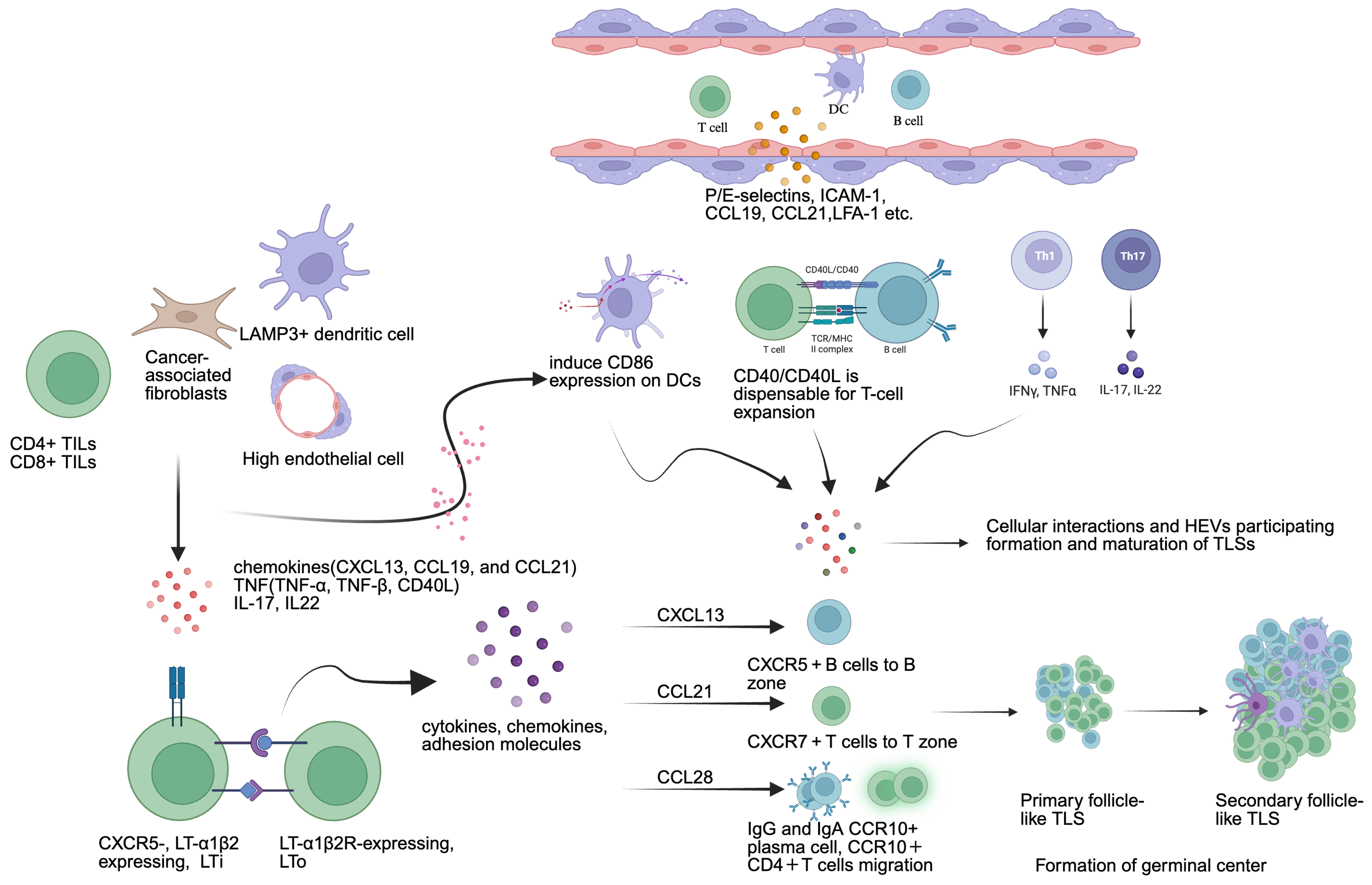
Figure 2. Mechanisms of formation and maturation of TLS in CRC. This diagram illustrates the cellular interactions, chemokine cascades (CXCL13, CCL19, CCL21), and TNF superfamily signaling pathways that drive the progressive development from early lymphocytic aggregates to mature SFL-TLS. Copyright permission link: https://BioRender.com/omsjn1g. TLS: Tertiary lymphoid structure; CRC: colorectal cancer; CXCL/CCL: CXC/CC chemokine ligand; TNF: tumor necrosis factor; SFL-TLS: secondary follicle-like tertiary lymphoid structure; DC: dendritic cell; ICAM-1: intercellular adhesion molecule 1; CD: cluster of differentiation; CD40L: CD40 ligand; IL: interleukin; CXCR: CXC chemokine receptor; LTi: lymphoid-tissue inducer cell; LTo: lymphoid-tissue organizer cell; HEVs: high endothelial venules.
CORRELATION OF TLS AND DRUG RESISTANCE IN CRC
Molecular heterogeneity and TLS as modulators of oncogenic resistance
The genomic and transcriptomic heterogeneity of CRC imposes a profound barrier to therapeutic efficacy, frequently steering tumors toward chemoresistance and post-adjuvant recurrence. Genomic stratification suggests that the prognostic weight and functional configuration of TLS are intimately linked with specific oncogenic drivers. For instance, tumors harboring oncogenic BRAF mutations (BRAFmut) typically present an aggressive clinical phenotype refractory to standard chemotherapy. However, clinical evidence indicates that robust TLS formation can significantly attenuate this genetic disadvantage; BRAFmut patients exhibiting high peritumoral or intratumoral TLS densities demonstrate OS rates comparable to their BRAFmt counterparts. A parallel paradigm emerges in the context of targeted therapies for KRAS-driven and EGFR-mutant colorectal malignancies. While combined EGFR and KRASG12C inhibition represents a validated approach to counter adaptive resistance mechanisms in primary CRC, the structural integrity of the local immune microenvironment remains an important contributor to long-term durable responses. In CRCLMs, a high TLS score strongly correlates with specific KRAS mutation subtypes, characterized by an enrichment of IgG+ plasma cells and robust production of antitumor antibodies. Furthermore, quantitative nomograms confirm that TLS density and higher HEV proportions stand as independent favorable prognosticators for disease-free survival (DFS) in both EGFR-mutant and KRAS-mutant gastrointestinal tumors, irrespective of baseline PD-L1 tumor proportion scores. These insights highlight a novel therapeutic strategy: combining oncogenic kinase inhibitors with TLS-inducing interventions to synergistically dismantle cell-intrinsic resistance and reinforce cell-extrinsic immune clearance[163-165].
BRAF mutations and TLS as a resistance modulator
BRAF mutations, prevalent in CRC, typically confer high-level drug resistance and poor clinical outcomes. However, compelling evidence shows that BRAF-mutant (BRAFMT) patients with high TLS expression and BRAF wild-type (BRAFWT) patients with low TLS expression share comparable OS, indicating that robust TLS formation may partially mitigate the adverse prognostic impact in BRAF-mutant CRC patients. Further analysis of TLS features - including location, number, and maturity - in relation to BRAF mutation status suggests that intratumoral TLS may enhance prognosis and sensitize BRAFMT CRC patients to therapeutic interventions[166].
EGFR signaling, KRAS mutations and TLS as a strategy to improve therapeutic sensitivity
Recent research identifies EGFR signaling as the dominant mechanism of CRC resistance to KRAS G12C inhibitors; correspondingly, dual targeting of EGFR and KRAS G12C effectively circumvents this resistance in CRC, reverting resistance to KRASG12C inhibition in CRC patients[167,168]. Similarly, in colorectal cancer liver metastases, the location and density of TLS are associated with molecular subtypes and clinical outcomes. The study found that the TLS score was positively correlated with specific KRAS mutation subtypes[30]. Meanwhile, TLS density has been confirmed as a potential prognostic biomarker for DFS in EGFR-mutant tumors, including CRC and lung adenocarcinoma, independent of PD-L1 TPS or EGFR mutation subtype, suggesting a potential therapeutic strategy to improve therapeutic sensitivity of patients with EGFR- or KRAS-mutation G12C CRC[160,169,170].
Dynamic single-cell insights into TLS functionality
Single-cell mapping of immune landscape dynamics during CRC progression reveals significant functional differences in CD4+ Tfh and germinal center B (GC-B) cells between early and advanced CRC. Notably, CXCL13 expression on CD8+ Tex cells, along with CD40–CD40L interactions between CD4+ Tfh and B~GC~ cells, emerges as a key regulator of TLS functionality. These interactions significantly influence the response to immunotherapy and the development of drug resistance[171].
Implications for overcoming resistance
Collectively, these findings underscore that TLS heterogeneity - shaped by molecular drivers such as BRAF mutations, immune cell interactions, and dynamic changes during disease progression - plays a pivotal role in determining therapeutic sensitivity. Strategies aimed at preserving TLS integrity, promoting B cell-mediated humoral immunity, or enhancing key interactions such as the CD40–CD40L axis may represent novel approaches to overcome drug resistance in CRC, particularly in molecularly defined subsets with poor baseline prognosis.
Interactions between TLS and neoadjuvant drug treatment resistance
Functional roles of TLS in MSS CRC neoadjuvant drug treatment resistance
In MSS CRC, which typically exhibits limited response to chemotherapy or immunotherapy, recent studies reveal that responders harbor a higher abundance of more activated and mature TLS compared to non-responders. Importantly, LAMP3+ DCs have been identified as negative regulators of TLS formation in MSS CRC, suggesting that suppressing this cell subset may reduce drug resistance[88]. Similar findings in ovarian carcinoma and non-small cell lung cancer demonstrate that chemotherapy-induced endoplasmic reticulum stress drives TLS formation and maturation in metastatic lesions, thereby increasing infiltration of ICI-responsive TCF1+CD8+ T cells[172]. Neoadjuvant chemotherapy also upregulates B-cell- and TLS-associated genes (JCHAIN, CXCR4, MS4A1), correlating with enhanced antigen presentation and deeper pathological remission, including complete remission[173]. Neoadjuvant chemoradiotherapy further promotes TLS maturity within tumors and improves OS. Radiotherapy modulates the TME to activate and proliferate immune cells within mature TLS - including CD20+Ki-67+ B cells, CD21+ DCs, CD8+Ki-67+ cytotoxic T cells, and CD4+Ki-67+ Th cells - thereby enhancing immunotherapy efficacy[174,175].
Functional roles of TLS in advanced CRC neoadjuvant drug treatment resistance
In advanced CRC, multivariate analysis confirms that the presence of TLS in primary tumors prior to systemic chemotherapy is significantly associated with favorable chemotherapy response and prolonged survival. Mature TLS correlates with MAdCAM-1+ vessels and higher CD3+/CD8+ T-cell infiltration in both primary and metastatic tumors[176]. Based on these insights, a multi-center TLS gene signature has been established. A high TLS score - characterized by increased proportions of CD8+ T cells, B cells, and DCs, along with activation of anti-tumoral signaling pathways - predicts better response to neoadjuvant FOLFOX chemotherapy and sintilimab (PD-1 blockade), even in MSS CRC[177]. Similarly, the presence of mature TLS with GCs, enriched for activated CD4+ memory cells, naïve B cells, and NK cells, forms in the context of high-quality tumor neoantigens, leading to enhanced humoral immunity that is sensitive to neoadjuvant therapy in gastrointestinal solid tumors[178,179].
Implications for drug resistance
These findings collectively demonstrate that TLS maturation status is strongly associated with sensitivity to neoadjuvant therapies. TLS deficiency or immaturity represents an actionable mechanism of drug resistance. Key resistance-reversing strategies include: (1) suppressing or modulating LAMP3+ DCs to block their IL-10/TGF-β signaling axes, thereby removing the molecular brakes on chemokine cascades to restore mature TLS formation in MSS CRC; (2) Harnessing chemotherapy-induced ER stress to promote TLS neogenesis; (3) Combining radiotherapy to enhance TLS maturity and immune cell activation; (4) Utilizing TLS gene signatures to identify resistant patients who may benefit from alternative or combination regimens. These approaches hold promise for overcoming intrinsic resistance, particularly in MSS CRC populations that are conventionally refractory to treatment.
MEASURES FOR INDUCING TLS TO OVERCOME DRUG RESISTANCE AND IMPROVE THERAPEUTIC EFFICACY OF CRC
As the presence of TLS is associated with improved ICI response and reduced cancer drug resistance, it is hypothesized that the enhancement of TILs represents a pivotal therapeutic strategy, which may be achieved via inducing the formation of TLS, by accelerating the maturation of TLS or by enriching the density of TLS. Increasing studies suggest that the presence of TLS within the TME is associated with favorable prognoses and a higher likelihood of responding to ICB therapy in several cancer types[180].
Inducing TLS to overcome drug resistance: enriching TILs and HEVs
Emerging evidence highlights that therapeutic strategies aimed at inducing TLS formation - particularly through enriching TILs and HEVs - hold promise for overcoming drug resistance in CRC.
Radiotherapy and immunotherapy combinations
In mouse solid tumor models, hypofractionated radiotherapy combined with lenalidomide (an immunomodulatory agent) induces TLS formation in a CD8+ T cell- and type I interferon-dependent manner. This combination enhances anti-tumor CD8+ T cell immunity, DC cross-presentation, upregulates TLS-associated genes (GZMB, IFN-γ, TNF-α), and increases tumor-associated HEV (TA-HEV) density. Importantly, it recruits and enriches stem-like (TCF1+ TIM3- PD1+) and transitory (TCF1- TIM3+ CD101- PD1+) tumor-specific T cell subsets, both of which are important for sustained responses to immunotherapy[181,182]. These findings suggest that radiotherapy-ICI combinations can overcome resistance by promoting TLS neogenesis and sustaining effector T cell pools.
Chemotherapy-induced TLS formation
Certain chemotherapeutic agents also demonstrate TLS-inducing potential. Combined administration of tanshinone IIA (TSA) and astragaloside IV (As) normalizes tumor blood vessels - including HEVs - while reducing immunosuppressive factors, thereby enhancing TIL abundance and activity[183]. This dual mechanism addresses two key resistance pathways: poor lymphocyte infiltration and an immunosuppressive microenvironment.
Nanovaccines and bio-nanoparticle strategies
Bio-nanoparticle-based λ phage vaccines targeting Aspartate β-hydroxylase (ASPH), a tumor-associated antigen, show promising TLS-inducing effects. When combined with PD-1 blockade, ASPH-targeting vaccines reduce immunosuppressive Treg infiltration and, upon PD-1/PD-L1 inhibition, promote CXCL13 production by ASPH+ cancer cells, which recruits CXCR5+ CD8+ T lymphocytes to TLS. This is accompanied by increased enrichment of effector and memory CTLs, Tfh cells, GC-B cells, and FDCs[184]. Similarly, lymph node-targeting nanovaccines enhance tumor immune responses and accelerate the tumor-immunity cycle[185,186]. These approaches directly counteract immune evasion mechanisms that underlie resistance to conventional immunotherapies.
3D-printed scaffold vaccines
Implantable 3D-printed scaffold vaccines represent an innovative strategy for in situ TLS induction. These scaffolds recruit antigen-presenting cells (APCs) and T cells, initiating TLS formation with mature features, including activation of NK cells, macrophages, and CD11c+ DCs. When combined with PD-L1 blockade, scaffolds further upregulate the proliferation (Ki67+), cytotoxic (granzyme B+), and IFN-γ-producing CD8+ T cells[187]. Other scaffold designs incorporating ovalbumin, GM-CSF, and sustained TLR2 agonists facilitate long-term DC activation and recruitment of activated CD8+ cytotoxic T lymphocytes and memory T cells[188,189].
Implications for drug resistance
Collectively, these TLS-inducing strategies address multiple mechanisms of drug resistance: (1) enhancing lymphocyte infiltration via HEV normalization and chemokine recruitment; (2) expanding stem-like and effector T cell populations that sustain anti-tumor immunity; (3) reducing immunosuppressive Tregs; and (4) generating durable memory responses. By converting immunologically “cold” tumors into “hot” tumors with organized TLS, these approaches hold potential to overcome intrinsic and acquired resistance to immunotherapy and other treatments in CRC and beyond.
Stimulator of interferon genes agonists as crucial triggers for TLS induction and overcoming drug resistance in CRC
Stimulator of interferon genes (STING) agonism has emerged as a promising strategy to induce TLS formation and overcome drug resistance in CRC. Activation of the STING pathway within the TME upregulates both anti-angiogenic factors (e.g., Tnfsf15/VEGI, CXCL10) and TLS-inducing chemokines (e.g., CCL19, CCL21, LTA, LTB, LIGHT), leading to enhanced infiltration of CD8+ T cells and CD11c+ DCs, as well as local TLS neogenesis[190,191].
Advanced STING-activating platforms
Innovative delivery systems have been developed to optimize STING-mediated TLS induction. A STING-activating hydrogel (ZCCG) incorporating Zn2+, chitosan (a STING pathway activator), and CpG (a TLR9 agonist) enables dual activation of cGAS-STING and TLR9 pathways. This dual-pathway approach effectively enhances immune cell infiltration and expression of lymphocyte-recruiting chemokines, thereby promoting TLS formation and potentiating tumoricidal immunity[192]. Mannose-modified stearic acid-grafted chitosan (M-CS-SA) micelles target the cGAS-STING pathway, increasing CD3+ CD8+ T cell infiltration and accelerating DC maturation[193].
Additional STING-oriented strategies include cyclic dinucleotides combined with Pam3CSK4 (Pam3CSK4-CDGSF), a conjugated adjuvant targeting both STING and TLR1/2, as well as immunostimulatory nanoparticles (immuno-NPs) containing monophosphoryl lipid A (MPLA) and cyclic diguanylate
| STING interventions | Mechanistic pathway | Induction roles in TLS and TME |
| Low-dose STING agonist ADU S-100 (5 µg/mouse) (in vivo)[190] | TCRβ-CDR3 on TILs, LIGHT/TNFSF14, TLR7/9 on DCs | Enhanced infiltration of CD8+ T cells and CD11c+ DCs, local TLS neogenesis |
| ZCCG plus Zn2+ with 4,5-imidazole dicarboxylic acid (in vivo)[192] | cGAS/STING, CpG/TLR9 | Enhanced infiltration of immune cells, expression of lymphocyte-recruiting chemokines, and induced formation of TLS |
| M-CS-SA micelles dose of 15 mg/kg plus oxaliplatin dose of 5 mg/kg (in vivo)[193] | cGAS/STING | Increased infiltration of CD3+ CD8+ T cell, maturation of DCs |
| Pam3CSK4-CDGSF (in vivo)[194] | cGAS/STING, Pam3CSK4-CDGSF/TLR1/2 | Increased production of Th1 cytokines (TNF-α, IL-6) and IFN-γ+CD8+ T cells, enhanced APC activation |
| Immunostimulatory nanoparticle (immuno-NP), (in vivo)[195] | cdGMP/STING, MLPA/TLR 4 | Increased amount of innate immune cell subpopulations and APC, extended perivascular space |
Implications for drug resistance
STING agonists address multiple mechanisms underlying therapeutic resistance. First, they promote vascular normalization via anti-angiogenic factors, counteracting the abnormal vasculature that limits immune cell infiltration in resistant tumors. Second, they upregulate chemokines that recruit T cells and DCs, overcoming immune exclusion - a hallmark of resistance to ICIs. Third, by inducing TLS formation, STING agonists establish sustained immune niches that support the generation of durable anti-tumor responses. Notably, tumors with low baseline TLS formation - such as MSS CRC - often exhibit poor responses to ICIs; STING activation represents a strategy to convert these immunologically “cold” tumors into “hot” tumors with organized TLS, thereby overcoming intrinsic resistance. Thus, STING-targeted therapies hold significant promise as a combinatorial approach to sensitize resistant CRC to immunotherapy and other treatments.
Clinical applications and trials targeting TLS in CRC
Several clinical trials investigating TLS-associated strategies in CRC have been registered on ClinicalTrials.gov. One completed single-arm, open-label phase II trial evaluated the combination of regorafenib and avelumab in patients with advanced or metastatic solid tumors[196]. Published results demonstrated antitumor activity in biliary tract cancers (A phase II trial), with patients exhibiting high PD-L1 expression showing greater treatment benefit[197]. However, clinical data specifically assessing TLS in CRC patients remain limited, highlighting an urgent need for dedicated, systematic clinical trials that incorporate TLS as a biomarker or therapeutic target.
Implications for drug resistance
The current lack of TLS-focused clinical trials in CRC represents a significant gap, particularly given the established association between TLS and response to ICIs. In resistant tumor types - including MSS CRC, which comprises the majority of cases - the absence of mature TLS correlates with immune exclusion and poor ICI efficacy. Therefore, future trials should prioritize: (1) prospective evaluation of TLS as a predictive biomarker for immunotherapy response and resistance; (2) investigation of combination strategies (e.g., radiotherapy, STING agonists, or targeted therapies) designed to induce TLS formation in resistant tumors; and (3) inclusion of TLS-related endpoints to assess whether TLS neogenesis translates into improved clinical outcomes. Table 7 summarizes registered clinical trials involving TLS+ CRC cohorts[196-200], underscoring the evolving but still nascent landscape of TLS-directed therapeutic strategies in CRC.
| Title | Conditions | Design and arms | Primary objective | Role of TLS | Phase | NCT number and results |
| Targeting MDMD and PD1 in tumors with TLSs (EMPIRE) | Solid tumors (includes MSS CRC) | Single-arm: Ezabenlimab + BI907828 | Disease control rate (DCR) within 24 weeks | Exploratory biomarker, roles in targeting PD1 and/or MDMD drug resistance | II | NCT06084689[198] Results: not applicable (11/06/2025) |
| Phase Regorafenib + Avelumab | Advanced digestive solid tumors (includes CRC not MSI-H or dMMR) | Single-arm: Phase I: Regorafenib +Avelumab Phase II: Regorafenib (80 mg/day) | Confirmed objective response rate (ORR) | Positive antitumor activity in heavily pretreated biliary tract cancer population[197] | Ib/II | NCT03475953[197] Results: not applicable (31/12/2025) |
| Combining epigenetic and immune therapy to beat cancer (CAIRE) | Metastatic solid tumors with TLS | Single-arm: Durvalumab + Tazemetostat | OS, PFS | Roles in drug resistance, enhanced immune therapy | II | NCT04705818[199] Results: no CRC-specific results reported |
| MEDI5752 in patients with mature TLSs solid tumors (TAYLOR) | Patients with TLS+ solid tumors | Single-arm: patients with TLS+ PD1/PDL1-experienced solid tumor | Two-stage three-outcome | Anti-tumor efficacy and treatment response of the presence of mature TLS | II | NCT05888857[200] Results: applicable (03/2026) |
Critical perspectives and future directions: risks of TLS targeting
Despite the therapeutic promise of inducing TLS to overcome drug resistance in CRC, potential risks and unanswered questions necessitate a balanced perspective.
Potential risks
First, TLS functionality is context-dependent. Immature or disorganized TLS may harbor regulatory T cells, M2 macrophages, or exhausted T cells, potentially fostering immunosuppression rather than anti-tumor immunity. Second, TLS-associated B cells can include IL-10-producing regulatory subsets that suppress effector T cell function. Third, therapeutic TLS induction - particularly with STING agonists or radiotherapy combined with ICIs - risks autoimmune toxicities such as colitis or pneumonitis. Fourth, TLS in metastatic sites has been linked to worse prognosis in some cancers, raising concern about unintended pro-metastatic effects.
Future research directions and toward a risk-benefit framework
Key open questions include: (1) How to selectively induce protective vs. immunosuppressive TLS? (2) What are the optimal timing and combination strategies for TLS neogenesis? (3) Can TLS be safely induced in patients with chronic inflammation or autoimmune predisposition? (4) Do TLS influence metastatic progression in CRC? (5) How can TLS assessment be standardized for clinical trials?
TLS-directed therapies should be considered context-dependent interventions. Future trials must incorporate safety endpoints addressing autoimmunity and metastasis, alongside efficacy measures. Patient stratification by baseline TLS status, molecular subtype (MSI-H vs. MSS), and immune landscape will be essential to maximize benefit and minimize risk.
Prospective cohort incident-tumor biobank method as a promising approach
Most current evidence on TLS in CRC derives from cross-sectional analyses of tumor specimens obtained at the time of diagnosis or surgery. While these studies have established important associations between TLS and clinical outcomes, they cannot address the temporal sequence of TLS development or distinguish whether TLS are a cause or consequence of effective anti-tumor immunity. Emerging research frameworks such as the prospective cohort incident-tumor biobank method (PCIBM) offer a promising approach to overcome these limitations[201]. By integrating long-term prospective follow-up of large populations with tumor tissue profiling of incident cancer cases, PCIBM enables the investigation of how decades of environmental exposures, lifestyle factors, and host characteristics influence the development and maturation of TLS within the TME[202]. For example, recent PCIBM-based studies have successfully linked long-term smoking habits with CRC incidence stratified by tumor mutational and neoantigen loads, providing unique etiological insights that would be impossible to obtain through traditional case-series designs[203]. We propose that future TLS research would greatly benefit from adopting similar prospective cohort approaches to examine the long-term factors of TLS formation and their causal role in modulating drug resistance.
CONCLUSION AND LIMITATIONS IN TLS OF CRC
Positioned directly within the malignant stroma and anatomically mirroring SLOs, mature TLS function as key frontline sentinels in the antitumor immune landscape. It is now well established that the presence of structurally organized TLS - characterized by BCL6+ GC-B cells, CD21+CD23+ FDC networks, functional MECA-79+ HEVs, and a balanced infiltration of cytotoxic CD8+ T cells and CXCR5+ Tfh cells - serves as a powerful contributor of therapeutic sensitivity and prolonged survival in CRC. Mechanistic insights into the chemokine grids (CXCL13, CCL19, CCL21) and TNF superfamily cues that is associated with TLS ontogeny have illuminated translational pathways to potentially modulate drug resistance. Novel engineering modalities, including STING-activating hydrogel systems, bio-nanovaccines, 3D-printed scaffolds, and hypofractionated radiotherapy combinations, hold tremendous promise to therapeutically induce TLS neogenesis and sensitize traditionally refractory MSS CRC to immunotherapy.
Nevertheless, several important knowledge gaps must be bridged before TLS-targeted strategies can be fully integrated into clinical practice. A primary limitation of the current literature is the predominance of retrospective, cross-sectional studies, which capture merely a static snapshot of the TME and fail to establish whether TLS are a true causal driver or merely a bystander consequence of effective immunity. The dynamic evolution of these structures over the natural history of CRC remains poorly understood. To address this issue, future research must leverage robust prospective frameworks, such as the PCIBM, to trace how long-term host exposures and longitudinal treatments reshape TLS functionality. Additionally, the field urgently requires a standardized, consensus-based scoring system to uniformly categorize TLS maturation grades and cellular compositions across pathological laboratories. Sequentially and systematically conducted multicenter clinical trials are mandatory to clarify the risk-benefit ratios of therapeutic TLS induction, ensuring that induced structures do not inadvertently harbor immunosuppressive subsets like LAMP3+ DCs or FoxP3+ Tregs that drive autoimmunity or accelerate metastasis. Resolving these challenges will undoubtedly elevate TLS from valuable prognostic biomarkers to powerful tools in the personalized armamentarium against CRC resistance.
Although accumulating evidence supports an important association between TLS and therapeutic responsiveness in CRC, most available studies remain retrospective or preclinical. Future multicenter prospective studies and mechanistic investigations are required to validate TLS-based biomarkers and therapeutic strategies before routine clinical implementation.
DECLARATIONS
Acknowledgement
The graphic abstract was created using biorender (Link: https://BioRender.com/nnbo144).
Authors’ contributions
Literature acquisition, formal analysis, methodology, visualization, writing - original draft, writing - review and editing: Li Z
Review design, data curation, formal analysis, methodology, visualization, writing - original draft: Jia W
Review design, data curation, formal analysis, methodology, visualization, writing - original draft, writing - review and editing: Guo Z
Data curation, formal analysis, methodology: Zheng F
Data curation, methodology, visualization: Xu Z, Huang C
Data curation, formal analysis, visualization: Zhang T
Writing - review and editing, visualization, data curation, supervision, methodology, funding acquisition: Feng H
Conceptualization, supervision, methodology, funding acquisition: Cheng X
Conceptualization, formal analysis, funding acquisition, methodology, supervision, writing - review and editing: Zhao R
Availability of data and materials
Not applicable.
AI and AI-assisted tools statement
Not applicable.
Financial support and sponsorship
This study was supported by the Shanghai Hospital Development Center, SHDC2020CR1026B (Zhao R); National Natural Science Foundation of China, 82271766 (Zhao R); National Natural Science Foundation of China, 82002475 (Cheng X); Shanghai Sailing Program, 20YF1427700 (Cheng X); Shanghai “Rising Stars of Medical Talents” Youth Development Program, Youth Medical Talents-Specialist Program (Cheng X); Special Project for Clinical Research in the Health Industry of Shanghai Municipal Health Commission [20254Y0113]; National Natural Science Foundation of China, 82202839 (Zhang T); National Natural Science Foundation of China, 82003169 (Feng H). China Postdoctoral Science Foundation (2025M782180, Guo Z); Shanghai Anticancer Association EYAS PROJECT (SACA-CY25C04, Guo Z).
Conflicts of interest
Cheng X is a Guest Editor of the Special Topic “Overcoming Drug Resistance in Solid Tumors: Nanotechnology, Precision Medicine, and Multimodal Therapies” of the journal Cancer Drug Resistance. Cheng X was not involved in any steps of the editorial process, including reviewers’ selection, manuscript handling, and decision-making. The other authors declare no conflicts of interest.
Ethical approval and consent to participate
Not applicable.
Consent for publication
Not applicable.
Copyright
© The Author(s) 2026.
REFERENCES
1. Morgan E, Arnold M, Gini A, et al. Global burden of colorectal cancer in 2020 and 2040: incidence and mortality estimates from GLOBOCAN. Gut. 2023;72:338-44.
2. Dekker E, Tanis PJ, Vleugels JLA, Kasi PM, Wallace MB. Colorectal cancer. Lancet. 2019;394:1467-80.
3. Biller LH, Schrag D. Diagnosis and treatment of metastatic colorectal cancer: a review. JAMA. 2021;325:669-85.
4. Le DT, Diaz LA Jr, Kim TW, et al. Pembrolizumab for previously treated, microsatellite instability-high/mismatch repair-deficient advanced colorectal cancer: final analysis of KEYNOTE-164. Eur J Cancer. 2023;186:185-95.
5. Chalabi M, Fanchi LF, Dijkstra KK, et al. Neoadjuvant immunotherapy leads to pathological responses in MMR-proficient and MMR-deficient early-stage colon cancers. Nat Med. 2020;26:566-76.
6. Ciardiello F, Ciardiello D, Martini G, Napolitano S, Tabernero J, Cervantes A. Clinical management of metastatic colorectal cancer in the era of precision medicine. CA Cancer J Clin. 2022;72:372-401.
7. Li J, Wu C, Hu H, et al. Remodeling of the immune and stromal cell compartment by PD-1 blockade in mismatch repair-deficient colorectal cancer. Cancer Cell. 2023;41:1152-69.e7.
8. Kornepati AVR, Vadlamudi RK, Curiel TJ. Programmed death ligand 1 signals in cancer cells. Nat Rev Cancer. 2022;22:174-89.
9. Sautès-Fridman C, Petitprez F, Calderaro J, Fridman WH. Tertiary lymphoid structures in the era of cancer immunotherapy. Nat Rev Cancer. 2019;19:307-25.
10. Dieu-Nosjean MC, Goc J, Giraldo NA, Sautès-Fridman C, Fridman WH. Tertiary lymphoid structures in cancer and beyond. Trends Immunol. 2014;35:571-80.
12. Hayase S, Miyoshi N, Fujino S, et al. Fibroblast activation protein and tertiary lymphoid structure in colorectal cancer recurrence. Anticancer Res. 2022;42:5897-907.
13. Cui X, Gu X, Li D, et al. Tertiary lymphoid structures as a biomarker in immunotherapy and beyond: advancing towards clinical application. Cancer Lett. 2025;613:217491.
14. Petitprez F, de Reyniès A, Keung EZ, et al. B cells are associated with survival and immunotherapy response in sarcoma. Nature. 2020;577:556-60.
15. Vanhersecke L, Brunet M, Guégan JP, et al. Mature tertiary lymphoid structures predict immune checkpoint inhibitor efficacy in solid tumors independently of PD-L1 expression. Nat Cancer. 2021;2:794-802.
16. Gao J, Navai N, Alhalabi O, et al. Neoadjuvant PD-L1 plus CTLA-4 blockade in patients with cisplatin-ineligible operable high-risk urothelial carcinoma. Nat Med. 2020;26:1845-51.
17. Germain C, Gnjatic S, Tamzalit F, et al. Presence of B cells in tertiary lymphoid structures is associated with a protective immunity in patients with lung cancer. Am J Respir Crit Care Med. 2014;189:832-44.
18. Noël G, Fontsa ML, Garaud S, et al. Functional Th1-oriented T follicular helper cells that infiltrate human breast cancer promote effective adaptive immunity. J Clin Invest. 2021;131:e139905.
19. Helmink BA, Reddy SM, Gao J, et al. B cells and tertiary lymphoid structures promote immunotherapy response. Nature. 2020;577:549-55.
20. Fridman WH, Meylan M, Petitprez F, Sun CM, Italiano A, Sautès-Fridman C. B cells and tertiary lymphoid structures as determinants of tumour immune contexture and clinical outcome. Nat Rev Clin Oncol. 2022;19:441-57.
21. Ware MB, Wolfarth AA, Goon JB, et al. The role of interleukin-7 in the formation of tertiary lymphoid structures and their prognostic value in gastrointestinal cancers. J Immunother Precis Oncol. 2022;5:105-17.
22. Ding GY, Ma JQ, Yun JP, et al. Distribution and density of tertiary lymphoid structures predict clinical outcome in intrahepatic cholangiocarcinoma. J Hepatol. 2022;76:608-18.
23. Xia J, Xie Z, Niu G, et al. Single-cell landscape and clinical outcomes of infiltrating B cells in colorectal cancer. Immunology. 2023;168:135-51.
24. Fridman WH, Meylan M, Pupier G, Calvez A, Hernandez I, Sautès-Fridman C. Tertiary lymphoid structures and B cells: an intratumoral immunity cycle. Immunity. 2023;56:2254-69.
25. Cipponi A, Mercier M, Seremet T, et al. Neogenesis of lymphoid structures and antibody responses occur in human melanoma metastases. Cancer Res. 2012;72:3997-4007.
26. Meylan M, Petitprez F, Becht E, et al. Tertiary lymphoid structures generate and propagate anti-tumor antibody-producing plasma cells in renal cell cancer. Immunity. 2022;55:527-41.e5.
27. Goc J, Germain C, Vo-Bourgais TKD, et al. Dendritic cells in tumor-associated tertiary lymphoid structures signal a Th1 cytotoxic immune contexture and license the positive prognostic value of infiltrating CD8+ T cells. Cancer Res. 2014;74:705-15.
28. Kroeger DR, Milne K, Nelson BH. Tumor-infiltrating plasma cells are associated with tertiary lymphoid structures, cytolytic T-cell responses, and superior prognosis in ovarian cancer. Clin Cancer Res. 2016;22:3005-15.
29. Shalapour S, Karin M. The neglected brothers come of age: B cells and cancer. Semin Immunol. 2021;52:101479.
30. Zhang C, Wang XY, Zuo JL, et al. Localization and density of tertiary lymphoid structures associate with molecular subtype and clinical outcome in colorectal cancer liver metastases. J Immunother Cancer. 2023;11:e006425.
31. Zou X, Lin X, Cheng H, et al. Characterization of intratumoral tertiary lymphoid structures in pancreatic ductal adenocarcinoma: cellular properties and prognostic significance. J Immunother Cancer. 2023;11:e006698.
32. Wu Z, Zhou J, Xiao Y, et al. CD20+CD22+ADAM28+ B cells in tertiary lymphoid structures promote immunotherapy response. Front Immunol. 2022;13:865596.
33. Zhu W, Wu C, Hu S, et al. Chemokine- and chemokine receptor-based signature predicts immunotherapy response in female colorectal adenocarcinoma patients. Sci Rep 2023;13:21358.
34. Hu C, You W, Kong D, et al. Tertiary lymphoid structure-associated B cells enhance CXCL13+CD103+CD8+ tissue-resident memory T-cell response to programmed cell death protein 1 blockade in cancer immunotherapy. Gastroenterology. 2024;166:1069-84.
35. Gallimore A. Tumor-associated high endothelial venules: inroads enabling immune control of cancer progression. Cancer Immunol Res. 2022;10:371.
36. Martinet L, Garrido I, Filleron T, et al. Human solid tumors contain high endothelial venules: association with T- and B-lymphocyte infiltration and favorable prognosis in breast cancer. Cancer Res. 2011;71:5678-87.
37. Duan Q, Zhang H, Zheng J, Zhang L. Turning cold into hot: firing up the tumor microenvironment. Trends Cancer. 2020;6:605-18.
38. Li Z, Jiang Y, Li B, et al. Development and validation of a machine learning model for detection and classification of tertiary lymphoid structures in gastrointestinal cancers. JAMA Netw Open. 2023;6:e2252553.
39. Li Q, Liu X, Wang D, et al. Prognostic value of tertiary lymphoid structure and tumour infiltrating lymphocytes in oral squamous cell carcinoma. Int J Oral Sci. 2020;12:24.
40. Posch F, Silina K, Leibl S, et al. Maturation of tertiary lymphoid structures and recurrence of stage II and III colorectal cancer. Oncoimmunology. 2018;7:e1378844.
41. Baxevanis CN, Sofopoulos M, Tsitsilonis OE, Gritzapis AD. Exploring the pivotal functions of tertiary lymphoid structures in cancer prognosis and immunotherapy outcomes. Cancers. 2025;17:3754.
42. Cheng N, Li P, Cheng H, et al. Prognostic value of tumor-infiltrating lymphocytes and tertiary lymphoid structures in epstein-barr virus-associated and -negative gastric carcinoma. Front Immunol. 2021;12:692859.
43. Ahmed A, Halama N. Tertiary lymphoid structures in colorectal cancer liver metastases: association with immunological and clinical parameters and chemotherapy response. Anticancer Res. 2020;40:6367-73.
44. Sofopoulos M, Fortis SP, Vaxevanis CK, et al. The prognostic significance of peritumoral tertiary lymphoid structures in breast cancer. Cancer Immunol Immunother. 2019;68:1733-45.
45. Nakamura S, Ohuchida K, Hayashi M, et al. Tertiary lymphoid structures correlate with enhancement of antitumor immunity in esophageal squamous cell carcinoma. Br J Cancer. 2023;129:1314-26.
46. Sun H, Liu Y, Cheng W, et al. The distribution and maturation of tertiary lymphoid structures can predict clinical outcomes of patients with gastric adenocarcinoma. Front Immunol. 2024;15:1396808.
47. Xu W, Lu J, Liu WR, et al. Heterogeneity in tertiary lymphoid structures predicts distinct prognosis and immune microenvironment characterizations of clear cell renal cell carcinoma. J Immunother Cancer. 2023;11:e006667.
48. Ribeiro V, Teillaud JL, Dieu-Nosjean MC, Lescaille G, Rochefort J. The prognostic significance of tertiary lymphoid structures in oral squamous cell carcinomas: a systematic review. Front Oral Health. 2024;5:1524313.
49. Meylan M, Petitprez F, Lacroix L, et al. Early hepatic lesions display immature tertiary lymphoid structures and show elevated expression of immune inhibitory and immunosuppressive molecules. Clin Cancer Res. 2020;26:4381-9.
50. Chen Y, Wang D, Li Y, et al. Spatiotemporal single-cell analysis decodes cellular dynamics underlying different responses to immunotherapy in colorectal cancer. Cancer Cell. 2024;42:1268-85.e7.
51. Wennhold K, Thelen M, Lehmann J, et al. CD86+ antigen-presenting B cells are increased in cancer, localize in tertiary lymphoid structures, and induce specific T-cell responses. Cancer Immunol Res. 2021;9:1098-108.
52. Wang Q, Shen X, An R, et al. Peritumoral tertiary lymphoid structure and tumor stroma percentage predict the prognosis of patients with non-metastatic colorectal cancer. Front Immunol. 2022;13:962056.
53. Mao Y, Wang X, Xi L, et al. Prediction values of tertiary lymphoid structures in the prognosis of patients with left- and right-sided colon cancer: a multicenter propensity score-matched study. Int J Surg. 2023;109:2344-58.
54. Calderaro J, Petitprez F, Becht E, et al. Intra-tumoral tertiary lymphoid structures are associated with a low risk of early recurrence of hepatocellular carcinoma. J Hepatol. 2019;70:58-65.
55. Wakasu S, Tagawa T, Haratake N, et al. Preventive effect of tertiary lymphoid structures on lymph node metastasis of lung adenocarcinoma. Cancer Immunol Immunother. 2023;72:1823-34.
56. Di Caro G, Bergomas F, Grizzi F, et al. Occurrence of tertiary lymphoid tissue is associated with T-cell infiltration and predicts better prognosis in early-stage colorectal cancers. Clin Cancer Res. 2014;20:2147-58.
57. Maibach F, Sadozai H, Seyed Jafari SM, Hunger RE, Schenk M. Tumor-infiltrating lymphocytes and their prognostic value in cutaneous melanoma. Front Immunol. 2020;11:2105.
58. Wang YQ, Chen WJ, Zhou W, et al. Integrated analysis of tertiary lymphoid structures and immune infiltration in ccRCC microenvironment revealed their clinical significances: a multicenter cohort study. J Immunother Cancer. 2024;12:e008613.
59. Johansson-Percival A, Ganss R. Therapeutic induction of tertiary lymphoid structures in cancer through stromal remodeling. Front Immunol. 2021;12:674375.
60. Zhang C, Feng H, Zhu J, Hong W. Application of spatial and single-cell omics in tumor immunotherapy biomarkers. LabMed Discov. 2025;2:100076.
61. Zhang Y, Liu G, Zeng Q, et al. CCL19-producing fibroblasts promote tertiary lymphoid structure formation enhancing anti-tumor IgG response in colorectal cancer liver metastasis. Cancer Cell. 2024;42:1370-85.e9.
62. Yu A, Fan Z, Ma L, et al. The relationship between the tertiary lymphoid structure and immune-infiltrating cells in gastrointestinal cancers: a systematic review and meta-analysis. Immun Inflamm Dis. 2024;12:e70003.
63. Mustapha R, Ng K, Monypenny J, Ng T. Insights into unveiling a potential role of tertiary lymphoid structures in metastasis. Front Mol Biosci. 2021;8:661516.
64. Karjula T, Niskakangas A, Mustonen O, et al. Tertiary lymphoid structures in pulmonary metastases of microsatellite stable colorectal cancer. Virchows Arch. 2023;483:21-32.
65. Schweiger T, Berghoff AS, Glogner C, et al. Tumor-infiltrating lymphocyte subsets and tertiary lymphoid structures in pulmonary metastases from colorectal cancer. Clin Exp Metastasis. 2016;33:727-39.
66. Yang R, Hu S, Li K, et al. Integrated single-cell and spatial transcriptomics reveal the colorectal cancer microenvironment. Crit Rev Oncol Hematol. 2026;222:105311.
67. Zhan Z, Shi-Jin L, Yi-Ran Z, et al. High endothelial venules proportion in tertiary lymphoid structure is a prognostic marker and correlated with anti-tumor immune microenvironment in colorectal cancer. Ann Med. 2023;55:114-26.
68. Ling Y, Zhong J, Weng Z, et al. The prognostic value and molecular properties of tertiary lymphoid structures in oesophageal squamous cell carcinoma. Clin Transl Med. 2022;12:e1074.
69. Ye L, Zhang T, Kang Z, et al. Tumor-infiltrating immune cells act as a marker for prognosis in colorectal cancer. Front Immunol. 2019;10:2368.
70. Tokunaga R, Nakagawa S, Sakamoto Y, et al. 12-Chemokine signature, a predictor of tumor recurrence in colorectal cancer. Int J Cancer. 2020;147:532-41.
71. Li X, Wan Z, Liu X, Ou K, Yang L. A 12-chemokine gene signature is associated with the enhanced immunogram scores and is relevant for precision immunotherapy. Med Oncol. 2022;39:43.
72. Coppola D, Nebozhyn M, Khalil F, et al. Unique ectopic lymph node-like structures present in human primary colorectal carcinoma are identified by immune gene array profiling. Am J Pathol. 2011;179:37-45.
73. Weinstein AM, Storkus WJ. Biosynthesis and functional significance of peripheral node addressin in cancer-associated TLO. Front Immunol. 2016;7:301.
74. Chaurio RA, Anadon CM, Lee Costich T, et al. TGF-β-mediated silencing of genomic organizer SATB1 promotes Tfh cell differentiation and formation of intra-tumoral tertiary lymphoid structures. Immunity. 2022;55:115-28.e9.
75. Alexander J, Watanabe T, Wu TT, Rashid A, Li S, Hamilton SR. Histopathological identification of colon cancer with microsatellite instability. Am J Pathol. 2001;158:527-35.
76. Zhang L, Yu X, Zheng L, et al. Lineage tracking reveals dynamic relationships of T cells in colorectal cancer. Nature. 2018;564:268-72.
77. Bugatti M, Bergamini M, Missale F, et al. A population of TIM4+FOLR2+ macrophages localized in tertiary lymphoid structures correlates to an active immune infiltrate across several cancer types. Cancer Immunol Res. 2022;10:1340-53.
78. Mei Y, Xiao W, Hu H, et al. Single-cell analyses reveal suppressive tumor microenvironment of human colorectal cancer. Clin Transl Med. 2021;11:e422.
79. Becht E, de Reyniès A, Giraldo NA, et al. Immune and stromal classification of colorectal cancer is associated with molecular subtypes and relevant for precision immunotherapy. Clin Cancer Res. 2016;22:4057-66.
80. Kim JH, Seo MK, Lee JA, et al. Genomic and transcriptomic characterization of heterogeneous immune subgroups of microsatellite instability-high colorectal cancers. J Immunother Cancer. 2021;9:e003414.
81. Wang H, Zhou Y, Zhang Y, et al. Subtyping of microsatellite stability colorectal cancer reveals guanylate binding protein 2 (GBP2) as a potential immunotherapeutic target. J Immunother Cancer. 2022;10:e004302.
82. Masuda K, Kornberg A, Miller J, et al. Multiplexed single-cell analysis reveals prognostic and nonprognostic T cell types in human colorectal cancer. JCI Insight. 2022;7:e154646.
83. Mlecnik B, Bindea G, Angell HK, et al. Integrative analyses of colorectal cancer show immunoscore is a stronger predictor of patient survival than microsatellite instability. Immunity. 2016;44:698-711.
84. Xu J, Dai S, Jiang K, Xiao Q, Yuan Y, Ding K. Combining single-cell sequencing to identify key immune genes and construct the prognostic evaluation model for colon cancer patients. Clin Transl Med. 2021;11:e465.
85. Yang Y, Huang S, Zhou H, Wang Y, Wu S. Integrative single-cell and spatial transcriptomics reveals the crosstalk of CTHRC1+ CAF and MMP7+ epithelial axis as a potential therapeutic target and predicts poor clinical outcomes in colorectal cancer. Mediators Inflamm. 2026;2026:e9314553.
86. Jia H, Liu X, Wang G, et al. Spatial and single-cell transcriptomic analysis reveals fibroblasts dependent immune environment in colorectal cancer. Biofactors. 2025;51:e70012.
87. Bhatt D, Kang B, Sawant D, et al. STARTRAC analyses of scRNAseq data from tumor models reveal T cell dynamics and therapeutic targets. J Exp Med. 2021;218:e20201329.
88. Wang M, Shi J, Xu K, et al. T cell exhaustion and dendritic cell-mediated tertiary lymphoid structures (TLSs) modulation affect response to neoadjuvant chemoradiotherapy in microsatellite stable rectal cancer. Adv Sci. 2026;13:e14332.
89. Kang J, Lee JH, Cha H, et al. Systematic dissection of tumor-normal single-cell ecosystems across a thousand tumors of 30 cancer types. Nat Commun. 2024;15:4067.
90. Xu Z, Wang Q, Zhang Y, et al. Exploiting tertiary lymphoid structures gene signature to evaluate tumor microenvironment infiltration and immunotherapy response in colorectal cancer. Front Oncol. 2024;14:1383096.
91. Liu X, Li D, Zhang Y, et al. Multi-algorithm-integrated tertiary lymphoid structure gene signature for immune landscape characterization and prognosis in colorectal cancer patients. Biomedicines. 2024;12:2644.
92. Lu H, Lou H, Wengert G, et al. Tumor and local lymphoid tissue interaction determines prognosis in high-grade serous ovarian cancer. Cell Rep Med. 2023;4:101092.
93. Leko V, Rosenberg SA. Identifying and targeting human tumor antigens for T cell-based immunotherapy of solid tumors. Cancer Cell. 2020;38:454-72.
94. Kalaora S, Nagler A, Nejman D, et al. Identification of bacteria-derived HLA-bound peptides in melanoma. Nature. 2021;592:138-43.
95. Pelka K, Hofree M, Chen JH, et al. Spatially organized multicellular immune hubs in human colorectal cancer. Cell. 2021;184:4734-52.e20.
96. Kinker GS, Vitiello GAF, Diniz AB, et al. Mature tertiary lymphoid structures are key niches of tumour-specific immune responses in pancreatic ductal adenocarcinomas. Gut. 2023;72:1927-41.
97. Yoshitomi H, Ueno H. Shared and distinct roles of T peripheral helper and T follicular helper cells in human diseases. Cell Mol Immunol. 2021;18:523-7.
98. Cui C, Craft J, Joshi NS. T follicular helper cells in cancer, tertiary lymphoid structures, and beyond. Semin Immunol. 2023;69:101797.
99. Crotty S. T follicular helper cell biology: a decade of discovery and diseases. Immunity. 2019;50:1132-48.
100. Bindea G, Mlecnik B, Tosolini M, et al. Spatiotemporal dynamics of intratumoral immune cells reveal the immune landscape in human cancer. Immunity. 2013;39:782-95.
101. Rodriguez AB, Peske JD, Woods AN, et al. Immune mechanisms orchestrate tertiary lymphoid structures in tumors via cancer-associated fibroblasts. Cell Rep. 2021;36:109422.
102. O’Connor RA, Martinez BR, Koppensteiner L, Mathieson L, Akram AR. Cancer-associated fibroblasts drive CXCL13 production in activated T cells via TGF-beta. Front Immunol. 2023;14:1221532.
103. Horeweg N, Workel HH, Loiero D, et al.; TransPORTEC consortium. Tertiary lymphoid structures critical for prognosis in endometrial cancer patients. Nat Commun. 2022;13:1373.
104. Thommen DS, Koelzer VH, Herzig P, et al. A transcriptionally and functionally distinct PD-1+ CD8+ T cell pool with predictive potential in non-small-cell lung cancer treated with PD-1 blockade. Nat Med. 2018;24:994-1004.
105. Workel HH, Lubbers JM, Arnold R, et al. A transcriptionally distinct CXCL13+CD103+CD8+ T-cell population is associated with B-cell recruitment and neoantigen load in human cancer. Cancer Immunol Res. 2019;7:784-96.
106. Remark R, Alifano M, Cremer I, et al. Characteristics and clinical impacts of the immune environments in colorectal and renal cell carcinoma lung metastases: influence of tumor origin. Clin Cancer Res. 2013;19:4079-91.
107. van de Pavert SA, Olivier BJ, Goverse G, et al. Chemokine CXCL13 is essential for lymph node initiation and is induced by retinoic acid and neuronal stimulation. Nat Immunol. 2009;10:1193-9.
108. Borelli A, Irla M. Lymphotoxin: from the physiology to the regeneration of the thymic function. Cell Death Differ. 2021;28:2305-14.
109. Ansel KM, Ngo VN, Hyman PL, et al. A chemokine-driven positive feedback loop organizes lymphoid follicles. Nature. 2000;406:309-14.
110. Barone F, Gardner DH, Nayar S, Steinthal N, Buckley CD, Luther SA. Stromal fibroblasts in tertiary lymphoid structures: a novel target in chronic inflammation. Front Immunol. 2016;7:477.
111. Nayar S, Campos J, Smith CG, et al. Immunofibroblasts are pivotal drivers of tertiary lymphoid structure formation and local pathology. Proc Natl Acad Sci U S A. 2019;116:13490-7.
112. de Chaisemartin L, Goc J, Damotte D, et al. Characterization of chemokines and adhesion molecules associated with T cell presence in tertiary lymphoid structures in human lung cancer. Cancer Res. 2011;71:6391-9.
113. Nayar S, Campos J, Chung MM, et al. Bimodal expansion of the lymphatic vessels is regulated by the sequential expression of IL-7 and lymphotoxin α1β2 in newly formed tertiary lymphoid structures. J Immunol. 2016;197:1957-67.
114. Drayton DL, Ying X, Lee J, Lesslauer W, Ruddle NH. Ectopic LT alpha beta directs lymphoid organ neogenesis with concomitant expression of peripheral node addressin and a HEV-restricted sulfotransferase. J Exp Med. 2003;197:1153-63.
115. Mempel TR, Lill JK, Altenburger LM. How chemokines organize the tumour microenvironment. Nat Rev Cancer. 2024;24:28-50.
116. Asrir A, Tardiveau C, Coudert J, et al. Tumor-associated high endothelial venules mediate lymphocyte entry into tumors and predict response to PD-1 plus CTLA-4 combination immunotherapy. Cancer Cell. 2022;40:318-34.e9.
117. Yang M, Che Y, Li K, et al. Detection and quantitative analysis of tumor-associated tertiary lymphoid structures. J Zhejiang Univ Sci B. 2023;24:779-95.
118. Vinuesa CG, Cyster JG. How T cells earn the follicular rite of passage. Immunity. 2011;35:671-80.
119. Yeh CH, Finney J, Okada T, Kurosaki T, Kelsoe G. Primary germinal center-resident T follicular helper cells are a physiologically distinct subset of CXCR5hiPD-1hi T follicular helper cells. Immunity. 2022;55:272-89.e7.
120. Yuan H, Mao X, Yan Y, et al. Single-cell sequencing reveals the heterogeneity of B cells and tertiary lymphoid structures in muscle-invasive bladder cancer. J Transl Med. 2024;22:48.
121. Zhang Q, Wu S. Tertiary lymphoid structures are critical for cancer prognosis and therapeutic response. Front Immunol. 2022;13:1063711.
122. Cha HR, Ko HJ, Kim ED, et al. Mucosa-associated epithelial chemokine/CCL28 expression in the uterus attracts CCR10+ IgA plasma cells following mucosal vaccination via estrogen control. J Immunol. 2011;187:3044-52.
123. Xiong N, Fu Y, Hu S, Xia M, Yang J. CCR10 and its ligands in regulation of epithelial immunity and diseases. Protein Cell. 2012;3:571-80.
124. Mergia Terefe E, Catalan Opulencia MJ, Rakhshani A, et al. Roles of CCR10/CCL27-CCL28 axis in tumour development: mechanisms, diagnostic and therapeutic approaches, and perspectives. Expert Rev Mol Med. 2022;24:e37.
125. deLuca L, Ng D, Gao Y, Wortzman ME, Watts TH, Gommerman JL. LTβR signaling in dendritic cells induces a type I IFN response that is required for optimal clonal expansion of CD8+ T cells. Proc Natl Acad Sci U S A. 2011;108:2046-51.
126. Summers-DeLuca LE, McCarthy DD, Cosovic B, et al. Expression of lymphotoxin-alphabeta on antigen-specific T cells is required for DC function. J Exp Med. 2007;204:1071-81.
127. Gommerman JL, Browning JL, Ware CF. The Lymphotoxin Network: orchestrating a type I interferon response to optimize adaptive immunity. Cytokine Growth Factor Rev. 2014;25:139-45.
128. León B, Ballesteros-Tato A, Browning JL, Dunn R, Randall TD, Lund FE. Regulation of T(H)2 development by CXCR5+ dendritic cells and lymphotoxin-expressing B cells. Nat Immunol. 2012;13:681-90.
129. Hashimoto M, Hikichi T, Honma R, et al. A single center study of genes involved in synchronous and metachronous multiple early-stage gastric cancers in Japanese patients with current or former helicobacter pylori infection. Cancers. 2025;17:464.
130. Ji Y, Yu B, Yin X, et al. Multi-omics profiling of sodium-overload (NECSO) programs identifies NEK8 as a central driver of colorectal cancer progression through single-cell and spatial transcriptomics. Front Immunol. 2026;17:1765055.
131. Shaul ME, Zlotnik A, Tidhar E, et al. Tumor-associated neutrophils drive B-cell recruitment and their differentiation to plasma cells. Cancer Immunol Res. 2021;9:811-24.
132. Liu W, Stachura P, Xu HC, et al. BAFF attenuates immunosuppressive monocytes in the melanoma tumor microenvironment. Cancer Res. 2022;82:264-77.
133. Johansson-Percival A, He B, Li ZJ, et al. De novo induction of intratumoral lymphoid structures and vessel normalization enhances immunotherapy in resistant tumors. Nat Immunol. 2017;18:1207-17.
134. Schmidleithner L, Feuerer M. Tfh cells induce intratumoral tertiary lymphoid structures. Trends Immunol. 2022;43:274-6.
135. Furtado GC, Pacer ME, Bongers G, et al. TNFα-dependent development of lymphoid tissue in the absence of RORγt+ lymphoid tissue inducer cells. Mucosal Immunol. 2014;7:602-14.
136. Peske JD, Thompson ED, Gemta L, Baylis RA, Fu YX, Engelhard VH. Effector lymphocyte-induced lymph node-like vasculature enables naive T-cell entry into tumours and enhanced anti-tumour immunity. Nat Commun. 2015;6:7114.
137. Guedj K, Khallou-Laschet J, Clement M, et al. M1 macrophages act as LTβR-independent lymphoid tissue inducer cells during atherosclerosis-related lymphoid neogenesis. Cardiovasc Res. 2014;101:434-43.
138. Liu X, Lv W, Huang D, Cui H. The predictive role of tertiary lymphoid structures in the prognosis and response to immunotherapy of lung cancer patients: a systematic review and meta-analysis. BMC Cancer. 2025;25:87.
139. Franciszkiewicz K, Le Floc’h A, Jalil A, et al. Intratumoral induction of CD103 triggers tumor-specific CTL function and CCR5-dependent T-cell retention. Cancer Res. 2009;69:6249-55.
140. Franciszkiewicz K, Le Floc’h A, Boutet M, Vergnon I, Schmitt A, Mami-Chouaib F. CD103 or LFA-1 engagement at the immune synapse between cytotoxic T cells and tumor cells promotes maturation and regulates T-cell effector functions. Cancer Res. 2013;73:617-28.
141. Qian J, Ding L, Wu Q, et al. Nanosecond pulsed electric field stimulates CD103+ DC accumulation in tumor microenvironment via NK-CD103+ DC crosstalk. Cancer Lett. 2024;593:216514.
142. Konkel JE, Maruyama T, Carpenter AC, et al. Control of the development of CD8αα+ intestinal intraepithelial lymphocytes by TGF-β. Nat Immunol. 2011;12:312-9.
143. Han J, Liu N, Jin W, et al. TGF-β controls development of TCRγδ+CD8αα+ intestinal intraepithelial lymphocytes. Cell Discov. 2023;9:52.
144. Vella G, Guelfi S, Bergers G. High endothelial venules: a vascular perspective on tertiary lymphoid structures in cancer. Front Immunol. 2021;12:736670.
145. Hiraoka N, Ino Y, Yamazaki-Itoh R, Kanai Y, Kosuge T, Shimada K. Intratumoral tertiary lymphoid organ is a favourable prognosticator in patients with pancreatic cancer. Br J Cancer. 2015;112:1782-90.
146. Arasa J, Collado-Diaz V, Halin C. Structure and immune function of afferent lymphatics and their mechanistic contribution to dendritic cell and T cell trafficking. Cells. 2021;10:1269.
147. Girard JP, Moussion C, Förster R. HEVs, lymphatics and homeostatic immune cell trafficking in lymph nodes. Nat Rev Immunol. 2012;12:762-73.
148. Gunn MD, Tangemann K, Tam C, Cyster JG, Rosen SD, Williams LT. A chemokine expressed in lymphoid high endothelial venules promotes the adhesion and chemotaxis of naive T lymphocytes. Proc Natl Acad Sci U S A. 1998;95:258-63.
149. Ager A. High endothelial venules and other blood vessels: critical regulators of lymphoid organ development and function. Front Immunol. 2017;8:45.
150. Held W, Luther SA, Petrova TV. Stem-cell-like T cells have a specific entry gate to the tumor. Cancer Cell. 2022;40:243-5.
151. Blanchard L, Girard JP. High endothelial venules (HEVs) in immunity, inflammation and cancer. Angiogenesis. 2021;24:719-53.
152. Sawada J, Hiraoka N, Qi R, et al. Molecular signature of tumor-associated high endothelial venules that can predict breast cancer survival. Cancer Immunol Res. 2022;10:468-81.
153. Chen H, Li G, Cui Y, Zhang Q, Li B, Liu X. High endothelial venules in intracranial germinomas: implications for lymphocytes infiltration. Cancer Med. 2023;12:5450-60.
154. Overacre-Delgoffe AE, Bumgarner HJ, Cillo AR, et al. Microbiota-specific T follicular helper cells drive tertiary lymphoid structures and anti-tumor immunity against colorectal cancer. Immunity. 2021;54:2812-24.e4.
155. Honda K, Littman DR. The microbiota in adaptive immune homeostasis and disease. Nature. 2016;535:75-84.
156. Li Z, Xiong W, Liang Z, et al. Critical role of the gut microbiota in immune responses and cancer immunotherapy. J Hematol Oncol. 2024;17:33.
157. Routy B, Le Chatelier E, Derosa L, et al. Gut microbiome influences efficacy of PD-1-based immunotherapy against epithelial tumors. Science. 2018;359:91-7.
158. Xu X, Lv J, Guo F, et al. Gut microbiome influences the efficacy of PD-1 antibody immunotherapy on MSS-type colorectal cancer via metabolic pathway. Front Microbiol. 2020;11:814.
159. Lynch KT, Young SJ, Meneveau MO, et al. Heterogeneity in tertiary lymphoid structure B-cells correlates with patient survival in metastatic melanoma. J Immunother Cancer. 2021;9:e002273.
160. Lee SK, Cho YH, Cha PH, et al. A small molecule approach to degrade RAS with EGFR repression is a potential therapy for KRAS mutation-driven colorectal cancer resistance to cetuximab. Exp Mol Med. 2018;50:1-12.
161. Yang F, Sheng X, Huang X, Zhang Y. Interactions between Salmonella and host macrophages - dissecting NF-κB signaling pathway responses. Microb Pathog. 2021;154:104846.
162. Cao Y, Cheng Y, Li T, Zhang Y, Li C, Li H. Review of metabolomics and microbiomics in esophageal cancer: from pathogenesis to prognosis, LabMed Discov 2025;2:100045.
163. Liu Z, Meng X, Tang X, Zou W, He Y. Intratumoral tertiary lymphoid structures promote patient survival and immunotherapy response in head neck squamous cell carcinoma. Cancer Immunol Immunother. 2023;72:1505-21.
164. Lei JX, Wang R, Hu C, et al. Deciphering tertiary lymphoid structure heterogeneity reveals prognostic signature and therapeutic potentials for colorectal cancer: a multicenter retrospective cohort study. Int J Surg. 2024;110:5627-40.
165. Tian N, Wang Q, Lv Y, et al. Mature tertiary lymphoid structures support B cell-mediated antitumour immunity and are disrupted by neoadjuvant therapy in rectal cancer: a multicentre, retrospective study. EBioMedicine. 2025;122:106030.
166. Qin C, Cheng S, Ma J, et al. A multi-omic analysis reveals a predictive value of tertiary lymphoid structures in improving the prognosis of colorectal cancer patients with BRAF mutation. Front Immunol. 2025;16:1662573.
167. Amodio V, Yaeger R, Arcella P, et al. EGFR blockade reverts resistance to KRASG12C inhibition in colorectal cancer. Cancer Discov. 2020;10:1129-39.
168. Krauß D, Moreno-Viedma V, Adachi-Fernandez E, et al. EGFR controls transcriptional and metabolic rewiring in KRASG12D colorectal cancer. EMBO Mol Med. 2025;17:1355-92.
169. Hsu WH, Hsu CC, Hsieh MS, Yang JC. The prognostic role of tertiary lymphoid structures and immune microenvironment signatures in early-stage EGFR-mutant lung adenocarcinoma. Cancers. 2025;17:2379.
170. Xie M, Gao J, Ma X, et al. The radiological characteristics, tertiary lymphoid structures, and survival status associated with EGFR mutation in patients with subsolid nodules like stage I-II LUAD. BMC Cancer. 2024;24:372.
171. Chen Z, Hu B, Cai K, et al. Dynamics of tertiary lymphoid structures and immune cross talk in early versus advanced colorectal cancer: potential implications for immunotherapy. Cancer Immunol Immunother. 2025;74:185.
172. Lanickova T, Hensler M, Kasikova L, et al. Chemotherapy drives tertiary lymphoid structures that correlate with ICI-responsive TCF1+CD8+ T cells in metastatic ovarian cancer. Clin Cancer Res. 2025;31:164-80.
173. Ángeles Gil González MDL, Molina-Alejandre M, Sierra-Rodero B, et al. 734 Tertiary lymphoid structures as hallmarks of response to neoadjuvant chemoimmunotherapy in locally advanced non-small cell lung cancer NADIM trial. J ImmunoTher Cancer. 2025;13.
174. Zhai K, Xie R, Ru K, Zhao M. Tertiary lymphoid structures correlate with the therapeutic efficacy and prognosis of resectable esophageal squamous cell carcinoma undergoing neoadjuvant chemoradiotherapy plus immunotherapy. Front Immunol. 2025;16:1616247.
175. Zhao K, Jiang L, Si Y, Zhou S, Huang Z, Meng X. TIGIT blockade enhances tumor response to radiotherapy via a CD103 + dendritic cell-dependent mechanism. Cancer Immunol Immunother. 2023;72:193-209.
176. Hosoi N, Komine C, Dorjkhorloo G, et al. Mature tertiary lymphoid structures indicate good chemotherapy response and prognosis in advanced colorectal cancer. Ann Gastroenterol Surg. 2026;10:710-21.
177. Guo Z, Feng H, Jia W, et al. Tertiary lymphoid structures signature predicts prognosis and clinical benefits from neoadjuvant chemotherapy and PD-1 blockade in colorectal adenocarcinoma. Int J Surg. 2025;111:5088-104.
178. Zhao Z, Ding H, Lin ZB, et al. Relationship between tertiary lymphoid structure and the prognosis and clinicopathologic characteristics in solid tumors. Int J Med Sci. 2021;18:2327-38.
179. Gunderson AJ, Rajamanickam V, Bui C, et al. Germinal center reactions in tertiary lymphoid structures associate with neoantigen burden, humoral immunity and long-term survivorship in pancreatic cancer. Oncoimmunology. 2021;10:1900635.
180. Onyshchenko K, Luo R, Rao X, et al. Hypofractionated radiotherapy combined with lenalidomide improves systemic antitumor activity in mouse solid tumor models. Theranostics. 2024;14:2573-88.
181. Petroni G, Pillozzi S, Antonuzzo L. Exploiting tertiary lymphoid structures to stimulate antitumor immunity and improve immunotherapy efficacy. Cancer Res. 2024;84:1199-209.
182. Oba T, Makino K, Kajihara R, et al. In situ delivery of iPSC-derived dendritic cells with local radiotherapy generates systemic antitumor immunity and potentiates PD-L1 blockade in preclinical poorly immunogenic tumor models. J Immunother Cancer. 2021;9:e002432.
183. Guo H, Li X, Mao D, et al. Homologous-magnetic dual-targeted metal-organic framework to improve the anti-hepatocellular carcinoma efficacy of PD-1 inhibitor. J Nanobiotechnology. 2024;22:206.
184. Bai X, Zhou Y, Yokota Y, et al. Adaptive antitumor immune response stimulated by bio-nanoparticle based vaccine and checkpoint blockade. J Exp Clin Cancer Res. 2022;41:132.
185. He A, Li X, Dai Z, et al. Nanovaccine-based strategies for lymph node targeted delivery and imaging in tumor immunotherapy. J Nanobiotechnology. 2023;21:236.
186. Li Y, Li S, Jiang Z, et al. Targeting lymph node delivery with nanovaccines for cancer immunotherapy: recent advances and future directions. J Nanobiotechnology. 2023;21:212.
187. Zhang Y, Xu J, Fei Z, et al. 3D printing scaffold vaccine for antitumor immunity. Adv Mater. 2021;33:e2106768.
188. Xie D, Han C, Chen C, et al. A scaffold vaccine to promote tumor antigen cross-presentation via sustained toll-like receptor-2 (TLR2) activation. Bioact Mater. 2024;37:315-30.
189. Sinha A, Choi Y, Nguyen MH, Nguyen TL, Choi SW, Kim J. A 3D macroporous alginate graphene scaffold with an extremely slow release of a loaded cargo for in situ long-term activation of dendritic cells. Adv Healthc Mater. 2019;8:e1800571.
190. Chelvanambi M, Fecek RJ, Taylor JL, Storkus WJ. STING agonist-based treatment promotes vascular normalization and tertiary lymphoid structure formation in the therapeutic melanoma microenvironment. J Immunother Cancer. 2021;9:e001906.
191. Lemos H, Ou R, McCardle C, et al. Overcoming resistance to STING agonist therapy to incite durable protective antitumor immunity. J Immunother Cancer. 2020;8:e001182.
192. Jin XK, Liang JL, Zhang SM, et al. Engineering metal-based hydrogel-mediated tertiary lymphoid structure formation via activation of the STING pathway for enhanced immunotherapy. Mater Horiz. 2023;10:4365-79.
193. Zhang S, Zeng Y, Wang K, et al. Chitosan-based nano-micelles for potential anti-tumor immunotherapy: synergistic effect of cGAS-STING signaling pathway activation and tumor antigen absorption. Carbohydr Polym. 2023;321:121346.
194. Hu HG, Wu JJ, Zhang BD, Li WH, Li YM. Pam3CSK4-CDGSF augments antitumor immunotherapy by synergistically activating TLR1/2 and STING. Bioconjug Chem. 2020;31:2499-503.
195. Lorkowski ME, Atukorale PU, Bielecki PA, et al. Immunostimulatory nanoparticle incorporating two immune agonists for the treatment of pancreatic tumors. J Control Release. 2021;330:1095-105.
196. A phase I/II study of Regorafenib plus Avelumab in solid tumors (REGOMUNE). ClinicalTrials.gov ID: NCT03475953. 2023. Available from: https://clinicaltrials.gov/study/NCT03475953?cond=tertiary%20lymphoid%20structure&limit=25&page=1&rank=12. [Last accessed on 21 Jul 2026].
197. Cousin S, Cantarel C, Guegan JP, et al. Regorafenib-avelumab combination in patients with biliary tract cancer (REGOMUNE): a single-arm, open-label, phase II trial. Eur J Cancer. 2022;162:161-9.
198. Targeting MDMD and PD1 in tumors with tertiary lymphoid structures (EMPIRE). ClinicalTrials.gov ID: NCT06084689. More Information. 2025. Available from: https://clinicaltrials.gov/study/NCT06084689?cond=tertiary%20lymphoid%20structure&limit=25&page=1&rank=2#more-information. [Last accessed on 21 Jul 2026].
199. Combining epigenetic and immune therapy to beat cancer. (CAIRE). ClinicalTrials.gov ID: NCT04705818. Collaborators and Investigators. 2026. Available from: https://clinicaltrials.gov/study/NCT04705818?cond=tertiary%20lymphoid%20structure&limit=25&page=1&rank=11#collaborators-and-investigators. [Last accessed on 21 Jul 2026].
200. MEDI5752 in patients with mature tertiary lymphoid structures solid tumors. (TAYLOR). ClinicalTrials.gov ID: NCT05888857. Study Plan. 2025. Available from: https://clinicaltrials.gov/study/NCT05888857?cond=tertiary%20lymphoid%20structure&limit=25&page=1&rank=1#study-plan. [Last accessed on 21 Jul 2026].
201. Ogino S, Patel AV, Ugai S, Giannakis M, Ugai T. Unique role of the prospective cohort incident-tumor biobank method in etiological research. Lancet Reg Health Am. 2026;53:101290.
202. Ogino S, Ugai S, Hamada T, Ugai T. The early-onset cancer epidemics: evidence synthesis using the prospective cohort incident-tumor biobank method. Eur J Epidemiol. 2025;40:1405-17.
Cite This Article
 Open Access
How to Cite
Download Citation
Export Citation File:
Type of Import
Tips on Downloading Citation
Citation Manager File Format
Type of Import
Direct Import: When the Direct Import option is selected (the default state), a dialogue box will give you the option to Save or Open the downloaded citation data. Choosing Open will either launch your citation manager or give you a choice of applications with which to use the metadata. The Save option saves the file locally for later use.
Indirect Import: When the Indirect Import option is selected, the metadata is displayed and may be copied and pasted as needed.
About This Article
Special Topic
Copyright

Data & Comments
Data
0











Comments
Comments must be written in English. Spam, offensive content, impersonation, and private information will not be permitted. If any comment is reported and identified as inappropriate content by OAE staff, the comment will be removed without notice. If you have any queries or need any help, please contact us at [email protected].