


fig1
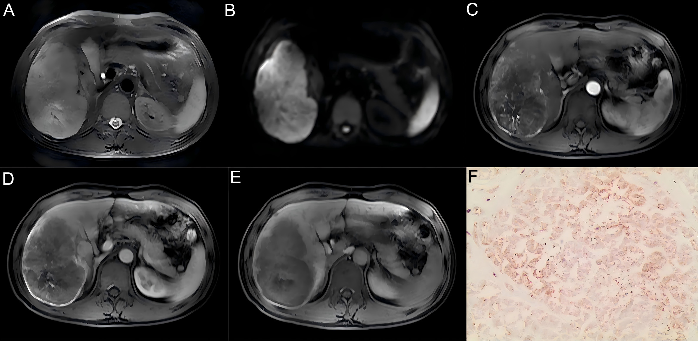
Figure 1. Representative MRI images with corresponding histopathological findings from a DPHCC case. (A) T2WI image showing slightly heterogeneous high signal intensity; (B) DWI image (b = 800 s/mm2) showing marginal ring-like high signal intensity; (C) Arterial phase image showing marginal circular enhancement with intratumoral vascularity and central low signal intensity; (D) Portal venous phase image showing persistent peripheral enhancement; (E) Hepatobiliary phase image showing low signal intensity; (F) Immunohistochemical staining (original magnification 100×) showing CK19 (+). MRI: Magnetic resonance imaging; DPHCC: dual-phenotype hepatocellular carcinoma; T2WI: T2-weighted imaging; DWI: diffusion-weighted imaging; CK19: cytokeratin 19.


