
fig1
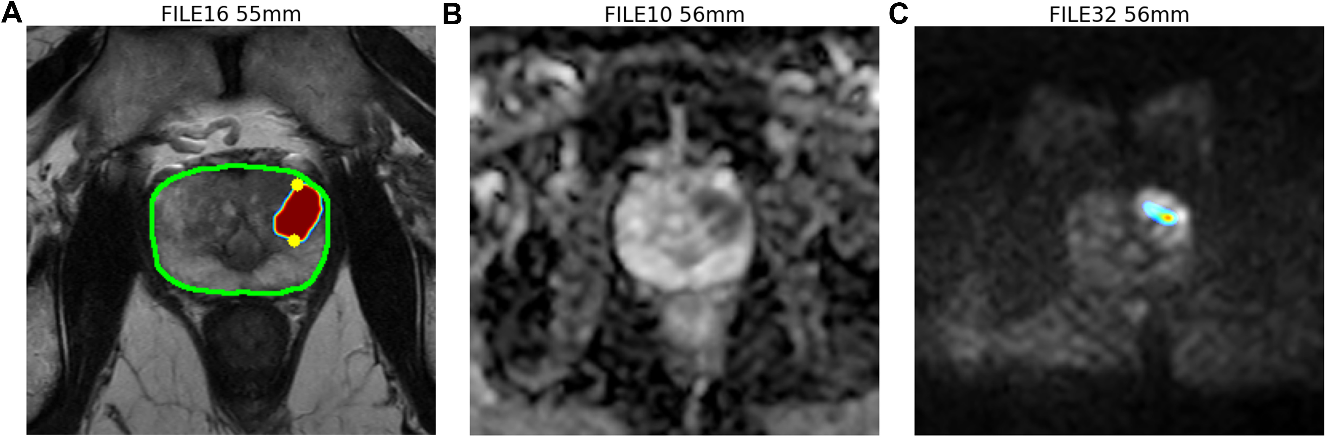
Figure 1. AI system analyzing prostate multiparametric MRI. (A) T2 weighted (T2W) phase of MRI showing whole-prostate segmentation outlined in green. The region of interest (ROI) is labeled with a heat map, with the highest possibility in red. The largest diameter of the ROI is automatically calculated and marked by two yellow dots, indicating an estimated lesion diameter of approximately 1.5 cm and a lesion proportion of 98.63% in the transition zone (TZ); (B) Apparent diffusion coefficient (ADC) phase of MRI; (C) Diffusion-weighted images (DWI) phase of MRI. The ROI is also labeled with heat map with highest possibility in red zone. The AI system will assemble every file from different phases, align each cut of prostate MRI image in order to evaluate PI-RADS and label the ROI. In this representative case, the AI system identified a 1.5 cm lesion in the TZ and assigned a PI-RADS score of 5 based on the lesion’s greatest diameter. The dominant abnormality is seen on T2W imaging, while a mild corresponding abnormality is also noted on DWI, consistent with PI-RADS v2.1, in which TZ lesions are primarily assessed on T2W imaging. AI: Artificial intelligence; MRI: magnetic resonance imaging; PI-RADS: Prostate Imaging Reporting and Data System; DWI: diffusion-weighted images; PI-RADS v2.1: PI-RADS version 2.1.