


fig2
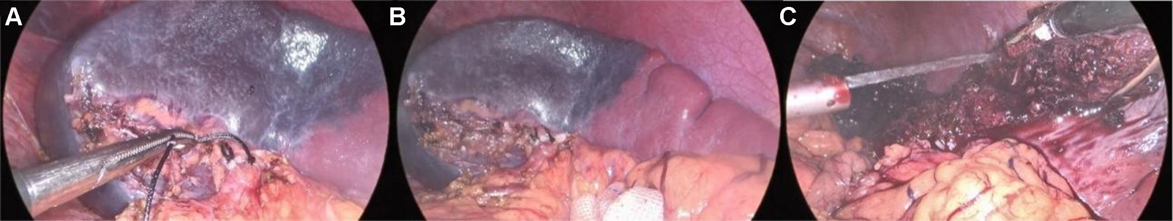
Figure 2. Key steps in vascular management and parenchymal transection. (A) Dissection of the secondary splenic pedicle; (B) Visualization of the splenic ischemic demarcation line; (C) Hemostasis of the splenic transection surface.
Figure 2. Key steps in vascular management and parenchymal transection. (A) Dissection of the secondary splenic pedicle; (B) Visualization of the splenic ischemic demarcation line; (C) Hemostasis of the splenic transection surface.


All published articles are preserved here permanently:
https://www.portico.org/publishers/oae/

