


fig2
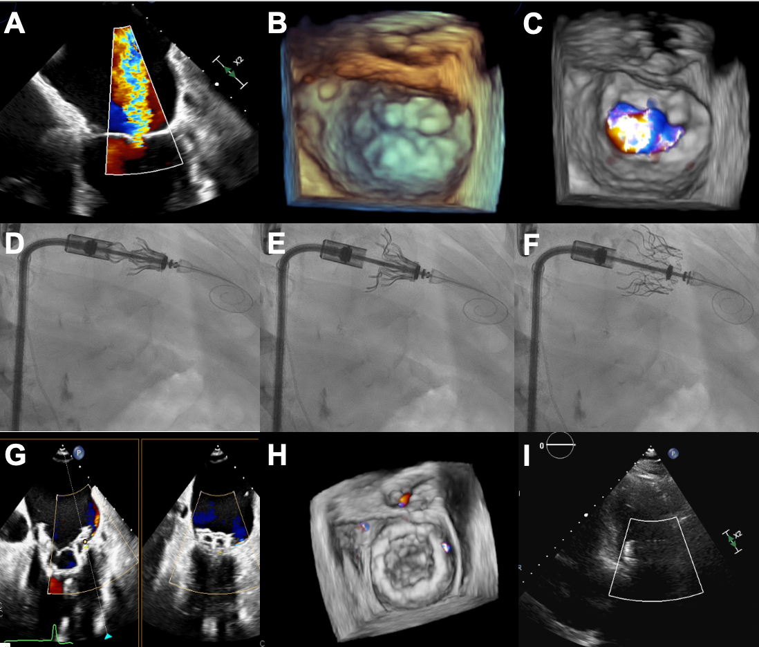
Figure 2. Transseptal Transcatheter Mitral Valve Replacement with Cardiovalve system. (A-C) Severe MR with MV annulus dilated and thickened leaflets; (D) Fluoroscopic image of the Cardiovalve device with open grasp legs into the LV; (E) Fluoroscopic image showing the atrial flange and ventricular part deployment; (F) Fully deployed Cardiovalve device; (G-I) Echocardiographic images showing good expansion and stable position with mild MR and trace PVL. Figure provided by the authors. MR: Mitral regurgitation; LV: left ventricle; MV: mitral valve; PVL: paravalvular leak.


