

Immune functions of vascular structural cells are endothelial communication, smooth muscle transdifferentiation and lymphatic immune regulation
 0
Graphical Abstract
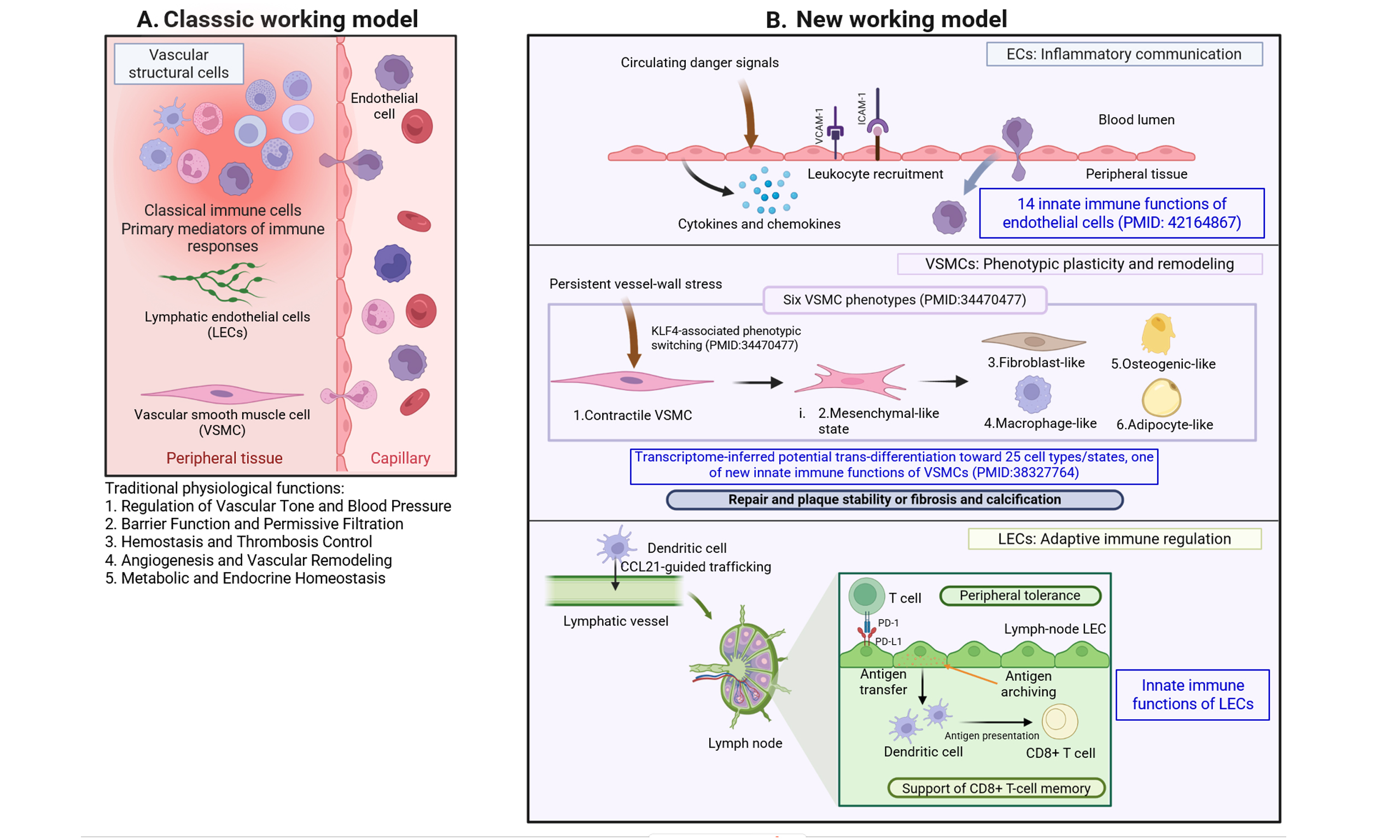
Abstract
The vascular wall maintains structural integrity, regulates blood flow, and supports tissue functions and homeostasis. Evidence also shows that its structural cells participate in immune sensing, inflammation and tissue remodeling. Vascular endothelial cells (ECs), vascular smooth muscle cells (VSMCs), and lymphatic endothelial cells (LECs) encounter different combinations of inflammatory, metabolic, mechanical, microbial, tissue and intracellular immune-derived signals. We propose here that their anatomical locations and cell-intrinsic programs shape distinct, context-dependent immune functions of vascular structural cell types. ECs act as immune communicators at the blood-tissue interface. In response to inflammatory cues, they produce cytokines and chemokines, express adhesion molecules, regulate vascular permeability, and direct leukocyte recruitment. VSMCs respond to persistent vessel wall stress through phenotypic plasticity, secretomes and transdifferentiation, adopting cell states that may support repair and plaque stability or contribute to fibrosis, calcification, and disease progression. LECs combine innate sensing with regulation of immune cell trafficking, antigen handling, peripheral tolerance, adaptive immune responses, and fluid balance. Single-cell transcriptomic studies also identify immune-associated signatures in other vascular structural cell populations, including fibroblasts and pericytes. However, these populations remain candidates whose immune functions require spatial lineage-resolved and functional validation. Together, ECs, VSMCs, and LECs illustrate complementary ways in which structural cells contribute to vascular immune regulation. Defining the novel mechanisms and disease contexts of these responses may reveal opportunities to moderate vascular inflammation and remodeling for development of new disease therapeutics.
Keywords


 Download PDF
INTRODUCTION
The vascular system has traditionally been regarded as a structural and blood transport network[1] that maintains tissue perfusion, regulates vascular tone, transports oxygen, nutrients, cytokines[2], secretomes[3], and hormones, removes waste products such as carbon dioxide, and preserves fluid and metabolic homeostasis. The vascular wall is composed of endothelial cells (ECs)[4-6], vascular smooth muscle cells (VSMCs)[7], fibroblasts[8], extracellular matrix, and associated immune cells including monocytes, macrophages, neutrophils, dendritic cells (DCs), T cells and B cells[8], while a related lymphatic endothelial network returns interstitial fluid to the circulation and connects peripheral tissues with draining lymph nodes. Increasing evidence demonstrates that all inflammatory processes are vascular responses with participation of migrated hematopoietic immune cells[6]; and these structural cells such as ECs, VSMCs and lymphatic endothelial cells (LECs) are not merely passive targets of inflammation: they actively sense disturbances in tissue homeostasis, initiate immune signaling, communicate with hematopoietic immune cells, and participate in tissue remodeling[9].
ECs (also termed vascular ECs or blood ECs[10]) are among the best-characterized nonhematopoietic innate immune cells. Positioned at the interface between circulating blood and peripheral tissues, they continuously encounter microbial products, circulating metabolites, inflammatory mediators, endogenous danger signals, and hemodynamic forces, and express multiple classes of pattern-recognition receptors including receptors for pathogen-associated molecular patterns (PAMPs) and damage-associated molecular patterns (DAMPs)[11-14] that allow them to activate cytokines, chemokines, secretomes[3], adhesion molecules, inflammasomes, and trained-immunity programs[15,16]. These findings established ECs as environment-supporting innate immune cells and expanded the traditional leukocyte-centered definition of innate immunity[4,5,15,17,18]. Building on this, our own work has shown that vascular tissues and ECs undergo broad immune and metabolic reprogramming in response to cardiovascular risk factors and pathological stress, including in diseased aortas that upregulate secreted mediators capable of promoting immune- and vascular-cell activation and transdifferentiation[3,19]. The dominant immune consequence of this activation is communication: activated ECs rapidly produce cytokines, chemokines, secretomes[3], lipid mediators, and extracellular vesicles, increase adhesion molecules, direct leukocyte recruitment and transendothelial migration[20], translating changes in the blood and surrounding tissue into coordinated inflammatory responses.
VSMCs occupy the medial vessel wall layers and are exposed to a different but overlapping set of stresses, including oxidized lipids, uremic toxins, oxidative and endoplasmic-reticulum stress, altered extracellular matrix, and mechanical forces. They express innate immune genes and produce cytokines and chemokines, but their most distinctive response to persistent stress is extensive cellular plasticity: during atherosclerosis, aneurysm formation, restenosis, and vascular calcification, contractile VSMCs can acquire synthetic/proliferative, mesenchymal, macrophage-like, foam cell-like, fibroblast-like, osteogenic, chondrogenic/osteogenic states and adipocytes[21]. Our previous integration of transcriptomic datasets across these disease models proposed that VSMCs possess transdifferentiation potential toward as many as 25 cell types or states, linking innate immune activation directly to this phenotypic diversification[7,22].
LECs represent a third form of vascular immune specialization. Traditionally regarded as passive conduits for tissue-fluid drainage, they are now recognized as active sensors and regulators of immune responses, expressing innate immune receptors such as Toll-like receptors[23], immune checkpoints (programmed cell death ligand 1, PD-L1, CD274)[24,25] and interferon receptors[24], remodeling lymphatic junctions, inducing immune tolerance via PD-L1[25], scavenging and archiving antigens[26], cross presenting antigens and priming CD8+ T cells[27], controlling DC and lymphocyte migration through C-C chemokine motif ligand 21 (CCL21)- C-C motif chemokine receptor 7 (CCR7) guidance[28] and functioning in periaortic lymphatic vessels to protect thoracic aortic dissection[29]. Beyond trafficking, compared with blood endothelial cells[10], lymph node LECs respond to tissue inflammation DAMPs/PAMPs via transcriptomic reprogramming[30], acquire antigen-presentation machinery from DCs[31], contribute to peripheral tolerance[25], and can archive and transfer antigens[26] for extended presentation to migratory dendritic cells—including through specialized transcriptional programs that generate long-lived, memory-like CD8+ T cells[32,33].
These findings suggest that vascular structural cells should not be viewed as executing identical immune programs. Although they share the innate immune sensor ability to detect disturbances in tissue homeostasis, their dominant signals and outputs as innate immune effectors are shaped by anatomical niches[34]: ECs at the blood-tissue interface primarily coordinate inflammatory communication and leukocyte recruitment; VSMCs within the vessel wall translate persistent stress into cellular plasticity/trans-differentiation and structural remodeling; and LECs at the tissue-lymph-lymph-node interface use innate sensing, innate immune effectors and antigen handling to regulate adaptive immunity. This raises a fundamental question: why do neighboring vascular cell types, often exposed to overlapping DAMP/PAMP signals, generate such different immune outputs?
In this review, we propose a new unified framework of immune specialization within the vascular wall to address this question — discussing vascular ECs as specialized immune communicators, VSMCs as structural immune effectors whose dominant strategy is cellular plasticity and transdifferentiation, and LECs as innate immune structural cells specialized in adaptive immune regulation. Rather than emphasizing only the shared sensing pathways of these populations, we focus on how anatomical locations, tissue and functional cues and cell-intrinsic/differentiation programming generate distinct biological outputs. This framework integrates rapid inflammatory communication, long-term vessel-wall remodeling, and adaptive immune regulation, and may help identify cell- and context-specific therapeutic strategies for cardiovascular, cerebrovascular, and inflammatory diseases.
NEW IMMUNE FUNCTIONS OF VASCULAR STRUCTURAL CELLS: ESTABLISHED EVIDENCE AND EMERGING CANDIDATES
Vascular structural cells contribute to immune regulation while retaining their physiological roles in tissue organization and homeostasis. In this review, we use “structural cells with innate immune functions” to describe non-hematopoietic cells that can sense microbial or tissue-derived danger signals and produce an immune response. Previously, we extensively characterized the responses triggered by immune cells[22]. These responses include cytokine or chemokine production, regulation of leukocyte recruitment, antimicrobial activity or changes in barrier function. Antigen handling and presentation provide additional evidence of immune regulation in some populations. The strength of a claim depends on the evidence available: expression of an immune-associated gene suggests a potential function, whereas cell-specific experiments that demonstrate an immune outcome provide stronger support. These functions can also depend on tissue locations and disease states; they do not necessarily make a structural equivalent to a leukocyte. ECs, VSMCs and LECs provide the strongest evidence among all vascular cell types for the framework proposed here. We discuss the supporting evidence for each population in detail below.
Single-cell RNA sequencing (scRNA-seq) studies of diseased vessels (in humans and mice) have identified additional structural populations and cell states, including fibroblast, pericyte, mesothelial, and neural-associated clusters, as well as proliferative VSMC and fibromyocyte populations[8,35-40]. Analysis of genes enriched in each cell population recovered established cell-type markers [Table 1] supporting the accuracy of the scRNA-seq cell annotations and illustrating this technique as a powerful tool to understand the transcriptome of each cell population. While these scRNA-seq datasets reveal cellular heterogeneity, it also enables us to identify immune-associated transcriptional programs in each cell population. They do not, by themselves, establish that every identified population is a distinct innate immune cell type. Interpretation of immune-related transcripts requires particular care in diseased vessels. Ambient RNA released from disrupted cells, contamination by infiltrating leukocytes, and doublets containing both structural cells and leukocytes can introduce misleading immune cell-associated signals. Disease-associated phenotypic transitions can also blur the distinction between cell identity and cell state. Therefore, findings from scRNA-seq require further functional validation through additional experiments that establish spatial localization, cell lineage, and immune functions of the above-mentioned cells.
Single-cell RNA sequencing identified new cell types and cell clusters in the vasculature in pathological conditions
| Category | Cell population | Reported markers or features | Reference |
| Structural | Fibroblasts | COL1A2+; PDGFRA+; APOE+; APOE+CXCL14+; COL3A1+SFRP2+ populations | [35] |
| Structural | Endothelial cells | ACE+CD34+CD93+ cells; clusters characterized by FABP4, CYTL1, or MMRN1, among other markers | [8,36] |
| Structural | VSMCs | ADK+ inflammatory VSMCs; NR1H3+ VSMCs; VSMCs associated with ATF3 suppression; clusters characterized by TNS1, MYL9, GPX3, CD52, or F13A1, among other markers | [36-39] |
| Structural | Proliferating VSMCs | Proliferating smooth muscle cell cluster expressing MGP, TPM4, and MYH10 | [40] |
| Structural | Fibromyocytes | Contractile genes ACTA2 and MYL9; extracellular matrix genes COL1A2 and COL8A1 | [40] |
| Structural | Pericytes | WIF1+; CHAD+; SULT1D1+ populations | [36] |
| Structural | Mesothelial cells | NKAIN4+; IGFBP5+; CD248+ populations | [36] |
| Structural | Neural cells | KCNA1+; PLP1+; MYL1+ populations | [36] |
| Immune | Proliferating immune cells | MKI67; STMN1 | [8] |
| Immune | Dendritic cells | ITGAX; FLT3 | [8] |
| Immune | Macrophages | CD68; FCGR1; CCR5; ADGRE1 | [8] |
| Immune | Monocytes | CD14; CD11B; CSF1R | [8] |
| Immune | B cells | CD79A; CD19; IGHD | [8] |
| Immune | T cells | CD3E; TRBC1; TRBC2 | [8] |
| Immune | Neutrophils | FCGR3; LY6G | [8] |
In the RNA-seq analysis, fibromyoblasts and VSMCs were identified as distinct cell populations, emphasizing the need to distinguish a cell state from a separate cell type. During vascular disease, VSMCs can lose aspects of their contractile phenotype and acquire proliferative, extracellular matrix-producing, or inflammatory features. In atherosclerotic lesions, lineage and perturbation studies have provided evidence for VSMC-derived fibromocytes and their contribution to the fibrous cap[41]. These findings establish the importance of VSMC plasticity in vascular remodeling, but fibromyocyte identity or expression of inflammatory genes alone does not demonstrate a distinct innate immune function. Similarly, proliferative VSMCs are best described here as a disease-associated VSMC state rather than an additional stable structural cell class.
The cell populations, other than ECs, VSMCs and LECs, are best described as emerging candidates for structural cells with innate immune functions. These cell populations have shown immune-related gene expression signatures in response to various disease conditions and stresses such as Coronavirus disease 2019 (COVID-19)[15], cigarette smoke exposure[42-44], hyperlipidemia[45-48], hyperglycemia[49], hyperhomocysteinemia[50-53], metabolic syndrome[54,55], obesity[47,56,57] and chronic kidney disease (CKD) or end-stage renal disease[16,58-63]. These findings suggest possible immune-related roles for some structural cells but do not, on their own, establish immune functions. Fibroblasts can perform immune-related functions, including danger sensing, mediator production and leukocyte regulation, although the extent to which findings from other tissues apply to specific vascular fibroblast populations needs to be defined[64]. Pericytes have been reported to respond to inflammatory stimuli and influence immune cell trafficking, but their functions require further cell-specific testing in vascular disease settings[65]. Evidence that mesothelial cells regulate inflammation comes largely from serosal tissues that should not be taken as direct evidence for an equivalent function with the vessel wall[66]. Neural-associated cells have been detected in vascular single-cell datasets, and broader neuroimmune research than previous ones provides reasons to investigate them further[36,67-69]. Evidence of immune functions of structural vascular cells is summarized in Table 2. Their vascular-specific immune functions, however, remain uncertain. Findings concerning microglia, which are innate immune cells, cannot establish an innate immune identity for vascular neural cells.
Reported evidence indicating immune functions of vascular structural cell populations and related cell states
| Cell population or state | Immune-associated feature | Supporting evidence type | Disease context | Remaining uncertainty |
| ECs | Sense inflammatory and danger signals; produce cytokines and chemokines; regulate adhesion, permeability, and leukocyte recruitment | Receptor and signaling studies, cell-specific experiments, and functional vascular studies[11-20] | Vascular inflammation, hyperlipidemia, and other cardiovascular stresses | Which responses are shared across vascular beds, and which depend on disease context? |
| VSMCs | Produce inflammatory mediators and adopt altered phenotypes during vessel-wall injury | Single-cell profiling, experimental perturbation, and lineage studies[7,22,37-41] | Atherosclerosis, aneurysm, restenosis, and calcification | Which immune-associated programs directly change immune-cell behavior rather than accompany structural remodeling? |
| LECs | Regulate immune-cell trafficking, antigen handling, and peripheral tolerance | Receptor and antigen-handling studies, cell-specific experiments, and functional studies[23-30,32,33] | Tissue inflammation, lymph nodes, and periaortic lymphatics | How well do findings from lymph node LECs extend to vascular-associated lymphatics and specific diseases? |
| Fibroblasts | Can sense danger signals and produce inflammatory mediators that influence leukocyte recruitment | Vascular single-cell profiles[35]; functional evidence primarily from fibroblasts in other tissues[64] | Aneurysmal aorta; infection and tissue injury in broader fibroblast literature | Direct, cell-specific evidence for these functions in vascular-wall fibroblasts is limited |
| Pericytes | Express inflammatory receptors and adhesion molecules; may influence immune-cell trafficking | Vascular cell identification by single-cell profiling[36]; broader functional literature summarized in[65] | Vascular inflammation and tumor-associated vessels | Their contribution to immune responses needs direct testing in defined vascular disease models |
| Mesothelial cells | Interact with immune cells and participate in inflammatory responses | Cell identification by single-cell profiling[36]; functional evidence from serosal tissues[66] | Serosal injury, infection, and mesothelioma | Whether mesothelial cells associated with vascular samples have comparable functions remains unclear |
| Vascular-associated neural cells | May detect inflammatory signals and communicate with immune cells | Vascular single-cell identification[36]; functional evidence largely from other neural settings[67-69] | Vascular samples; infection and neuroinflammation in other tissues | Vascular-specific neural immune functions have not been established by the cited evidence |
| Proliferative VSMCs-VSMC state | Express proliferation and synthetic-state markers; may coexist with inflammatory remodeling | Single-cell identification of a proliferating smooth muscle cell cluster[40] | Vascular remodeling | Proliferation alone does not establish an immune-effector function; lineage and functional studies are needed |
| Fibromyocytes—VSMC-derived state | Acquire fibroblast-like features during VSMC phenotypic modulation; contribute to lesion and fibrous-cap biology | Single-cell analysis and VSMC-specific TCF21 perturbation[41] | Mouse and human atherosclerotic lesions | Their direct immune-effector role remains uncertain. Their presence in a protective fibrous cap does not show that they promote disease |
Together, these observations support ECs, VSMCs and LECs as examples of structural cells with context-dependent immune functions. The immune roles of other structural cell populations remain less established and require validation through cell-specific functional assays and in vivo experiments. The following sections examine how ECs, VSMCs, and LECs contribute to vascular immune regulation.
ECs ARE SPECIALIZED IMMUNE COMMUNICATORS
In 2013, we proposed a new concept that ECs can function as conditional innate immune cells[4,5,9]. This concept was based on evidence that ECs perform several innate immune functions associated with innate immune cells such as macrophages[4], including sensing PAMPs/DAMPs, producing cytokines, regulating leukocyte recruitment and in some contexts, participating in antigen presentation and immune tolerance[19,20]. Subsequent studies by our group and others have reported endothelial immune responses under inflammatory and cardiometabolic stresses, including exposure to cigarette smoke, hyperlipidemia, hyperglycemia, and hyperhomocysteinemia [Figures 1 and 2][6,15,17,18].
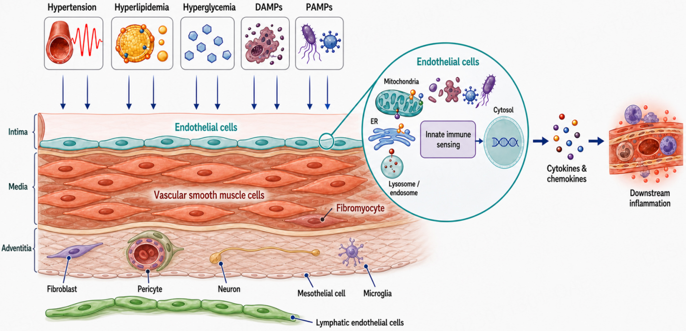
Figure 1. Endothelial sensing of vascular stress and coordination of inflammatory responses. Hypertension, hyperlipidemia, hyperglycemia, damage-associated molecular patterns (DAMPs), and pathogen-associated molecular patterns (PAMPs) stimulate endothelial immune responses. The enlarged endothelial cell illustrates intracellular compartments involved in stress sensing, and signaling, including mitochondria, the endoplasmic reticulum (ER), lysosomes/ endosomes, and the cytosol. Activation of these pathways promotes cytokine and chemokine production, contributing to downstream vascular inflammation. The schematic also depicts vascular structural cells associated cell populations to provide anatomical context; their inclusion does not imply equivalent or established innate immune functions.
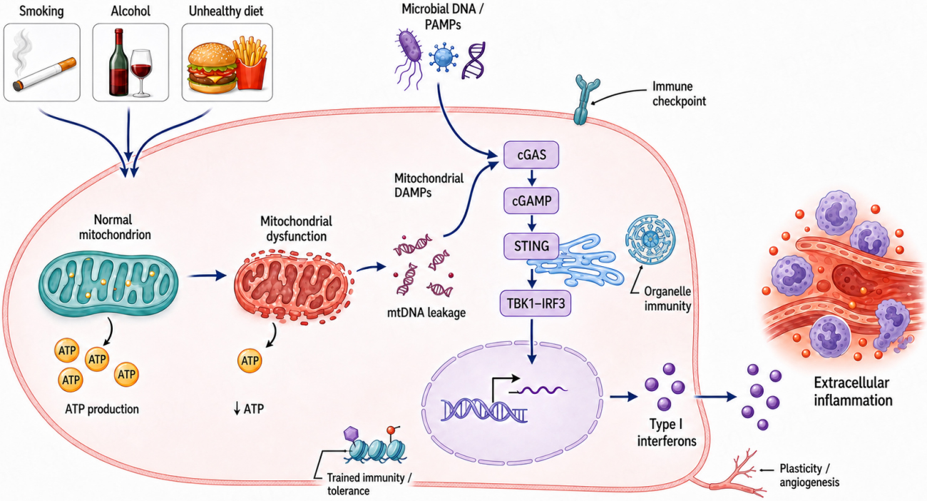
Figure 2. Mitochondrial dysfunction and innate immune signaling in endothelial inflammation. Lifestyle exposures, including smoking, alcohol consumption, and an unhealthy diet, together with microbial DNA and pathogen-associated molecular patterns (PAMPs), can activate endothelial stress responses. Mitochondrial dysfunction reduces adenosine triphosphate (ATP) production and promotes mitochondrial DNA (mtDNA) leakage into the cytosol. Cytosolic DNA activates the cyclic GMP-AMP synthase (cGAS)-stimulator of interferon genes (STING) pathway through cyclic GMP-AMP (cGAMP), engaging TANK binding kinase 1 (TBK1) and interferon regulatory factor 3 (IRF3) to induce type 1 interferons and inflammatory responses. The schematic also illustrates immune checkpoint signaling, organelle-associated immune responses, trained immunity or tolerance, and changes in endothelial plasticity and angiogenesis as context-dependent components of endothelial immune regulation.
Different forms of stress engage distinct endothelial inflammatory pathways. For example, PAMPs and DAMPs can engage pattern recognition pathways, while metabolic injury can activate inflammasome-associated signaling. In experimental models, endothelial caspase-1 activation has been linked to inflammatory responses via the release of initial cytokines and secretomes in a gasdermin D-dependent manner during early hyperlipemia and to impaired vascular repair[45,70-72]. Further, secondary inflammatory cytokines activate transcriptional programs, including nuclear factor kappa-light-chain-enhancer of activated B cells (NF-κB) signaling, that increase the expression of adhesion molecules and inflammatory mediators. Hemodynamic forces modify this response[73,74]. Laminar shear stress induces Krüppel-like Factor 2 (KLF2), a transcription factor that restrains cytokine-induced endothelial activation, whereas disturbed flow is associated with increased adhesion molecule expression and leukocyte adhesion. Thus, the same inflammatory cue may produce different endothelial responses depending on the local flow environment[75].
Organelle stress can also shape endothelial immune signaling. Mitochondrial reactive oxygen species (ROS) have been shown to promote NF-κB-dependent Intercellular Adhesion Molecule 1 (ICAM-1) expression and leukocyte adhesion in experimental EC models. Our studies further identified a role for mitochondrial ROS in lysophosphatidylcholine-induced EC activation and showed that suppression of ROS-associated histone H3 lysine 14 (H3K14) acetylation reduces endothelial activation[48,76-78]. Endoplasmic reticulum stress and metabolic reprogramming may further modify these responses[16,79,80]. These observations support the view that organelles influence endothelial immunity through identifiable signaling and transcriptional pathways, rather than acting as interchangeable immune sensors[81].
DNA sensing pathways provide another route in specific disease settings. When mitochondrial injury leads to the release of mitochondrial DNA, cytosolic Cyclic GMP-AMP synthase (cGAS)- stimulator of interferon genes (STING) signaling activates interferon regulatory factor 3 (IRF3) and NF-κB and increases endothelial inflammatory gene expression. Endothelial expression of a STING gain-of-function mutation has also been linked to pulmonary lymphocytic infiltration[82]. In experimental models of diabetes, hyperglycemia triggered mitochondrial DNA release and activated cGAS-STING signaling, contributing to aortic endothelial injury[83]. These findings support a role for STING in particular forms of vascular inflammation, but its activation should not be assumed to explain all endothelial responses to stress. Metabolic signals may act through other mechanisms: for example, experimental work has linked succinate- succinate receptor 1 (SUCNR1) signaling to a hypoxia-inducible factor 1-alpha (HIF-1α)/interleukin-1b (IL-1b) inflammatory programs in ECs. The importance of this pathway across vascular beds and disease contexts remains to be established[84].
Among 14 innate immune functions outlined in Table 5A of our recent paper[22], one of the most significant innate immune functions identified in ECs is trained immunity (also termed innate immune memory for inflammation enhancement)[5,9,16,85]. We reported for the first time that proinflammatory cytokine TNF-a production will be significantly increased when human aortic endothelial cells are first primed by beta-glucan and further activated by gut microbiota-generated uremic toxin trimethylamine N-oxide (TMAO)[16]. Trained immunity is a new mechanism underlying inflammatory synergy between various disease risk factors and chronic inflammation stimulated with consistent presence of disease risk factors via metabolic reprogramming and epigenetic memory.
Although ECs can undergo phenotypic remodeling, including endothelial-to-mesenchymal transition under pathological conditions[86], their dominant immune response is the rapid dissemination and coordination of inflammatory information. This communication-centered strategy distinguishes them from neighboring VSMCs, whose response to persistent inflammatory and metabolic stress is characterized more prominently by extensive cellular plasticity and long-term vessel-wall remodeling. We therefore propose that inflammatory communication represents the defining immune specialization of vascular ECs, reflecting their anatomical position and their capacity to coordinate leukocyte recruitment, vascular permeability, and immune interactions within the vascular microenvironment[87].
VSMCS ARE SPECIALIZED STRUCTURAL IMMUNE EFFECTORS
Beyond contractility: VSMCs as dynamic immune structural cells
VSMCs have traditionally been defined by their contractile function in regulating vascular tone and supporting vessel-wall structure. This view has expanded considerably with evidence that VSMCs actively sense inflammatory, metabolic, and mechanical disturbances and participate directly in vascular immune responses. VSMCs respond to DAMPs and cardiovascular risk factors—including oxidized lipids, CKD-derived uremic toxins[61], hyperglycemia, hyperhomocysteinemia, inflammatory cytokines, and altered mechanical forces—by activating innate immune genes, cytokine and chemokine pathways, secretomes, adhesion molecules, stress-response systems, and cell-death programs. These properties support their classification as tissue-resident innate immune structural cells rather than passive cellular targets of vascular inflammation[7,22].
VSMCs can produce inflammatory mediators and communicate with neighboring cells; therefore, their immune behavior should not be portrayed as entirely separate from endothelial inflammatory communication. However, their most distinctive response to persistent vascular stress is the extensive reprogramming of cellular identity. In contrast to ECs, whose dominant immune output is the rapid coordination of leukocyte recruitment and intercellular signaling, VSMCs frequently translate innate immune activation into transcriptional, metabolic, and phenotypic remodeling. We propose that this capacity for cellular plasticity represents the defining immune strategy of VSMCs.
Cellular plasticity and pathological transdifferentiation are the dominant immune responses
Mature VSMCs are not terminally fixed in a contractile state. During atherosclerosis, aneurysm formation[34], vascular calcification, restenosis, and other chronic vascular disorders, they can reduce expression of canonical contractile markers and acquire diverse phenotypes, including synthetic, mesenchymal, macrophage-like, foam cell-like, fibroblast-like, osteogenic, chondrogenic states and adipocytes[7,21]. These transitions alter extracellular-matrix production, lipid handling, inflammatory signaling, proliferation, migration, calcification, and tissue mechanics. Their consequences are context-dependent: some VSMC-derived states may support repair or plaque stabilization, whereas others promote fibrosis, calcification, vessel-wall weakening, or disease progression.
Previously, we integrated transcriptomic data from atherosclerotic and abdominal aortic aneurysm models with datasets from DAMP-stimulated mouse and human VSMCs. We found stage-dependent waves of innate immune activation and proposed that VSMCs possess transdifferentiation potential toward as many as 25 cellular states. The associated programs included innate immune genes, cytokines and chemokines, adhesion and plasma-membrane proteins, nuclear stress-sensing pathways, transcription factors, cell-death pathways, and regulators of trained immunity. Our published new working model therefore links innate immune activation directly with VSMC phenotypic diversification and pathological transdifferentiation rather than treating inflammation and plasticity as unrelated pathological processes such as neointimal hyperplasia[7,59,60].
This interpretation shifts attention away from cataloging individual VSMC phenotypes. Synthetic, macrophage-like, osteogenic, fibroblast-like, and other states[21] can instead be viewed as different manifestations of a common response: reprogramming of VSMC identity under sustained stress. Whereas ECs primarily disseminate inflammatory information, VSMCs alter themselves and, through those changes, reshape extracellular-matrix composition, vessel architecture, and local immune interactions. Cellular plasticity thus provides a mechanistic bridge between innate immune sensing and long-term structural remodeling of the vascular wall.
Mechanisms linking immune activation to VSMC reprogramming
VSMC plasticity/transdifferentiation is controlled by interacting transcriptional, epigenetic, metabolic, and stress-response networks. Inflammatory and metabolic stresses can alter the expression of contractile genes and activate regulators of alternative cell states. Loss of the contractile program and acquisition of alternative phenotypes involve lineage-modifying transcription factors such as KLF Transcription Factor 4 (KLF4), Transcription Factor 21 (TCF21), and RUNX Family Transcription Factor 2 (RUNX2), together with context-specific inflammatory and developmental regulators. KLF4 is strongly associated with broad phenotypic modulation[21], including the loss of contractile markers and acquisition of macrophage-like features in lineage-traced VSMC-derived cells. TCF21 promotes modulation toward fibromyocyte-like states that contribute to the fibrous cap, whereas RUNX2 directs osteogenic differentiation and vascular calcification. These examples show that VSMC reprogramming can produce either potentially protective or maladaptive outcomes, depending on the resulting cell state. These transcription factors operate within chromatin landscapes shaped by histone-modifying enzymes, pioneer transcription factors, and persistent inflammatory signaling[7,88,89].
Metabolic and organelle stress further reinforce these transitions[90,91]. Mitochondrial dysfunction, ROS, endoplasmic-reticulum stress, altered substrate utilization and innate immune memory can modify signaling pathways that regulate VSMC gene expression. For example, oxidative stress has been shown to promote VSMC calcification through phosphoinositide 3-kinase (PI3K)-protein kinase B (AKT)-dependent regulation of RUNX2. Mechanical cues such as pressure, stiffness, stretch, and altered flow also influence VSMC phenotype, linking local vessel-wall mechanics to inflammatory and developmental gene programs. These processes should not be considered isolated pathways; together they form an integrated reprogramming network through which VSMCs interpret persistent injury and adopt context-dependent cellular states. Whether particular states persist after the initiating stimulus has resolved, and whether this persistence reflects innate immune memory, requires further investigation[92].
Human vein transcriptomic studies provide clinical evidence that immune activation and structural remodeling occur together during chronic systemic disease. Previously, we compared native upper-extremity veins from 48 patients with advanced CKD/end-stage renal disease with veins from 20 non-CKD donors. CKD altered 1,290 transcripts and increased expression of cytokine and chemokine genes, canonical and noncanonical secretomes, innate immune-response genes, and plasma-membrane proteins associated with intercellular communication. These immune changes occurred alongside endoplasmic-reticulum and mitochondrial stress, immunometabolic reprogramming, fibroblast-enriched and fibrogenic programs, and altered cell-death and survival pathways. The analysis also identified changes involving the developmental and stress-responsive regulators such as MYCN Proto-Oncogene, bHLH Transcription Factor (MYCN) and Fos Proto-Oncogene, AP-1 Transcription Factor Subunit (AP-1). These findings suggest that CKD-associated conditions, potentially including exposure to uremic toxins[61] such as TMAO[16,93], may prime veins for inflammatory and structural remodeling leading to arteriovenous fistula failure[94,95].
The MYCN- and AP-1-associated changes identified in CKD veins are particularly relevant to the proposed link between chronic stress and vascular plasticity. These transcriptional regulators participate in developmental growth, cell-state regulation, and muscle-structure programs, raising the possibility that chronic disease engages pathways involved in changes of vascular cell state. Their reactivation during chronic disease suggests that pathological vascular remodeling may involve partial reuse of developmental programs for pathological transdifferentiation. Rather than restoring normal vessel structure, such reprogramming may generate adaptive or maladaptive states depending on the duration of stress, the surrounding inflammatory environment, and the phenotype acquired.
A novel conceptual shift: cellular plasticity and pathological transdifferentiation are a structural immune strategy
Collectively, these findings support a model in which VSMCs execute an immune strategy distinct from, but complementary to, that of ECs. VSMCs can secrete cytokines and secretomes and influence neighboring cells, but their defining responses to persistent inflammatory, metabolic, and mechanical stress are the capacity to alter cellular identity[22]. Through this plasticity, VSMCs change matrix production, lipid metabolism, calcification, proliferation, migration, and immune-cell interactions, thereby reshaping the vessel wall over prolonged periods.
We therefore propose that cellular plasticity and transdifferentiation are the dominant structural immune strategy of VSMCs[22]. The resulting phenotypes should not be viewed as uniformly protective or pathological. Depending on disease stage and local context, VSMC reprogramming may support repair and plaque stabilization or contribute to fibrosis, calcification, neointimal hyperplasia, aneurysm formation, and vascular failure. This framework avoids assuming that structural adaptation necessarily preserves vascular integrity. Instead, it positions VSMC plasticity as a context-dependent response through which sustained immune and metabolic stress is translated into long-term vessel-wall remodeling.
In this model, ECs primarily communicate vascular danger, whereas VSMCs embody and perpetuate its structural consequences. Recognizing innate immune activation and cellular plasticity as interconnected processes provides a more unified interpretation of VSMC heterogeneity and identifies the redirection of maladaptive cell states—not the indiscriminate suppression of all VSMC responses—as a potential therapeutic objective.
LECs: SPECIALIZED REGULATORS OF ADAPTIVE IMMUNITY
Compared with vascular ECs, the immune functions of LECs have received limited attention[96-98]. Traditionally, LECs were regarded as structural cells of lymphatic vessels that maintain tissue-fluid balance and lymphatic drainage. However, many recent publications demonstrate that LECs actively sense inflammatory changes, remodel lymphatic vessels, direct leukocyte trafficking and antigen handling. These findings support LECs as structural cells with context-dependent innate immune functions that also influence adaptive immune responses. However, unlike vascular ECs, innate immune activities of LECs may closely integrate with the regulation of adaptive immune activity.
Single-cell transcriptomic studies have revealed substantial heterogeneity among LECs across anatomical locations and disease stages. During atherosclerosis, two LEC subpopulations increased in abundance during early disease and declined at later stages. Early disease LECs exhibited gene-expression patterns suggesting reduced support for T-cell tolerance, increased IL-7/IL-7R-associated signaling, and decreased expression of the lipid handling genes low-density lipoprotein receptor and ATP-binding cassette transporter subfamily A1 (ABCA1)[99]. These findings suggest changes in immune regulation and lipid metabolism but require functional validation. Computational trajectory analysis also suggested possible transitions from ECs and, specifically during early disease, fibroblast-associated populations toward LEC-like states. These inferred trajectories do not establish cellular origins or transdifferentiation without lineage-tracing evidence[98]. Other studies have identified anatomically distinct LEC subsets in lymph-node compartments and peripheral lymphatic vessels[100,101], as well as six transcriptionally defined populations distinguished by combinations of markers including Caveolin 1 (CAV1), Atypical Chemokine Receptor 4 (ACKR4), TNF Receptor Superfamily Member 9 (TNFRSF9), Macrophage Receptor With Collagenous Structure (MARCO), Microfibril Associated Protein 4 (MFAP4), Lymphatic Vessel Endothelial Hyaluronan Receptor 1 (LYVE1) and Claudin 11 (CLDN11)[102]. Together these studies provide a framework for investigating LEC specialization, while emphasizing that transcriptional differences alone do not establish distinct immune functions.
An innate immune foundation
LECs express innate immune receptors and directly respond to PAMPs and DAMPs. Human LECs express multiple Toll-like receptors and, following receptor activation, induce chemokine and adhesion molecule expression that regulate DC recruitment and migration. This suggests that LECs can directly sense the changes in their environment including PAMPs and DAMPs instead of relying on inflammatory signals sent indirectly through leukocytes and initiate immune responses that are distinct to LECs[23].
The innate immune responses of LECs depend on the lymphatic microenvironment in which they reside. A recent publication on specialized pulmonary LECs suggests that they play the role of both sensors and responders for inflammation and trigger immune responses by promoting lymphatic permeabilization through a G-protein-coupled receptor called Protease-activated receptor 1 (PAR1). This response enhances the drainage of tissue fluid and may facilitate the clearance of infiltrating leukocytes. Pulmonary LECs also upregulate C-C Motif Chemokine Ligand 21 (CCL21) chemokine release during inflammation, which promotes the migration of CCR7 (a receptor for CCL21 chemokine)-dependent leukocytes from peripheral tissues to the lymph nodes[103].
Lymph node LECs similarly respond rapidly to inflammatory stimulation. They induce interferon-responsive genes and chemokines such as C-X-C Motif Chemokine Ligand 9 (CXCL9), C-C Motif Chemokine Ligand 5 (CCL5) and CCL21, while dynamically regulating their proliferation, survival, and interactions with DCs, macrophages, and lymphocytes. These responses influence lymph-node expansion during immune activation and contraction during resolution[24]. Together, these studies demonstrate that LECs perform several innate immune functions, including inflammatory sensing, chemokine production, barrier remodeling and regulation of immune trafficking.
Translating innate immune signals to adaptive immune regulation
The anatomical location of LECs enables their innate immune activity to directly shape adaptive immunity[104]. Under physiological conditions, LECs mediate the crosstalk between the innate and adaptive immune systems by secreting chemokines such as C-C Motif Chemokine Ligand 2 (CCL2), modulating metabolic environment and expression of adhesion molecules. These innate immune cues guide DCs that carry peripheral antigens to the lymph nodes and present them to naïve T-cells. By controlling the delivery of antigen-bearing dendritic cells to lymphoid tissues, LECs connect local tissue surveillance with the initiation of adaptive immune responses[105].
LECs are now recognized as multifaceted gatekeepers in the immune system[106] and active regulators of adaptive immunity that influence antigen transport, immune tolerance, and long-term immune memory. LECs influence T-cell trafficking and function. Lymph node LECs can directly present endogenous peripheral tissue antigens through major histocompatibility complex class I to CD8+ T cells contributing to peripheral tolerance[107]. Under homeostatic conditions, antigen presentation together with high programmed death ligand-1 (PD-L1) expression and limited conventional co-stimulation promotes peripheral tolerance by deleting, restraining, or functionally modifying autoreactive T cells[25,108-111]. During infection, inflammation, or vaccination, however, LEC antigen handling supports protective immunity by facilitating antigen archiving and memory responses. Thus, LECs are not exclusively tolerogenic; rather, their immunological functions are shaped by antigen context, inflammatory cues, tissue location, and the immune microenvironment[24,105]. In addition, CD4+Foxp3+ regulatory T cells (Tregs)[112] interact with LECs under homeostasis and inflammation and dictate endothelial permissiveness and gating mechanisms for subsequent leukocyte migration through endothelial barriers (transendothelial migration, TEM). Activation of Toll-like receptor 2 (TLR2) on Tregs during inflammation specifically augments Lymphotoxin(LT)α1β2 (LTα1β2)-Lymphotoxin Beta Receptor (LTβR) signaling, which further enhances the permissiveness of LECs to facilitate TEM[112].
A particularly distinctive LEC function is antigen archiving. Lymph node LECs can retain foreign protein antigens for weeks after immunization, with ceiling and floor LECs of the subcapsular sinus displaying especially durable retention. This is not merely passive accumulation. Single-cell and spatial transcriptomic analyses identified specialized gene-expression programs associated with endocytosis, endosomal-lysosomal processing, intracellular trafficking, and protein homeostasis that predict the antigen-archiving capacity of individual LEC subsets. Archived antigen can later be transferred to migratory DCs, extending antigen availability and strengthening memory CD8+ T-cell responses[32].
LECs may also act directly on adaptive immune cells. Vokali et al. showed that LECs can cross-present exogenous antigens to naïve CD8+ T cells under steady-state conditions. Although many proliferating cells underwent early apoptosis, a surviving population of LEC-educated T cells acquired central-memory and stem cell-like memory characteristics. These cells persisted, homed preferentially to lymph nodes, rapidly differentiated after antigen recall, and conferred protection during subsequent infection. This finding indicates that LECs can influence not only immune tolerance but also the formation of long-lived, antigen-experienced adaptive immune populations[33].
Collectively, these findings suggest a two-layer model of LEC immunity. LECs possess an innate immune foundation, consisting of inflammatory sensing, chemokine production, junctional remodeling, antigen uptake, and leukocyte trafficking. These functions feed into their dominant specialization: adaptive immune regulation through antigen presentation, tolerance, antigen archiving, and immune-memory formation. Although LECs share inflammatory sensing and leukocyte regulatory functions with vascular ECs and VSMCs, their anatomical position and antigen handling capacity give them a distinctive role in regulating adaptive immunity.
Cardiac LECs and the resolution of inflammation after myocardial infarction (MI)
The relevance of LEC immune functions extends beyond lymph nodes. Following MI, the injured heart develops edema, extensive leukocyte infiltration, and a lymphangiogenic response[113]. Experimental enhancement of vascular endothelial growth factor C (VEGF-C)-vascular endothelial growth factor receptor 3 (VEGFR3) signaling increases cardiac lymphangiogenesis and has been reported to reduce myocardial edema and fibrosis and improve cardiac function after MI[114].
The immune mechanism is particularly relevant to the present framework. VEGF-C-induced cardiac lymphangiogenesis facilitates the removal of inflammatory cells from injured myocardium and their transport to mediastinal lymph nodes through a LYVE-1-dependent process, accelerating resolution of the acute inflammatory response. Lymphatic endothelia l Sphingosine-1-Phosphate Receptor 1 (S1PR1) signaling has likewise been implicated in macrophage clearance through cardiac lymphatics following MI. These findings indicate that cardiac LECs actively regulate the duration and resolution of post-infarction inflammation rather than serving solely as passive drainage conduits[115].
LECs may additionally provide tissue-supportive signals independently of immune-cell transport. LEC-derived lymphoangiocrine signals can promote cardiomyocyte survival, growth, and repair[116]. Separately, a T-Box Transcription Factor 1 (Tbx1)-dependent program in cardiac LECs has been shown to promote a locally immunosuppressive environment during post-infarction repair[117].
Nevertheless, cardiac literature requires careful interpretation. These studies demonstrate that lymphatic dysfunction can impair inflammatory clearance or worsen post-infarction remodeling; they do not establish that LEC dysfunction increases the incidence of the coronary occlusion that causes MI. Furthermore, genetic blockade of lymphangiogenesis did not significantly worsen long-term cardiac function in one experimental model, indicating that the benefits of lymphangiogenesis may depend on the timing, degree of lymphatic alteration, experimental model, and outcomes assessed[118].
LECs as specialized regulators of adaptive immunity
LECs therefore share important characteristics with vascular ECs: both are nonhematopoietic structural cells capable of sensing inflammatory cues, altering their secretome and surface molecules, and directing immune-cell behavior. Their dominant immune outputs, however, differ according to anatomical niche. Vascular ECs operate at the blood-tissue interface and primarily convert vascular stress into inflammatory communication and leukocyte recruitment. LECs operate at the tissue-lymph-lymph-node interface and use innate sensing, fluid transport, chemokine gradients, and antigen uptake to control the initiation, restraint, persistence, and recall of adaptive immune responses.
We therefore propose that LECs are structural cells with context-dependent innate immune functions that contribute to adaptive immune regulation. Their inflammatory sensing, chemokine production, junctional remodeling and antigen handling connect local tissue responses with immune cell trafficking, peripheral tolerance and immune memory. The relative contribution of these functions varies across lymphatic compartments and disease settings.
NEW CONCEPTUAL FRAMEWORK FOR IMMUNE SPECIALIZATION WITHIN THE VASCULAR WALL
The vascular wall is increasingly recognized as an active participant in immune regulation rather than a passive structural framework. Although ECs, VSMCs, and LECs are often studied independently because of their anatomical location, they may function in a coordinated manner and may perform distinct physiological roles in response to stress. Instead of responding uniformly to PAMPs and DAMPs, these structural cells exhibit distinct immune strategies that reflect their local microenvironment and biological function. We propose that understanding these distinct specialized responses, rather than focusing solely on shared signaling pathways, provides new insights into vascular immunology[119,120].
A common feature shared by all vascular structural cells mentioned above is their ability to detect and respond to alterations in tissue homeostasis, yet the nature of these signals may differ based on their anatomical location. ECs continuously encounter circulating PAMPs and DAMPs while VSMCs that are found within the vessel wall primarily respond to persistent inflammatory and metabolic stresses. In contrast, LECs are mostly exposed to tissue-derived antigens, immune cells, inflammatory cytokines and various other lymph-borne immune signals. These different stress signals that vascular structural cells get exposed to likely trigger distinct immune functions resulting in specific immune outputs specific to the structural cell type.
Because of their anatomical location[22], vascular ECs may be adapted to constantly monitor stress signals and evolved as specialized immune communicators to the neighboring cells by cytokine and chemokine secretion, expressing adhesion molecules, regulating vascular permeability and recruiting circulating leukocytes. In contrast, rather than responding to acute inflammatory triggers, VSMCs may respond to chronic stresses through cellular plasticity. Chronic inflammatory and metabolic stresses may trigger VSMCs to undergo extensive transcriptional, epigenetic, metabolic and phenotypic reprogramming. Depending on the duration and severity of injury, these responses may contribute to tissue repair, vascular remodeling, plaque stabilization, calcification, fibrosis, or disease progression. Cellular plasticity therefore represents the defining biological strategy through which VSMCs adapt to a changing vascular environment.
Emerging evidence suggests that LECs represent a distinct vascular immune specialization centered on adaptive immune regulation. Positioned at the interface between peripheral tissues and secondary lymphoid organs, LECs regulate antigen transport, antigen presentation, immune cell trafficking, peripheral tolerance, and long-term immune memory. Recent studies demonstrating specialized transcriptional programs for antigen archiving and the generation of LEC-educated adaptive immune cells further highlight the unique capacity of LECs to shape adaptive immune responses. Unlike vascular ECs or VSMCs, whose dominant responses are inflammatory communication and cellular plasticity, respectively, LECs function primarily to regulate the quality, magnitude, and duration of adaptive immunity.
Taken together, these observations suggest that ECs, VSMCs, and LECs share some immune functions but differ in their dominant responses, reflecting their anatomical locations and physiological roles. ECs primarily coordinate inflammatory communication and leukocyte recruitment; VSMCs translate persistent stress into vessel wall remodeling through cellular plasticity; and LECs regulate immune cell trafficking, antigen handling, peripheral tolerance, and immune memory. Other vascular structural populations remain emerging candidates whose immune functions require further validation. This framework links shared inflammatory sensing to complementary, context-dependent roles in vascular immune regulation.
The distinct but complementary immune strategies of ECs, VSMCs, and LECs, including their triggers, regulatory programs, effectors, cellular interactions, and disease consequences, are summarized in Table 3. This novel conceptual framework also has highly significant therapeutic implications. Current anti-inflammatory strategies generally target systemic inflammatory mediators or immune pathways without considering the specialized functions of individual vascular structural cell populations. Recognizing immune specialization of vascular structural cell types within the vascular wall raises the possibility of developing therapies that selectively modulate endothelial inflammatory communication, redirect maladaptive VSMC plasticity, or enhance beneficial LEC-mediated adaptive immune regulation according to disease stage and pathological context. Such an approach may provide greater therapeutic precision while preserving essential immune functions required for vascular homeostasis.
Complementary immune functions, signaling pathways and disease implications of endothelial cells, vascular smooth muscle cells and lymphatic endothelial cells
| Component | Ecs | VSMCs | LECs |
| Anatomical context | Blood-tissue interface[5] | Medial vessel wall and remodeled vascular tissue[7,41] | Tissue-lymph-lymph-node interface[24,28] |
| Dominant immune strategy | Inflammatory communication and leukocyte recruitment[5] | Cellular plasticity and pathological transdifferentiation[7,41] | Adaptive immune regulation supported by innate sensing and trafficking[24,25,32,33] |
| Key triggers | Circulating microbial products, metabolites, inflammatory mediators and DAMPs; hemodynamic forces[5] | DAMPs; oxidized lipids; uremic toxins; hyperglycemia; homocysteine; cytokines; mechanical stress[7,37-39] | PAMPs/DAMPs; inflammatory cytokines; tissue-derived antigens; lymph-borne immune signals[23,24,32] |
| Signaling and regulatory programs | Pattern-recognition receptors; inflammatory signaling; inflammasome and trained-immunity programs[5,48,75,82] | KLF4, TCF21 and RUNX2; epigenetic and metabolic reprogramming; mitochondrial and ER stress[37-39,41,89] | TLRs; interferon responses; PAR1 (pulmonary); VEGF-C-VEGFR3 (cardiac); PD-L1-associated tolerance*[23-25,103,114] |
| Effectors and responses | Cytokines, chemokines, secretomes, lipid mediators and extracellular vesicles; adhesion molecules; permeability regulation[5,20] | Cytokines and chemokines; altered matrix production and lipid handling; synthetic, macrophage-like, fibroblast-like and osteogenic states[7,41,89] | CCL21-CCR7 trafficking; junctional remodeling; antigen uptake, presentation and archiving*; tolerance and CD8+ memory formation*[25,27,28,32,33] |
| Cell-cell interactions | Signaling to circulating and tissue-resident immune cells; leukocyte recruitment and transendothelial migration[5,20] | Communication with neighboring vascular and immune cells; cell-state changes reshape local immune interactions[7,41] | DC guidance and archived-antigen transfer*; T-cell regulation*; Treg-LEC LTα1β2-LTβR signaling enhances migration[28,32,33,112] |
| Disease consequences | Coordination of vascular inflammation through leukocyte recruitment, inflammatory communication and permeability changes[5] | Context-dependent repair or plaque stabilization; fibrosis, calcification, neointimal hyperplasia, aneurysmal remodeling or vascular failure[7,38,41,89] | Context-dependent adaptive immunity*; cardiac inflammatory-cell clearance, reduced edema/fibrosis and post-MI repair in experimental models[25,32,33,114,115] |
CONCLUSIONS
Vascular structural cells actively participate in immune regulation through responses shaped by their anatomical locations, local environments, and intrinsic cellular programs. ECs coordinate inflammatory communication and leukocyte recruitment, VSMCs translate persistent stress into phenotypic changes and vessel-wall remodeling, and LECs connect inflammatory sensing and immune-cell trafficking with antigen handling, peripheral tolerance, and immune memory. These complementary functions provide a framework for understanding how vascular tissues integrate immune responses with structural adaptations and tissue homeostasis.
The consequences of these responses depend on disease context, stage, and duration. VSMC plasticity may support plaque stabilization or contribute to maladaptive remodeling, while LEC responses may promote tolerance, protective immunity, or inflammatory resolution. Defining these functions requires evidence that extends beyond immune-associated gene expression to establish cell lineage, spatial localization, and functional outcomes. Further investigation of these mechanisms, including the less well-defined roles of other vascular structural populations, may identify opportunities to selectively limit harmful inflammation and remodeling while preserving vascular integrity and beneficial immune functions.
DECLARATIONS
Acknowledgments
The Graphical Abstract was created with BioRender.com.
Authors’ contributions
Validation, investigation, formal analysis, data curation, conceptualization, writing: Nanayakkara GK
Resources, investigation, formal analysis, writing: Xi R (Rongwei Xi), Xi R (Rongming Xi), Hung SY
Resources, and editing: Xu K
Visualization: Wang EY, Shao Y, Saaoud F, Yuan B
Resources: Lu Y, Zhang W, Mohsin S, Wu S, Wei J, Saiman Y, Martinez L, Vazquez-Padron R, Autieri M, Wang H, Liu X
Conceptualization, writing - review, editing, and supervision: Yang X
All authors have read and approved the final version of the manuscript.
Availability of data and materials
Not applicable.
AI and AI-assisted tools statement
Not applicable.
Financial support and sponsorship
This work was partially supported by National Institutes of Health (NIH) grants to Yang X (HL163570-01A1) and Wang H (DK113775), and American Heart Association (AHA) Career Development Awards to Xu K (Grant 26CDA1609032) and to Lu Y (26CDA1610013).
Conflicts of interest
All authors declared that there are no conflicts of interest.
Ethical approval and consent to participate
Not applicable.
Consent for publication
Not applicable.
Copyright
© The Author(s) 2026.
REFERENCES
1. SEER training modules: physiology of circulation. U.S. National Institutes of Health, National Cancer Institute, 2026. Available from: https://training.seer.cancer.gov [Last accessed on 11 Nov 2026].
2. Shao Y, Cheng Z, Li X, Chernaya V, Wang H, Yang XF. Immunosuppressive/anti-inflammatory cytokines directly and indirectly inhibit endothelial dysfunction-a novel mechanism for maintaining vascular function. J Hematol Oncol 2014;7:80.
3. Lu Y, Sun Y, Xu K, et al. Aorta in pathologies may function as an immune organ by upregulating secretomes for immune and vascular cell activation, differentiation and trans-differentiation—early secretomes may serve as drivers for trained immunity. Front Immunol 2022;13:858256.
4. Mai J, Virtue A, Shen J, Wang H, Yang X. An evolving new paradigm: endothelial cells - conditional innate immune cells. J Hematol Oncol 2013;6:61.
5. Shao Y, Saredy J, Yang WY, et al. Vascular endothelial cells and innate immunity. Arterioscler Thromb Vasc Biol 2020;40:e138-52.
6. Yang X, Yin Y, Wang H. Vascular inflammation and atherogenesis are activated via receptors for PAMPs and suppressed by regulatory T cells. Drug Discov Today Ther Strateg 2008;5:125-42.
7. Yang Q, Saaoud F, Lu Y, et al. Innate immunity of vascular smooth muscle cells contributes to two-wave inflammation in atherosclerosis, twin-peak inflammation in aortic aneurysms and trans-differentiation potential into 25 cell types. Front Immunol 2024;14:1348238.
8. Yuan Z, Shu L, Fu J, et al. Single-cell RNA sequencing deconstructs the distribution of immune cells within abdominal aortic aneurysms in mice. Arterioscler Thromb Vasc Biol 2024;44:1986-2003.
9. Drummer C, Saaoud F, Shao Y, et al. Trained immunity and reactivity of macrophages and endothelial cells. Arterioscler Thromb Vasc Biol 2021;41:1032-46.
10. Berendam SJ, Koeppel AF, Godfrey NR, et al. Comparative transcriptomic analysis identifies a range of immunologically related functional elaborations of lymph node associated lymphatic and blood endothelial cells. Front Immunol 2019;10:816.
11. Yin Y, Pastrana JL, Li X, et al. Inflammasomes: sensors of metabolic stresses for vascular inflammation. Front Biosci 2013;18:638-49.
12. Yin Y, Yan Y, Jiang X, et al. Inflammasomes are differentially expressed in cardiovascular and other tissues. Int J Immunopathol Pharmacol 2009;22:311-22.
13. Wang X, Li Y, Nanayakkara G, et al. Lysophospholipid receptors, as novel conditional danger receptors and homeostatic receptors modulate inflammation—novel paradigm and therapeutic potential. J Cardiovasc Transl Res 2016;9:343-59.
14. Shao Y, Nanayakkara G, Cheng J, et al. Lysophospholipids and their receptors serve as conditional DAMPs and DAMP receptors in tissue oxidative and inflammatory injury. Antioxid Redox Signal 2018;28:973-86.
15. Shao Y, Saredy J, Xu K, et al. Endothelial immunity trained by coronavirus infections, DAMP stimulations and regulated by anti-oxidant NRF2 may contribute to inflammations, myelopoiesis, COVID-19 cytokine storms and thromboembolism. Front Immunol 2021;12:653110.
16. Saaoud F, Liu L, Xu K, et al. Aorta- and liver-generated TMAO enhances trained immunity for increased inflammation via ER stress/mitochondrial ROS/glycolysis pathways. JCI Insight 2023;8:e158183.
17. Li X, Wang L, Fang P, et al. Lysophospholipids induce innate immune transdifferentiation of endothelial cells, resulting in prolonged endothelial activation. J Biol Chem 2018;293:11033-45.
18. Xu K, Shao Y, Saaoud F, et al. Novel knowledge-based transcriptomic profiling of lipid lysophosphatidylinositol-induced endothelial cell activation. Front Cardiovasc Med 2021;8:773473.
19. Yang WY, Ben Issa M, Saaoud F, et al. Perspective: pathological transdifferentiation—a novel therapeutic target for cardiovascular diseases and chronic inflammation. Front Cardiovasc Med 2024;11:1500775.
20. Xu K, Saaoud F, Yu S, et al. Monocyte adhesion assays for detecting endothelial cell activation in vascular inflammation and atherosclerosis. In: Ramji D, editors. Atherosclerosis. New York, NY: Springer; 2022. pp. 169-82.
21. Yap C, Mieremet A, De Vries CJ, Micha D, De Waard V. Six shades of vascular smooth muscle cells illuminated by KLF4 (Krüppel-like factor 4). Arterioscler Thromb Vasc Biol 2021;41:2693-707.
22. Liu J, Nanayakkara GK, Lu Y, et al. A new paradigm: 46 structural cell types function as environment-supporting innate immune cells, with endothelial and vascular smooth muscle cells as key prototypes. iNew Med 2026;2:e70050.
23. Pegu A, Qin S, Fallert Junecko BA, Nisato RE, Pepper MS, Reinhart TA. Human lymphatic endothelial cells express multiple functional TLRs. J Immunol 2008;180:3399-405.
24. Lucas ED, Tamburini BAJ. Lymph node lymphatic endothelial cell expansion and contraction and the programming of the immune response. Front Immunol 2019;10:36.
25. Tewalt EF, Cohen JN, Rouhani SJ, et al. Lymphatic endothelial cells induce tolerance via PD-L1 and lack of costimulation leading to high-level PD-1 expression on CD8 T cells. Blood 2012;120:4772-82.
26. Kedl RM, Tamburini BA. Antigen archiving by lymph node stroma: a novel function for the lymphatic endothelium. Eur J Immunol 2015;45:2721-9.
27. Hirosue S, Vokali E, Raghavan VR, et al. Steady-state antigen scavenging, cross-presentation, and CD8+ T cell priming: a new role for lymphatic endothelial cells. J Immunol 2014;192:5002-11.
29. Pei Y, Lin W, Zhang T, et al. Periaortic lymphatic vessels protect against thoracic aortic dissection through mobilizing immune response. Cardiovasc Res 2025;121:2594-609.
30. Vigl B, Aebischer D, Nitschké M, et al. Tissue inflammation modulates gene expression of lymphatic endothelial cells and dendritic cell migration in a stimulus-dependent manner. Blood 2011;118:205-15.
31. Rouhani SJ, Eccles JD, Riccardi P, et al. Roles of lymphatic endothelial cells expressing peripheral tissue antigens in CD4 T-cell tolerance induction. Nat Commun 2015;6:6771.
32. Sheridan RM, Doan TA, Lucas CJ, et al. A specific gene expression program underlies antigen archiving by lymphatic endothelial cells in mammalian lymph nodes. Nat Commun 2025;16:8375.
33. Vokali E, Yu SS, Hirosue S, et al. Lymphatic endothelial cells prime naïve CD8+ T cells into memory cells under steady-state conditions. Nat Commun 2020;11:538.
34. Lu Y, Sun Y, Saaoud F, et al. ER stress mediates Angiotensin II-augmented innate immunity memory and facilitates distinct susceptibilities of thoracic from abdominal aorta to aneurysm development. Front Immunol 2023;14:1268916.
35. Davis FM, Tsoi LC, Ma F, et al. Single-cell transcriptomics reveals dynamic role of smooth muscle cells and enrichment of immune cell subsets in human abdominal aortic aneurysms. Ann Surg 2022;276:511-21.
36. Kan H, Zhang K, Mao A, et al. Single-cell transcriptome analysis reveals cellular heterogeneity in the ascending aortas of normal and high-fat diet-fed mice. Exp Mol Med 2021;53:1379-89.
37. Xu J, Liu Z, Yang Q, et al. Adenosine kinase inhibition protects mice from abdominal aortic aneurysm via epigenetic modulation of VSMC inflammation. Cardiovasc Res 2024;120:1202-17.
38. Guo X, Zhong J, Zhao Y, et al. LXRα promotes abdominal aortic aneurysm formation through UHRF1 epigenetic modification of miR-26b-3p. Circulation 2024;150:30-46.
39. Wen Y, Liu Y, Li Q, et al. Spatiotemporal ATF3 expression determines VSMC fate in abdominal aortic aneurysm. Circ Res 2024;134:1495-511.
40. Li Y, Ren P, Dawson A, et al. Single-cell transcriptome analysis reveals dynamic cell populations and differential gene expression patterns in control and aneurysmal human aortic tissue. Circulation 2020;142:1374-88.
41. Wirka RC, Wagh D, Paik DT, et al. Atheroprotective roles of smooth muscle cell phenotypic modulation and the TCF21 disease gene as revealed by single-cell analysis. Nat Med 2019;25:1280-9.
42. Shao Y, Saaoud F, Cornwell W, et al. Cigarette smoke and morphine promote treg plasticity to Th17 via enhancing trained immunity. Cells 2022;11:2810.
43. Shao Y, Cornwell W, Xu K, et al. Chronic exposure to the combination of cigarette smoke and morphine decreases CD4+ regulatory T cell numbers by reprogramming the treg cell transcriptome. Front Immunol 2022;13:887681.
44. Saaoud F, Shao Y, Cornwell W, Wang H, Rogers TJ, Yang X. Cigarette smoke modulates inflammation and immunity via reactive oxygen species-regulated trained immunity and trained tolerance mechanisms. Antioxid Redox Signal 2023;38:1041-69.
45. Yin Y, Li X, Sha X, et al. Early hyperlipidemia promotes endothelial activation via a caspase-1-sirtuin 1 pathway. Arterioscler Thromb Vasc Biol 2015;35:804-16.
46. Mai J, Nanayakkara G, Lopez-Pastrana J, et al. Interleukin-17A promotes aortic endothelial cell activation via transcriptionally and post-translationally activating p38 mitogen-activated protein kinase (MAPK) pathway. J Biol Chem 2016;291:4939-54.
47. Virtue A, Johnson C, Lopez-Pastraña J, et al. MicroRNA-155 deficiency leads to decreased atherosclerosis, increased white adipose tissue obesity, and non-alcoholic fatty liver diseasee: a novel mouse model of obesity paradox. J Biol Chem 2017;292:1267-87.
48. Li X, Shao Y, Sha X, et al. IL-35 (interleukin-35) suppresses endothelial cell activation by inhibiting mitochondrial reactive oxygen species-mediated site-specific acetylation of H3K14 (histone 3 lysine 14). Arterioscler Thromb Vasc Biol 2018;38:599-609.
49. Yan Y, Xiong Z, Zhang S, et al. CD25highT cells with a prolonged survival inhibit development of diabetes. Int J Immunopathol Pharmacol 2008;21:767-80.
50. Fang P, Zhang D, Cheng Z, et al. Hyperhomocysteinemia potentiates hyperglycemia-induced inflammatory monocyte differentiation and atherosclerosis. Diabetes 2014;63:4275-90.
51. Xi H, Zhang Y, Xu Y, et al. Caspase-1 inflammasome activation mediates homocysteine-induced pyrop-apoptosis in endothelial cells. Circ Res 2016;118:1525-39.
52. Zhang D, Fang P, Jiang X, et al. Severe hyperhomocysteinemia promotes bone marrow-derived and resident inflammatory monocyte differentiation and atherosclerosis in LDLR/CBS-deficient mice. Circ Res 2012;111:37-49.
53. Zhang D, Jiang X, Fang P, et al. Hyperhomocysteinemia promotes inflammatory monocyte generation and accelerates atherosclerosis in transgenic cystathionine β-synthase-deficient mice. Circulation 2009;120:1893-902.
54. Drummer CIV, Saaoud F, Sun Y, et al. Hyperlipidemia may synergize with hypomethylation in establishing trained immunity and promoting inflammation in NASH and NAFLD. J Immunol Res 2021;2021:1-35.
55. Drummer C, Saaoud F, Jhala NC, et al. Caspase-11 promotes high-fat diet-induced NAFLD by increasing glycolysis, OXPHOS, and pyroptosis in macrophages. Front Immunol 2023;14:1113883.
56. Johnson C, Drummer C, Virtue A, et al. Increased expression of resistin in MicroRNA-155-deficient white adipose tissues may be a possible driver of metabolically healthy obesity transition to classical obesity. Front Physiol 2018;9:1297.
57. Johnson C, Drummer Iv C, Shan H, et al. A novel subset of CD95+ pro-inflammatory macrophages overcome miR155 deficiency and may serve as a switch from metabolically healthy obesity to metabolically unhealthy obesity. Front Immunol 2021;11:619951.
58. Sun Y, Lu Y, Liu L, et al. Caspase-4/11 promotes hyperlipidemia and chronic kidney disease-accelerated vascular inflammation by enhancing trained immunity. JCI Insight 2024;9:e177229.
59. Monroy MA. Chronic kidney disease alters vascular smooth muscle cell phenotype. Front Biosci 2015;20:784-95.
60. Ferrer LM, Monroy AM, Lopez-Pastrana J, et al. Caspase-1 plays a critical role in accelerating chronic kidney disease-promoted neointimal hyperplasia in the carotid artery. J Cardiovasc Transl Res 2016;9:135-44.
61. Li R. Uremic toxins are conditional danger- or homeostasis-associated molecular patterns. Front Biosci 2018;23:348-87.
62. Zhang R, Saredy J, Shao Y, et al. End-stage renal disease is different from chronic kidney disease in upregulating ROS-modulated proinflammatory secretome in PBMCs - A novel multiple-hit model for disease progression. Redox Biol 2020;34:101460.
63. Lu Y, Sun Y, Saaoud F, et al. Uremic toxin receptor NR1H3 contributes to hyperlipidemia- and chronic kidney disease-accelerated vascular inflammation, which is partially suppressed by novel YBX2 anti-ROS pathway. Redox Biol 2025;85:103724.
64. Cavagnero KJ, Gallo RL. Essential immune functions of fibroblasts in innate host defense. Front Immunol 2022;13:1058862.
65. Dabravolski SA, Andreeva ER, Eremin II, et al. The role of pericytes in regulation of innate and adaptive immunity. Biomedicines 2023;11:600.
66. Mutsaers SE, Pixley FJ, Prêle CM, Hoyne GF. Mesothelial cells regulate immune responses in health and disease: role for immunotherapy in malignant mesothelioma. Curr Opin Immunol 2020;64:88-109.
67. Kim SW, Seok SH. Beyond barriers: when neurons act immune and immunity acts neural. Sig Transduct Target Ther 2025;10:392.
68. Yu D, Jain S, Wangzhou A, et al. Microglia regulate GABAergic neurogenesis in prenatal human brain through IGF1. Nature 2025;646:676-86.
69. Wheeler MA, Quintana FJ. The neuroimmune connectome in health and disease. Nature 2025;638:333-42.
70. Lopez-Pastrana J, Ferrer LM, Li Y, et al. Inhibition of caspase-1 activation in endothelial cells improves angiogenesis: a novel therapeutic potential for ischemia. J Biol Chem 2015;290:17485-94.
71. Li YF, Huang X, Li X, et al. Caspase-1 mediates hyperlipidemia-weakened progenitor cell vessel repair. Front Biosci 2016;21:178-91.
72. Li Y, Nanayakkara G, Sun Y, et al. Analyses of caspase-1-regulated transcriptomes in various tissues lead to identification of novel IL-1β-, IL-18- and sirtuin-1-independent pathways. J Hematol Oncol 2017;10:40.
73. Chen L, Li JY, Nguyen P, et al. Pulsatile flow induces chromatin interaction with lamin-associated proteins to enrich H3K9 methylation in endothelial cells. Proc Natl Acad Sci USA 2025;122:e2424566122.
74. Sriram K, Qi Z, Yuan D, et al. Regulation of nuclear transcription by mitochondrial RNA in endothelial cells. eLife 2024;13:e86204.
75. Senbanerjee S, Lin Z, Atkins GB, et al. KLF2 is a novel transcriptional regulator of endothelial proinflammatory activation. J Exp Med 2004;199:1305-15.
76. Li X, Fang P, Yang WY, et al. Mitochondrial ROS, uncoupled from ATP synthesis, determine endothelial activation for both physiological recruitment of patrolling cells and pathological recruitment of inflammatory cells. Can J Physiol Pharmacol 2017;95:247-52.
77. Nanayakkara GK, Wang H, Yang X. Proton leak regulates mitochondrial reactive oxygen species generation in endothelial cell activation and inflammation - A novel concept. Arch Biochem Biophys 2019;662:68-74.
78. Li X, Fang P, Li Y, et al. Mitochondrial reactive oxygen species mediate lysophosphatidylcholine-induced endothelial cell activation. Arterioscler Thromb Vasc Biol 2016;36:1090-100.
79. Xu K, Saaoud F, Shao Y, et al. Early hyperlipidemia triggers metabolomic reprogramming with increased SAH, increased acetyl-CoA-cholesterol synthesis, and decreased glycolysis. Redox Biol 2023;64:102771.
80. Saaoud F, Lu Y, Xu K, et al. Protein-rich foods, sea foods, and gut microbiota amplify immune responses in chronic diseases and cancers - targeting PERK as a novel therapeutic strategy for chronic inflammatory diseases, neurodegenerative disorders, and cancer. Pharmacol Ther 2024;255:108604.
81. Hawkins BJ, Solt LA, Chowdhury I, et al. G protein-coupled receptor Ca2+-linked mitochondrial reactive oxygen species are essential for endothelial/leukocyte adherence. Mol Cell Biol 2023;27:7582-93.
82. Gao KM, Chiang K, Jiang Z, et al. Endothelial cell expression of a STING gain-of-function mutation initiates pulmonary lymphocytic infiltration. Cell Rep 2024;43:114114.
83. An Y, Geng K, Wang H, et al. Hyperglycemia-induced STING signaling activation leads to aortic endothelial injury in diabetes. Cell Commun Signal 2023;21:365.
84. Xu J, Zheng Y, Zhao Y, et al. Succinate/IL-1β signaling axis promotes the inflammatory progression of endothelial and exacerbates atherosclerosis. Front Immunol 2022;13:817572.
85. Lu Y, Sun Y, Drummer C, et al. Increased acetylation of H3K14 in the genomic regions that encode trained immunity enzymes in lysophosphatidylcholine-activated human aortic endothelial cells - novel qualification markers for chronic disease risk factors and conditional DAMPs. Redox Biol 2019;24:101221.
86. Van Kesteren S, Smeehuijzen L, Stevenson R, Kroon J. Endothelial cells modulate immune cell responses during atherosclerosis. Trends Immunol 2026;47:746-65.
87. Kotlyarov S. Immune function of endothelial cells: evolutionary aspects, molecular biology and role in atherogenesis. Int J Mol Sci 2022;23:9770.
88. Lu Y, Sun Y, Xu K, et al. Editorial: Endothelial cells as innate immune cells. Front Immunol 2022;13:1035497.
89. Shankman LS, Gomez D, Cherepanova OA, et al. KLF4-dependent phenotypic modulation of smooth muscle cells has a key role in atherosclerotic plaque pathogenesis. Nat Med 2015;21:628-37.
90. Xu K, Hung SY, Saaoud F, et al. Novel organelle-based intracellular immunity with mechanistic and therapeutic implications. Barrier Immun 2026;2:68-94.
91. Xu K, Saaoud F, Shao Y, et al. A new paradigm in intracellular immunology: mitochondria emerging as leading immune organelles. Redox Biol 2024;76:103331.
92. Byon CH, Javed A, Dai Q, et al. Oxidative stress induces vascular calcification through modulation of the osteogenic transcription factor Runx2 by AKT signaling. J Biol Chem 2008;283:15319-27.
93. Chan MM, Yang X, Wang H, Saaoud F, Sun Y, Fong D. The microbial metabolite trimethylamine N-oxide links vascular dysfunctions and the autoimmune disease rheumatoid arthritis. Nutrients 2019;11:1821.
94. Saaoud F, Martinez L, Lu Y, et al. Chronic kidney disease transdifferentiates veins into a specialized immune-endocrine organ with increased MYCN-AP1 signaling. Cells 2023;12:1482.
95. Martinez L, Stoyell-Conti FF, Tabbara M, et al. The single-cell landscape of the human vein after arteriovenous fistula creation and implications for maturation failure. Kidney Int 2026;109:160-77.
96. Peluzzo AM, Bkhache M, Do LNH, Autieri MV, Liu X. Differential regulation of lymphatic junctional morphology and the potential effects on cardiovascular diseases. Front Physiol 2023;14:1198052.
97. Liu X, Oliver G. The lymphatic vasculature in cardiac development and ischemic heart disease. Circ Res 2023;132:1246-53.
98. Liu X, Cui K, Wu H, et al. Promoting lymphangiogenesis and lymphatic growth and remodeling to treat cardiovascular and metabolic diseases. Arterioscler Thromb Vasc Biol 2023;43:e1-10.
99. Shen Q, Gu G, Yang D, Zheng Y. Dysfunction and pathological origins of lymphatic endothelial cells in atherosclerosis revealed by single-cell transcriptomics. Genes 2025;16:1398.
100. Jalkanen S, Salmi M. Lymphatic endothelial cells of the lymph node. Nat Rev Immunol 2020;20:566-78.
101. Den Braanker H, Van Stigt A, Kok M, Lubberts E, Bisoendial R. Single-cell RNA sequencing reveals heterogeneity and functional diversity of lymphatic endothelial cells. Int J Mol Sci 2021;22:11976.
102. Takeda A, Hollmén M, Dermadi D, et al. Single-cell survey of human lymphatics unveils marked endothelial cell heterogeneity and mechanisms of homing for neutrophils. Immunity 2019;51:561-72.e5.
103. Rivers AK, Migues Ramirez C, Cleary SJ. Lymphatic endothelial cells actively shape immune responses in the lungs. Curr Opin Immunol 2026;101:102802.
104. Card CM, Yu SS, Swartz MA. Emerging roles of lymphatic endothelium in regulating adaptive immunity. J Clin Investig 2014;124:943-52.
105. Chen W, Wang Y, Zhang D, Zhang Y, Qin X, Wang X. The crosstalk between lymphatic endothelial cells and immune cells in Physiological and Pathological conditions. Cell Mol Life Sci 2026;83:309.
106. Takeda A, Salmi M, Jalkanen S. Lymph node lymphatic endothelial cells as multifaceted gatekeepers in the immune system. Trends Immunol 2023;44:72-86.
107. Cohen JN, Guidi CJ, Tewalt EF, et al. Lymph node-resident lymphatic endothelial cells mediate peripheral tolerance via Aire-independent direct antigen presentation. J Exp Med 2010;207:681-8.
108. Shao Y, Yang WY, Nanayakkara G, et al. Immune checkpoints are new therapeutic targets in regulating cardio-, and cerebro-vascular diseases and CD4+Foxp3+ regulatory T cell immunosuppression. Int J Drug Discov Pharm 2024;3:100022.
109. Shao Y, Saaoud F, Xu K, et al. A comprehensive analysis of immune checkpoint receptor-ligand pairs in aortic diseases highlights the immunosuppressive roles of CD155 and CD274. Genes Dis 2025;13:101724.
110. Shen H, Wu N, Nanayakkara G, et al. Co-signaling receptors regulate T-cell plasticity and immune tolerance. Front Biosci 2019;24:96-132.
111. Han B, Xu K, Saaoud F, et al. Discovery of seven ROS-sensitive immune checkpoints and 46 ligands mediating immune suppression through T cell-APC networks. J Cancer 2026;17:439-56.
112. Piao W, Xiong Y, Li L, et al. Regulatory T cells condition lymphatic endothelia for enhanced transendothelial migration. Cell Rep 2020;30:1052-62.e5.
113. Liu X, Yang X, Autieri MV. Editorial: Exploring lymphatic vasculature's role in cardiovascular and metabolic diseases. Front Cardiovasc Med 2026;13:1805707.
114. Henri O, Pouehe C, Houssari M, et al. Selective stimulation of cardiac lymphangiogenesis reduces myocardial edema and fibrosis leading to improved cardiac function following myocardial infarction. Circulation 2016;133:1484-97.
115. Vieira JM, Norman S, Villa Del Campo C, et al. The cardiac lymphatic system stimulates resolution of inflammation following myocardial infarction. J Clin Investig 2018;128:3402-12.
116. Liu X, De La Cruz E, Gu X, et al. Lymphoangiocrine signals promote cardiac growth and repair. Nature 2020;588:705-11.
117. Wang W, Li X, Ding X, et al. Lymphatic endothelial transcription factor Tbx1 promotes an immunosuppressive microenvironment to facilitate post-myocardial infarction repair. Immunity 2023;56:2342-57.e10.
118. Keller TS, Lim L, Shewale SV, et al. Genetic blockade of lymphangiogenesis does not impair cardiac function after myocardial infarction. J Clin Investig 2021;131:e147070.
119. Peluzzo AM, St. Paul A, Corbett CB, et al. IL-19 is a novel lymphangiocrine factor inducing lymphangiogenesis and lymphatic junctional regulation. Arterioscler Thromb Vasc Biol 2025;45:1244-65.
Cite This Article
 Open Access
How to Cite
Nanayakkara GK, Xi R, Xi R, Hung SY, Xu K, Wang EY, Lu Y, Shao Y, Saaoud F, Yuan B, Zhang W, Mohsin S, Wu S, Wei J, Saiman Y, Martinez L, Vazquez-Padron R, Autieri M, Wang H, Liu X, Yang X. Immune functions of vascular structural cells are endothelial communication, smooth muscle transdifferentiation and lymphatic immune regulation. Vessel Plus. 2026;10:59. https://dx.doi.org/10.20517/2574-1209.2026.81
Download Citation
If you have the appropriate software installed, you can download article citation data to the citation manager of your choice. Simply select your manager software from the list below and click on download.
Export Citation File
Type of Import
Tips on Downloading Citation
Citation Manager File Format
Type of Import
Direct Import: When the Direct Import option is selected (the default state), a dialogue box will give you the option to Save or Open the downloaded citation data. Choosing Open will either launch your citation manager or give you a choice of applications with which to use the metadata. The Save option saves the file locally for later use.
Indirect Import: When the Indirect Import option is selected, the metadata is displayed and may be copied and pasted as needed.
About This Article
Special Topic
Copyright

Data & Comments
Data
0









Comments
Comments must be written in English. Spam, offensive content, impersonation, and private information will not be permitted. If any comment is reported and identified as inappropriate content by OAE staff, the comment will be removed without notice. If you have any queries or need any help, please contact us at [email protected].