

Multimodality noninvasive imaging in the evaluation of coronary artery disease: a narrative review
 0
 ,
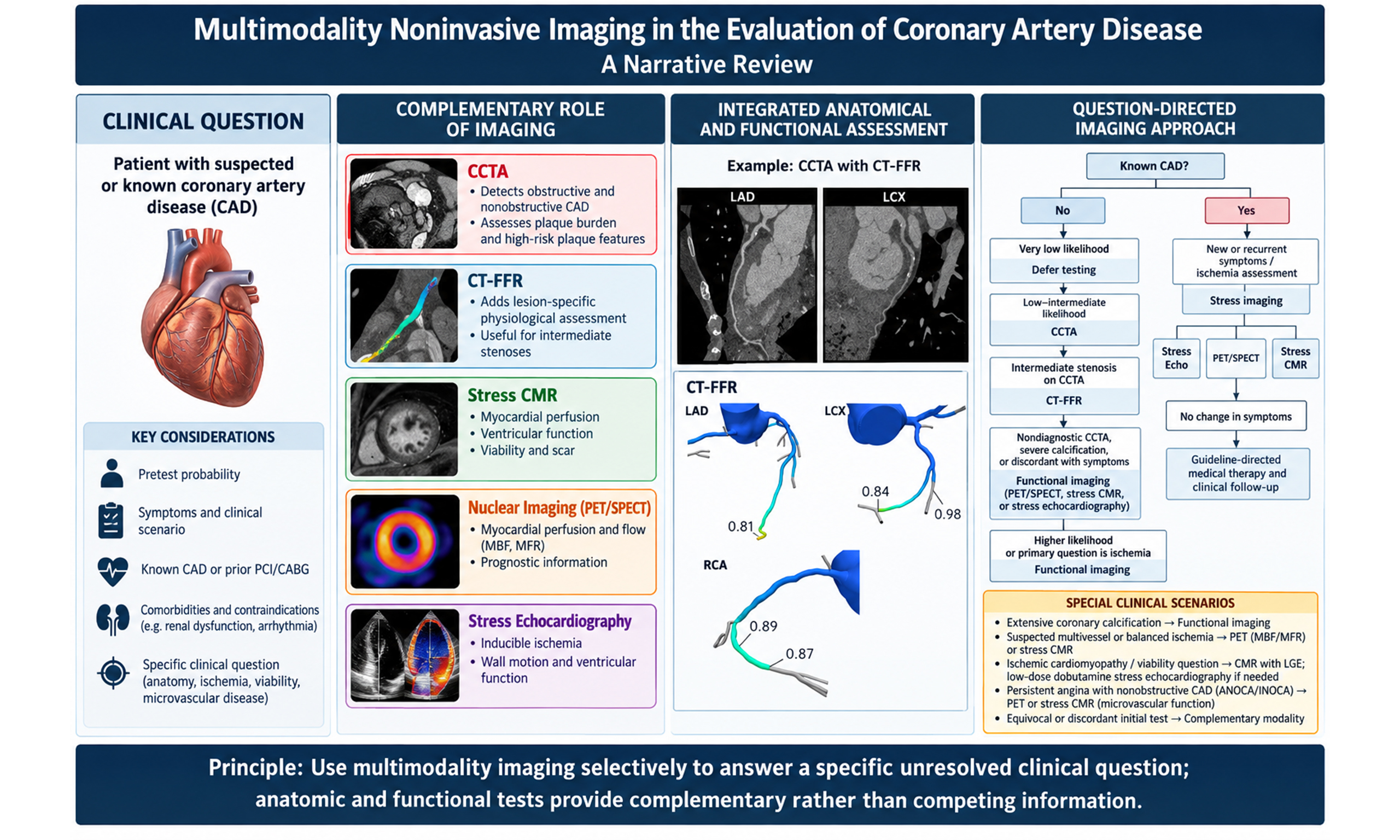
Graphical Abstract
Abstract
Coronary artery disease (CAD) remains a leading cause of cardiovascular morbidity and mortality, and its evaluation has evolved beyond the identification of obstructive epicardial stenosis alone. Contemporary imaging increasingly emphasizes an integrated assessment of coronary anatomy, plaque burden and composition, lesion-specific ischemia, myocardial viability, scar, ventricular remodeling, and coronary microvascular dysfunction. Anatomic and functional imaging provide complementary information regarding coronary atherosclerosis, lesion-specific physiology, inducible ischemia, myocardial blood flow, viability, scar, and microvascular disease, with test selection guided by the clinical question and patient characteristics. Randomized trials of specific imaging-guided strategies have demonstrated differences in diagnostic efficiency, downstream invasive angiography, preventive therapy, revascularization, and procedural complications. In contrast, effects on symptoms and hard cardiovascular outcomes vary according to the population, comparator, and duration of follow-up. Emerging technologies, including artificial intelligence, radiomics, photon-counting computed tomography, molecular imaging, and hybrid imaging, are at different stages of clinical readiness and require varying degrees of further validation before routine implementation. This review summarizes the current role, evidence base, limitations, and future directions of multimodality noninvasive imaging in CAD, providing a practical framework for selecting the appropriate test according to the clinical question and patient profile.
Keywords


 Download PDF
INTRODUCTION
Coronary artery disease (CAD) remains a leading cause of cardiovascular morbidity and mortality despite major advances in prevention, diagnosis, and treatment. Traditionally, diagnostic evaluation focused on identifying obstructive epicardial coronary lesions. Contemporary assessment has shifted beyond stenosis detection toward an integrated evaluation of luminal narrowing, plaque burden and composition, ischemia relevant to revascularization, myocardial viability, microvascular dysfunction, and adverse cardiovascular remodeling[1-3].
Modern cardiovascular imaging has evolved to mirror this shift. Coronary computed tomography angiography (CCTA) has become a key anatomic test for detecting obstructive CAD with high sensitivity while also identifying nonobstructive atherosclerosis and high-risk plaque features that may be missed by functional testing alone[1,4-6]. computed tomography-derived fractional flow reserve (CT-FFR) further extends the value of CCTA by adding lesion-specific physiologic assessment, particularly for intermediate stenoses in which anatomic severity may not reflect ischemic significance[7]. Functional imaging provides complementary assessment of ischemia, myocardial perfusion, ventricular function, viability, scar, and microvascular disease, with the choice of modality determined by the clinical question and patient characteristics[8-11]. These modalities should therefore be viewed as complementary rather than competing. Randomized trials have shown that specific noninvasive testing strategies can alter downstream invasive angiography, preventive therapy, revascularization, and procedural complications, although effects on symptoms and hard cardiovascular outcomes differ across trial designs and follow-up durations[11-15].
This review summarizes the role of multimodality noninvasive imaging in CAD, focusing on CCTA, CT-FFR, plaque analysis, stress echocardiography, nuclear imaging, cardiac magnetic resonance (CMR), randomized trial evidence, guideline recommendations, limitations, and future directions. The goal is to provide an integrated framework for selecting the appropriate imaging strategy according to the clinical question and patient profile. To operationalize this aim, the review culminates in a question-directed clinical framework and visual decision flowchart that synthesize the indications, strengths, and limitations of the major imaging modalities discussed. The scope of this review is focused on clinically relevant noninvasive cardiovascular imaging modalities rather than all noninvasive diagnostic tests for CAD. Exercise electrocardiography (ECG) is therefore discussed as an important non-imaging comparator because it remains appropriate in selected patients who can exercise adequately and have an interpretable resting ECG[1,2,16].
METHODS
This article was designed as a narrative review of contemporary noninvasive imaging strategies for the evaluation of CAD. PubMed/MEDLINE was the principal source used to identify relevant publications through targeted literature searches conducted between January and June 2026, with the final literature update performed in June 2026. The principal search concepts included CAD, chronic coronary syndromes, noninvasive and multimodality cardiovascular imaging, Coronary artery calcium scoring (CACS), CCTA, CT-FFR, coronary plaque characterization, CT myocardial perfusion (CTP), stress echocardiography, Single-photon emission computed tomography (SPECT), positron emission tomography (PET), CMR, and coronary microvascular dysfunction (CMD). Additional search concepts included diagnostic accuracy, prognostic performance, myocardial blood flow (MBF), randomized clinical trials, clinical outcomes, and professional-society guidelines. Search terms were adapted to the imaging modality and clinical question being addressed. Additional relevant publications were identified from the reference lists of key studies, guidelines, and consensus documents. Literature searches and publication selection were undertaken by Karim Hoyek, Anthony Bedran, Elio Haroun, Joseph Hajj, Ziad Zalaquett, Ankit Agrawal, and Daniella Aro Arockiam. Each author was responsible for identifying, reviewing, and selecting relevant publications for their assigned section(s) of the manuscript. Authors prioritized contemporary professional-society guidelines and consensus statements, landmark randomized controlled trials, systematic reviews and meta-analyses, and major prospective or multicenter observational studies with direct relevance to the diagnostic, prognostic, or management roles of noninvasive imaging in CAD. Particular emphasis was placed on evidence informing comparisons between imaging modalities, randomized imaging strategies, and current guideline recommendations.
As this was a narrative rather than systematic review, no formal systematic search protocol, study-level risk-of-bias assessment, or quantitative evidence grading was performed. When findings differed across studies or recommendations varied among professional societies, greater emphasis was placed on contemporary guidelines, randomized trials, larger prospective studies, and evidence most directly applicable to the clinical question under discussion. Areas of uncertainty or disagreement were described within the corresponding sections.
EVOLUTION OF CAD IMAGING
Historically, CAD assessment centered on identifying obstructive coronary stenoses and determining whether they were associated with myocardial ischemia. Accordingly, invasive coronary angiography and stress testing, including exercise ECG and stress echocardiography, were used primarily to identify flow-limiting disease. However, studies including the Scottish Computed Tomography of the Heart (SCOT-HEART) and the Prospective Multicenter Imaging Study for Evaluation of Chest Pain (PROMISE) demonstrated that coronary plaque burden and adverse plaque characteristics provide prognostic information beyond stenosis severity alone. In addition, some patients experience angina despite the absence of obstructive epicardial CAD[15,17]. These findings have shifted cardiovascular imaging toward a broader assessment that incorporates coronary plaque burden and composition, coronary inflammation, microvascular dysfunction, myocardial viability, scar burden, and adverse ventricular remodeling.
CORONARY ARTERY CALCIUM SCORE
CACS, quantified using the Agatston method on non-contrast electrocardiographically gated CT, measures calcified coronary atherosclerotic burden. In asymptomatic individuals undergoing primary prevention risk assessment, increasing coronary artery calcium (CAC) burden is associated with progressively higher cardiovascular event rates and can refine risk estimation beyond conventional risk factors[18,19]. A CACS of 0 indicates the absence of detectable calcified coronary plaque, but it does not establish the absence of coronary atherosclerosis because noncalcified plaque may still be present[4]. Accordingly, the prognostic implications of a CACS of 0 or a low CAC score depend on age, symptoms, cardiovascular risk factors, and duration of follow-up rather than representing a universal “low-risk” category[2,18]. Table 1 below shows the CACS categories.
Agatston coronary artery calcium score categories and calcified plaque burden
| CAC score | Interpretation |
| 0 | No detectable calcified coronary plaque |
| 1-99 | Mild calcified plaque burden |
| 100-399 | Moderate calcified plaque burden |
| 400 | Extensive calcified plaque burden |
In symptomatic patients with suspected chronic coronary syndrome, CAC has a distinct role as a modifier of the clinical likelihood of obstructive CAD, rather than solely as a long-term preventive risk marker. The 2024 European Society of Cardiology (ESC) Chronic Coronary Syndromes guideline incorporates CACS into the risk factor-weighted clinical likelihood model (CACS-CL), allowing CAC burden to refine the estimated likelihood of obstructive CAD and, in selected patients initially classified as having low clinical likelihood, potentially reclassify them to a very-low-likelihood group in whom additional testing may be deferred. However, a CACS of 0 does not exclude noncalcified atherosclerosis or obstructive CAD; this limitation is particularly relevant in younger symptomatic patients, in whom obstructive disease is less likely to be calcified. Therefore, CAC findings should be interpreted together with age, symptoms, risk factors, and the overall clinical likelihood rather than used as an isolated rule-out test[2]. CAC burden should also not be considered equivalent to total coronary plaque burden. CCTA directly depicts both calcified and noncalcified atherosclerosis and can identify adverse plaque characteristics that are not captured by calcium scoring alone[4,5]. In SCOT-HEART, low-attenuation noncalcified plaque burden was independently associated with subsequent myocardial infarction, illustrating the additional prognostic information available from direct plaque characterization beyond calcium burden alone[6].
CCTA, CT-FFR, PLAQUE ANALYSIS, AND CTP
CCTA is increasingly used as a first-line anatomic test in patients with suspected CAD, particularly in those without known CAD. Its clinical value lies in its high diagnostic sensitivity for obstructive stenosis and its ability to detect nonobstructive atherosclerosis that may be missed by functional testing alone[1,2,4]. Consistent with contemporary American Heart Association/American College of Cardiology (AHA/ACC) and ESC guidance, CCTA is an important initial anatomic test in appropriately selected patients without known CAD, with test selection determined by clinical likelihood, patient characteristics, anticipated image quality, and the primary diagnostic question[1,2]. CT-FFR expands the role of CCTA by adding lesion-specific physiologic assessment from the same anatomic dataset, helping distinguish anatomically intermediate stenoses that are flow-limiting from those that are not[7,20]. This is especially relevant for 40%-90% stenoses, in which visual stenosis severity alone may overestimate ischemic significance and lead to unnecessary invasive coronary angiography. Contemporary guidance supports selective use of CT-FFR when CCTA demonstrates stenosis of uncertain functional significance. A recent international expert consensus reported pooled per-vessel diagnostic accuracy of 71%-91%, sensitivity of 76%-98%, and specificity of 61%-94%, supporting CT-FFR as both a gatekeeper to invasive angiography and a prognostic tool[1,7]. CT-FFR should be interpreted as a physiologic measurement rather than as a reassessment of anatomic stenosis severity. Lesion-specific values are generally interpreted at a location distal to the stenosis of interest. A value > 0.80 is generally considered not to demonstrate lesion-specific ischemia, whereas values ≤ 0.75 provide stronger evidence of hemodynamic significance; values between 0.76 and 0.80 represent a borderline range in which lesion location, symptoms, translesional pressure loss, image quality, and the overall clinical context should be considered. Accordingly, a CT-FFR value > 0.80 does not mean that an anatomically identified stenosis is “nonobstructive”; rather, it indicates that the stenosis is not physiologically significant according to the CT-FFR assessment[5,7].
CT-FFR is not a single computational implementation. Established approaches include externally processed methods that use patient-specific coronary anatomy and computational fluid dynamics to estimate pressure loss under simulated hyperemia, whereas newer platforms may perform on-site analysis using machine learning, deep learning, or reduced-order computational approaches. These approaches differ in processing location and time, computational methodology, degree of automation, validation evidence, and regulatory status. Diagnostic performance and clinical evidence obtained with one CT-FFR implementation should therefore not automatically be generalized to all platforms[7,21-23].
Plaque analysis is a major strength of CCTA because it allows noninvasive characterization of atherosclerotic burden and composition, rather than luminal narrowing alone. CAD-Reporting and Data System version 2.0 (CAD-RADS 2.0) standardized this approach by incorporating stenosis severity, plaque burden, and modifiers such as ischemia and high-risk plaque features into structured reporting[5]. High-risk plaque features include low-attenuation plaque, positive remodeling, spotty calcification, and the napkin-ring sign; these features are associated with increased cardiovascular risk, even in the absence of obstructive stenosis[5,6,17]. In PROMISE, high-risk plaque was associated with increased major adverse cardiovascular events, and in SCOT-HEART, low-attenuation plaque burden was the strongest independent predictor of myocardial infarction, outperforming stenosis severity and coronary calcium score[6,17]. Pericoronary adipose tissue attenuation has shown associations with cardiovascular risk and may provide information regarding coronary inflammatory activity; however, evidence that its use improves clinical decision-making or patient outcomes remains limited. It should therefore be considered a selectively available, emerging biomarker rather than an established component of routine CAD assessment[24].
CTP extends coronary CT from anatomic assessment to evaluation of myocardial perfusion during pharmacologic stress. Static and dynamic techniques can be combined with CCTA, allowing coronary anatomy and myocardial ischemia to be assessed within an integrated CT-based examination. Dynamic CTP additionally permits quantitative assessment of myocardial perfusion and may provide complementary information when anatomic stenosis severity alone does not establish functional significance[25,26].
The principal limitations of CTP include additional ionizing radiation and iodinated contrast exposure compared with CCTA alone, susceptibility to motion and technical artifacts, and the requirement for dedicated acquisition protocols, post-processing expertise, and appropriate scanner capability. Its availability therefore remains more limited than established stress imaging modalities, and PET, stress CMR, or stress echocardiography may be preferred when they can answer the functional question without additional CT contrast or radiation exposure[16,25,26]. An example of integrated CCTA and CT-FFR assessment is shown in Figure 1.
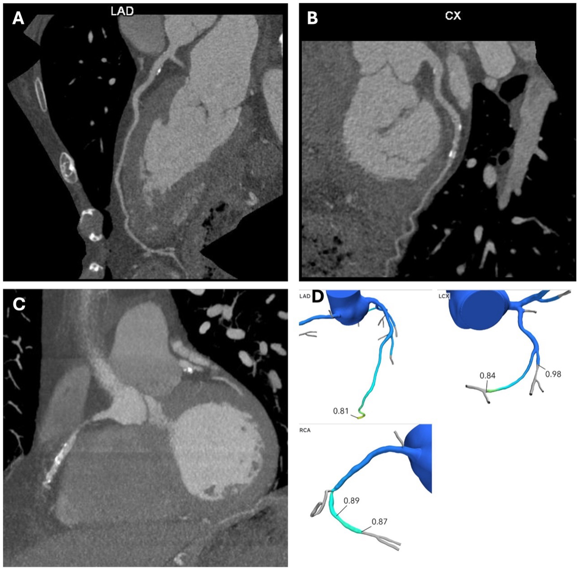
Figure 1. Coronary computed tomography angiography (CCTA) in a 72-year-old man presenting with exertional dyspnea and intermittent chest pain. (A) shows a curved planar reconstruction of the left anterior descending artery (LAD), demonstrating calcified plaque in the proximal LAD with minimal stenosis. (B) shows a curved planar reconstruction of the left circumflex artery (LCx), demonstrating calcified plaque in the proximal and mid LCx segments with mild stenosis. (C) shows a curved planar reconstruction of the right coronary artery (RCA), demonstrating calcified plaque in the mid RCA with moderate stenosis. (D) shows the CT-FFR analysis, with lesion-specific values > 0.80 measured distal to the corresponding stenoses, indicating no lesion-specific physiologic evidence of ischemia according to the CT-FFR assessment. The anatomic stenosis severity remains defined by the CCTA findings shown in (A-C).
EXERCISE ECG AS A NON-IMAGING COMPARATOR
Exercise ECG remains a widely available, low-cost functional test that provides information on exercise capacity, symptom reproduction, hemodynamic response, and exercise-induced electrocardiographic changes without ionizing radiation or contrast administration[1,2]. However, its diagnostic performance for obstructive CAD is lower than that of stress imaging, and interpretation requires both adequate exercise capacity and an interpretable resting ECG[1,16]. Contemporary guidelines therefore reserve exercise ECG for appropriately selected patients; when the resting ECG is uninterpretable, adequate exercise cannot be achieved, or greater diagnostic accuracy and localization of ischemia are required, stress imaging or CCTA is generally preferred according to the clinical question[1,2,16].
STRESS ECHOCARDIOGRAPHY
Stress echocardiography is an established functional imaging modality for the diagnosis and risk stratification of known or suspected CAD. Positive studies and greater ischemic burden are associated with higher long-term risk of myocardial infarction and other adverse cardiovascular events[27]. Its clinical utility derives from its ability to identify inducible myocardial ischemia through new or worsening regional wall-motion abnormalities during stress, while also providing information on ventricular function, hemodynamic response, valvular disease, and exercise capacity when exercise stress is used[8,28,29].
Exercise stress echocardiography
When patients can exercise adequately, exercise stress echocardiography is generally preferred, particularly in the evaluation of known or suspected CAD and chronic coronary syndromes. Because exercise is a physiologic stressor, this modality provides information beyond ischemia detection, including symptom reproduction, exercise capacity, heart rate and blood-pressure response, ECG changes, global and regional left ventricular systolic response, and, when image quality permits, deformation parameters such as global longitudinal strain. These features make exercise stress echocardiography particularly useful when the clinical question involves both diagnosis and functional risk stratification. High-risk stress echocardiographic features include extensive inducible wall-motion abnormalities, ischemia occurring at a low workload, and an abnormal global left ventricular response, such as stress-induced ventricular dilation or failure of left ventricular systolic function to augment appropriately[8,28,29]. A reduction in left ventricular ejection fraction of ≥ 10% has been used as a high-risk feature in some contemporary studies, particularly for identifying left main or multivessel CAD, but this threshold is not universal and should be interpreted together with regional wall-motion abnormalities, ventricular volumes, achieved workload, symptoms, and hemodynamic response[29]. Compared with exercise ECG alone, exercise stress echocardiography provides higher diagnostic accuracy for CAD because it evaluates stress-induced mechanical dysfunction rather than electrical changes alone[28]. However, exercise stress echocardiography has several limitations. It requires adequate exercise capacity and may be nondiagnostic when submaximal stress is achieved. Image acquisition at peak stress may be challenging because of tachycardia, respiratory motion, and limited acoustic windows. Treadmill protocols do not allow continuous imaging during exercise, and interpretability may be reduced in patients with arrhythmias. Sensitivity may also be reduced when anti-ischemic therapy, particularly beta-blockers, is continued[8,28,29].
Dobutamine stress echocardiography
Dobutamine stress echocardiography is the main pharmacologic alternative when patients cannot exercise adequately, when exercise testing is not feasible, or when exercise stress produces a submaximal or nondiagnostic study. It is also guideline-supported for assessing myocardial viability and contractile reserve, particularly when low-dose dobutamine protocols are used[8,28,29]. Dobutamine increases heart rate, myocardial contractility, and oxygen demand, thereby inducing ischemia in myocardial territories supplied by flow-limiting coronary stenoses. Ischemia is detected as new or worsening regional wall-motion abnormalities[8,28,30]. One commonly used protocol begins with dobutamine at 5 μg/kg/min, with dose escalation every 3 min up to 40 μg/kg/min; atropine may be added when necessary to achieve the target heart rate. For ischemia assessment, the principal echocardiographic criterion is the development of new or worsening regional wall-motion abnormalities during stress. The number of segments required to define a positive examination varies among protocols and interpretive standards; some criteria use abnormalities in at least two adjacent segments to improve specificity, whereas other standards recognize inducible abnormality in one or more segments. Accordingly, a requirement for at least two adjacent abnormal segments should not be considered a universal definition of a positive dobutamine stress echocardiogram[8,28].
The timing and dose at which an ischemic response develops also provide information regarding ischemic burden. Development of new or worsening wall-motion abnormalities early in the protocol or at relatively low dobutamine doses reflects a low ischemic threshold and may indicate more extensive disease; however, this should be distinguished from the use of low-dose dobutamine for myocardial viability assessment[8,29]. In dysfunctional myocardium, improvement in contractility during low-dose dobutamine indicates contractile reserve and supports myocardial viability. When this initial improvement is followed by deterioration in wall motion at higher dobutamine doses, the resulting biphasic response indicates viable myocardium with inducible ischemia[8,28]. In one contemporary study using its prespecified interpretive criteria, dobutamine stress echocardiography demonstrated 78% sensitivity, 75% specificity, and 76% overall accuracy for detecting obstructive CAD[30].
The diagnostic performance of dobutamine stress echocardiography depends on the reference standard and clinical population. In symptomatic patients with intermediate-to-low pretest probability, positive CCTA, and invasive fractional flow reserve (FFR) or instantaneous wave-free ratio (iFR) assessment, dobutamine stress echocardiography demonstrated only moderate diagnostic performance and limited incremental value for identifying hemodynamically significant CAD[31]. Discordance between stress-induced wall-motion abnormalities and invasive pressure-derived indices is physiologically plausible because these tests interrogate different components of the ischemic cascade. FFR and iFR assess the hemodynamic significance of an epicardial coronary stenosis, whereas stress echocardiography detects the downstream mechanical consequence of ischemia as new or worsening regional systolic dysfunction. A pressure-significant lesion may therefore not produce a visible wall-motion abnormality when ischemia is limited in extent or predominantly subendocardial, when adequate stress is not achieved or is attenuated by anti-ischemic therapy, or when collateral perfusion limits regional dysfunction. Conversely, abnormal or equivocal wall-motion responses may be influenced by pre-existing regional dysfunction, myocardial scar, image quality, and interpretive variability and therefore need not correspond directly to lesion-specific invasive physiology[8,28,31].
An additional limitation of conventional wall-motion stress echocardiography is its sensitivity for CMD. In patients with angina with no obstructive coronary arteries/ischemia with no obstructive coronary arteries (ANOCA/INOCA), microvascular ischemia may be diffuse or predominantly subendocardial rather than producing the focal or sufficiently extensive ischemia required to generate a discrete regional wall-motion abnormality. Consequently, a normal dobutamine stress echocardiogram does not exclude impaired coronary microvascular vasodilator function. When CMD is clinically suspected, PET with quantitative MBF and myocardial flow reserve (MFR) or quantitative stress CMR can provide more direct assessment of impaired vasodilator reserve, whereas invasive coronary function testing may be required when definitive mechanistic endotyping, including assessment for microvascular or epicardial vasospasm, is clinically necessary, as discussed in Section "CMD AND VASOMOTOR DISORDERS"[32-36].
The strengths of dobutamine stress echocardiography include its applicability in patients with limited exercise capacity, its ability to evaluate inducible wall-motion abnormalities, its assessment of global left ventricular response, and its value in characterizing viability and contractile reserve[8,28,29]. Its limitations include the need for pharmacologic infusion, continuous monitoring, appropriate expertise, and resuscitation capability; dependence on achieving target heart rate; potential need for atropine; and cardiovascular adverse effects, including angina, hypotension, and arrhythmias. It should therefore be used cautiously or avoided in patients with serious or recent arrhythmias. Additional limitations include subjectivity of visual wall-motion interpretation and technical challenges with image acquisition or strain analysis at high heart rates[8,28,30].
From a practical perspective, stress echocardiography is widely available, relatively inexpensive, and does not require ionizing radiation. Exercise stress echocardiography is particularly useful when exercise capacity and hemodynamic response are clinically relevant, whereas pharmacologic stress provides an alternative when adequate exercise cannot be achieved[8,28,29]. An alternative modality may be preferred when acoustic windows are inadequate, sufficient stress cannot be achieved, or the clinical question requires coronary anatomic assessment, absolute myocardial blood-flow quantification, or more detailed tissue and scar characterization[1,16,28].
NUCLEAR IMAGING
Nuclear myocardial perfusion imaging provides functional assessment of myocardial ischemia and has an established role in the diagnosis and risk stratification of patients with suspected or known CAD[1,37,38]. SPECT and PET should be viewed as complementary modalities, each with distinct clinical advantages and limitations. SPECT is widely used in routine clinical practice because of its broad availability, established prognostic value, and relative cost-effectiveness. Its ability to be performed with either exercise or pharmacologic stress is a practical advantage, allowing assessment of exercise capacity and hemodynamic response when patients are able to exercise. However, conventional SPECT relies primarily on relative perfusion imaging. This can underestimate disease severity when MBF is globally or diffusely reduced, as occurs in multivessel disease, balanced ischemia, diffuse atherosclerosis, and CMD. In these settings, perfusion may appear relatively homogeneous despite clinically significant disease[37]. Newer cadmium zinc telluride SPECT systems can perform dynamic imaging and estimate MBF and MFR, partially addressing this limitation. However, these approaches require further validation and standardization and remain less established than PET-based flow quantification[25].
PET provides superior diagnostic performance, improved image quality, shorter protocols, and generally lower radiation exposure, depending on the tracer and protocol used[38]. A major advantage of PET is its ability to quantify absolute MBF and MFR. PET-derived flow quantification is particularly useful for detecting multivessel, diffuse, or microvascular disease that may be missed by conventional relative perfusion imaging. Reduced MFR can identify impaired coronary vasodilator capacity but does not distinguish structural CMD from vasospastic mechanisms or directly diagnose coronary vasospasm[32,33]. For this reason, PET is often considered the noninvasive reference standard for quantitative myocardial perfusion assessment[9].
Hybrid imaging can combine complementary anatomic, metabolic, perfusion, and tissue-characterization information within a single diagnostic framework. PET/CT is clinically established for several applications and can provide anatomic coregistration for PET imaging; however, advanced coronary plaque and molecular-imaging applications remain specialized. Hybrid PET/magnetic resonance (MR) offers the potential to integrate PET-derived molecular or perfusion information with CMR-based ventricular and tissue characterization, but its application to routine CAD assessment remains limited to specialized centers and is not supported by evidence demonstrating improved clinical outcomes or routine management utility[39,40]. PET also has important limitations, including higher cost, limited availability, technical complexity, infrastructure requirements, and the need for specialized personnel[39]. In addition, protocol heterogeneity and the need for more outcome-driven evidence have limited the widespread routine implementation of quantitative PET[9]. Overall, SPECT remains an accessible, cost-effective, and prognostically validated modality for routine clinical use, whereas PET is preferred when available for higher diagnostic accuracy and more precise characterization of diffuse, multivessel, or microvascular coronary disease.
Both SPECT and PET involve ionizing radiation, although radiation exposure varies according to tracer and acquisition protocol[37,38]. Neither requires iodinated or gadolinium-based contrast, which may be advantageous when these agents are undesirable. When avoidance of ionizing radiation is a priority, stress echocardiography or stress CMR may be preferred[1,16], whereas CCTA may be more appropriate when the primary clinical question concerns coronary anatomy and plaque characterization[1,2]. When nuclear myocardial perfusion imaging is selected, and PET is available, contemporary guidelines favor PET over SPECT because of its greater diagnostic accuracy and lower rate of nondiagnostic studies[1].
CMR
CMR provides comprehensive noninvasive assessment of myocardial ischemia, viability, ventricular function, and tissue characterization without ionizing radiation. Stress CMR is guideline-supported for evaluation of myocardial ischemia and risk stratification in patients with suspected or known CAD, particularly when functional assessment is needed or when prior testing is equivocal[1,41,42]. Using vasodilator stress first-pass perfusion imaging, stress CMR identifies inducible perfusion defects with high spatial resolution and is less affected by attenuation artifacts than SPECT. In a 2023 meta-analysis, stress CMR demonstrated sensitivity of 81%, specificity of 86%, and an area under the receiver operating characteristic curve of 0.84 for detecting functionally obstructive CAD using FFR as the reference standard[43]. Head-to-head studies also support its diagnostic performance. Clinical evaluation of magnetic resonance imaging in coronary heart disease (CE-MARC) demonstrated higher sensitivity for CMR compared with SPECT, whereas CE-MARC 2 showed that a CMR-guided strategy reduced unnecessary invasive coronary angiography compared with guideline-directed care[44,45]. Stress CMR has also been validated as a management-guiding test. In MR-imaging to guide management of patients with stable coronary artery disease (INFORM), a stress perfusion CMR-guided strategy was noninferior to an invasive angiography and fractional flow reserve-guided strategy for 1-year major adverse cardiac events, while reducing coronary revascularization and invasive angiography[11]. Beyond diagnosis, CMR adds prognostic value through combined assessment of inducible ischemia and late gadolinium enhancement (LGE). A normal stress CMR is associated with a low subsequent event rate, whereas inducible ischemia and LGE are independently associated with increased mortality and major adverse cardiovascular events[10,43]. LGE-CMR is particularly useful in ischemic cardiomyopathy because it identifies myocardial scar and viability. The transmural extent of LGE helps predict the likelihood of contractile recovery after revascularization, with more extensive scar indicating a lower probability of functional recovery[10,46].
More recently, quantitative stress CMR perfusion has expanded the role of CMR beyond visual ischemia assessment by measuring absolute MBF and myocardial perfusion reserve. This may improve detection of multivessel or balanced ischemia and can identify impaired myocardial perfusion reserve suggestive of CMD in patients with ischemia and nonobstructive coronary arteries[35,47]. However, quantitative stress CMR does not directly diagnose epicardial or microvascular vasospasm, which requires assessment of coronary vasomotor responses[32]. Stress CMR also has important limitations, including longer acquisition time, dependence on local expertise and scanner protocols, need for gadolinium contrast, susceptibility to arrhythmia or motion artifacts, claustrophobia, and restrictions in patients with some implanted devices or severe renal dysfunction[42,46]. Therefore, CMR is best integrated into a multimodality strategy. It is particularly useful when ischemia, viability, scar burden, and ventricular function need to be evaluated together, whereas CCTA, PET, SPECT, stress echocardiography, or invasive angiography may be preferred depending on the clinical question, patient factors, and local availability[1,41,42,46]. An example of stress CMR demonstrating an inducible perfusion abnormality in a patient without obstructive epicardial CAD is shown in Figure 2. Table 2 summarizes the principal clinical indications, strengths, limitations, contraindications, radiation and contrast considerations, and myocardial blood-flow capabilities of contemporary noninvasive imaging modalities.
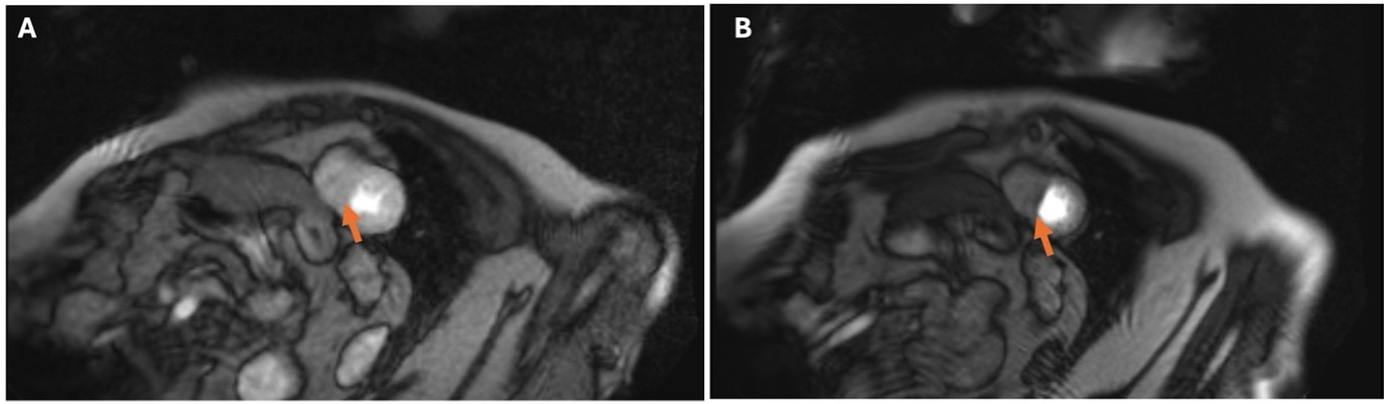
Figure 2. Stress CMR of a 64-year-old woman with exertional anginal symptoms despite no angiographic evidence of obstructive epicardial coronary artery disease. First-pass myocardial perfusion imaging was performed following intravenous administration of gadolinium-based contrast during pharmacologic stress, with resting perfusion images obtained for comparison. Adenosine was administered at 140 μg/kg/min for 2 min, with stress perfusion acquisition performed according to a protocol requiring an increase in heart rate of ≥ 15 beats/min. Perfusion assessment included evaluation of inducible and reversible versus persistent perfusion defects, the ratio of stress to resting myocardial blood flow (MBF), and the ratio of subendocardial to subepicardial stress MBF. (A) shows the resting perfusion image, with the orange arrow identifying the septal myocardial region that demonstrates no perfusion defect at rest. (B) shows the corresponding stress perfusion image, with the orange arrow indicating an inducible septal perfusion defect. In the absence of obstructive epicardial coronary artery disease, this finding may suggest impaired microvascular perfusion but does not independently establish coronary microvascular dysfunction; imaging artifacts and other potential causes should also be considered.
Practical comparison of contemporary noninvasive imaging modalities for CAD
| Modality | Principal indications | Key strengths | Limitations/contraindications | Ionizing radiation | Contrast/tracer | Quantitative MBF |
| Coronary artery calcium scoring (CACS)[2,18] | Preventive risk stratification; refinement of clinical likelihood in selected symptomatic patients | Rapid, noncontrast assessment of calcified plaque burden; established prognostic value | Does not detect noncalcified plaque or directly assess stenosis/ischemia; interpretation depends on age and clinical context | Yes | None | No |
| CCTA[1,2,4,48] | First-line anatomic assessment of suspected CAD, particularly in patients without known CAD | High sensitivity; defines stenosis, plaque burden, distribution, and high-risk plaque features | Severe calcification, motion, high/irregular heart rate, obesity, renal dysfunction, or iodinated-contrast allergy may reduce feasibility or accuracy | Yes | Iodinated contrast | No |
| CT-FFR[1,5,7,20] | Physiologic assessment of intermediate stenoses identified on CCTA | Adds lesion-specific physiologic information without a second image acquisition | Requires analyzable CCTA; borderline values require contextual interpretation; diffuse pressure loss and platform-specific differences may complicate interpretation | No additional radiation beyond CCTA | No additional contrast beyond CCTA | No |
| CTP[25,26] | Assessment of inducible ischemia, particularly when integrated with CCTA | Combined anatomic and functional CT assessment; dynamic techniques can quantify perfusion | Additional radiation and contrast; motion/artifact susceptibility; specialized protocols and limited availability; pharmacologic-stress contraindications apply | Yes | Iodinated contrast | Yes with dynamic CTP |
| Exercise stress echocardiography[8,28,29] | Suspected or known CAD in patients able to exercise adequately | No radiation; evaluates inducible wall-motion abnormalities, exercise capacity, symptoms, and hemodynamic response | Dependent on acoustic windows and adequate exercise; may be nondiagnostic with submaximal workload, tachycardia, motion, or arrhythmia | No | No iodinated/gadolinium contrast; ultrasound contrast may be used when needed | No |
| Dobutamine stress echocardiography[8,28-30] | Ischemia assessment when exercise is not feasible; assessment of contractile reserve/viability | No radiation; useful when exercise cannot be performed; evaluates wall motion and contractile reserve | Pharmacologic infusion and monitoring required; arrhythmias, hypotension, or angina may occur; interpretation remains image-quality and operator dependent | No | No iodinated/gadolinium contrast; ultrasound contrast may be used when needed | No |
| SPECT myocardial perfusion imaging[25,37] | Diagnosis and risk stratification of suspected or known CAD | Broad availability; established prognostic evidence; exercise or pharmacologic stress possible | Attenuation and motion artifacts; relative perfusion may underestimate balanced or diffuse ischemia | Yes | Radiotracer; no iodinated/gadolinium contrast | Usually no; dynamic CZT-SPECT can estimate MBF/MFR but is less established |
| PET myocardial perfusion imaging[9,33,38] | Ischemia assessment; diffuse/multivessel disease; suspected microvascular dysfunction | High diagnostic performance; attenuation correction; absolute MBF and MFR quantification | Cost, tracer availability, infrastructure, and specialized expertise limit access | Yes | Radiotracer; no iodinated/gadolinium contrast | Yes |
| Stress CMR/CMR with LGE[10,35,41-47] | Ischemia and risk stratification; ventricular function; scar and viability; microvascular perfusion assessment | No ionizing radiation; high spatial resolution; integrates perfusion, ventricular function, scar, viability, and tissue characterization | Longer acquisition; claustrophobia, arrhythmia/motion, device compatibility, gadolinium considerations, and vasodilator-stress contraindications | No | Gadolinium generally required for stress perfusion and LGE | Yes with quantitative perfusion CMR |
CMD AND VASOMOTOR DISORDERS
Patients may experience angina or objective evidence of ischemia despite the absence of obstructive epicardial CAD. This heterogeneous group encompasses several distinct coronary vascular endotypes, including impaired coronary vasodilator capacity, structural CMD, microvascular spasm, epicardial vasospasm, and combinations of these abnormalities. These mechanisms should not be considered interchangeable because they require different diagnostic approaches and may have different therapeutic implications[32,36].
PET is the most established noninvasive reference technique for quantitative assessment of MBF and MFR. Its principal role in suspected CMD is the identification and quantification of impaired coronary vasodilator capacity in patients with persistent symptoms or objective ischemia despite nonobstructive epicardial coronary arteries. Reduced MFR reflects impaired global or regional vasodilator reserve and is associated with adverse cardiovascular outcomes independent of angiographic stenosis severity[33,34]. PET is therefore particularly useful when the principal clinical question is whether coronary vasodilator function is impaired and when absolute flow quantification is required. However, reduced MFR is not specific for a single CMD endotype and does not distinguish structural microvascular dysfunction from microvascular or epicardial vasospasm[32,33].
Quantitative stress CMR provides complementary assessment of myocardial perfusion and perfusion reserve and may similarly identify impaired microvascular vasodilator function[35,47]. Its principal advantage over PET is the ability to combine perfusion assessment with high-resolution evaluation of ventricular function and myocardial tissue characterization within the same examination. LGE can identify focal scar or fibrosis, while parametric mapping techniques can provide additional information regarding diffuse myocardial abnormalities when clinically relevant. CMR may therefore be particularly useful when the differential diagnosis extends beyond isolated CMD to concomitant myocardial disease. However, as with PET, reduced perfusion reserve does not establish a specific vasomotor endotype and does not directly diagnose epicardial or microvascular vasospasm[10,32,35].
Invasive coronary function testing is appropriate when noninvasive testing demonstrates or suggests impaired vasodilator reserve, but more precise mechanistic endotyping is required, or when symptoms persist despite nonobstructive coronary anatomy and vasospastic disease remains a clinical consideration. Adenosine-based measurements of coronary flow reserve and microvascular resistance, including the index of microcirculatory resistance (IMR) or hyperaemic microvascular resistance (HMR), primarily assess endothelium-independent microvascular vasodilator function. In contrast, acetylcholine testing evaluates endothelium-dependent coronary vasomotor responses and can identify epicardial or microvascular spasm. Reduced coronary flow reserve with increased microvascular resistance supports structural microvascular dysfunction, whereas reduced flow reserve with preserved resistance may indicate a functional microvascular endotype. Acetylcholine-induced epicardial constriction with angina and ischemic ECG changes supports epicardial vasospasm, while reproduction of angina and ischemic ECG changes without significant epicardial constriction supports microvascular spasm.
Accordingly, PET and quantitative stress CMR are complementary noninvasive approaches for identifying impaired myocardial flow or perfusion reserve, whereas invasive coronary function testing provides the greatest mechanistic specificity when distinction among structural CMD, functional microvascular dysfunction, microvascular spasm, and epicardial vasospasm is clinically important[32,36].
SUMMARY OF RANDOMIZED TRIALS
The randomized trials summarized below evaluated different patient populations, comparators, primary endpoints, and follow-up durations and should not be interpreted as testing a single “multimodality imaging” intervention. Some trials primarily assessed diagnostic efficiency or downstream invasive angiography, whereas others examined procedural complications, preventive therapy, revascularization, symptoms, or hard cardiovascular outcomes. These outcome domains are therefore considered separately.
Noninvasive anatomic imaging: CCTA
The major CCTA trials addressed distinct clinical questions. SCOT-HEART randomized 4,146 patients with suspected stable angina to standard care with or without CCTA and therefore evaluated the incremental effect of adding CCTA to routine assessment. At 10 years, the primary composite of coronary heart disease death or nonfatal myocardial infarction was lower with CCTA-guided care, driven predominantly by fewer nonfatal myocardial infarctions; preventive therapy remained more frequently prescribed, whereas overall coronary revascularization rates were similar between groups[15].
PROMISE instead compared initial CCTA with initial functional testing in 10,003 symptomatic patients with suspected CAD. Over a median follow-up of approximately 25 months, the primary composite of death, myocardial infarction, hospitalization for unstable angina, or major procedural complications did not differ between strategies. CCTA resulted in fewer invasive angiograms showing no obstructive CAD, but more patients underwent invasive angiography within 90 days; thus, the main differences related to downstream diagnostic utilization rather than hard cardiovascular outcomes[12].
Diagnostic Imaging Strategies for Patients with Stable Chest Pain and Intermediate Risk of Coronary Artery Disease trial (DISCHARGE) evaluated a different population: 3,561 patients with stable chest pain who had already been referred for invasive coronary angiography were randomized to a CT-first or direct invasive angiography strategy. At a median of 3.5 years, the primary composite of cardiovascular death, nonfatal myocardial infarction, or stroke was similar between groups. However, major procedure-related complications were lower with the CT-first strategy, while angina outcomes were similar; revascularization was also less frequent in the CT-first group[13].
These trials therefore should not be interpreted as showing a uniform outcome benefit of CCTA. SCOT-HEART demonstrated a long-term reduction in its coronary event endpoint, whereas PROMISE primarily demonstrated differences in downstream diagnostic testing, and DISCHARGE primarily demonstrated fewer procedure-related complications without a significant difference in hard cardiovascular events.
Noninvasive functional imaging
Randomized trials of functional imaging have likewise evaluated different strategies and endpoints. CE-MARC 2 randomized 1,202 patients with suspected stable angina to CMR-guided care, myocardial perfusion scintigraphy-guided care, or National Institute for Health and Care Excellence (NICE) guideline-directed care. Its primary endpoint was unnecessary invasive coronary angiography within 12 months, rather than a hard cardiovascular outcome. Both CMR- and myocardial perfusion scintigraphy-guided strategies reduced unnecessary angiography compared with NICE-guided care, while major adverse cardiovascular events were infrequent and similar between groups[45].
MR-INFORM addressed a different question by comparing a stress perfusion CMR-guided revascularization strategy with invasive angiography plus fractional flow reserve in 918 patients with stable angina. At 1 year, CMR-guided management was noninferior for the composite of death, nonfatal myocardial infarction, or target-vessel revascularization and resulted in fewer index revascularization procedures. Freedom from angina at 12 months did not differ significantly between groups[11].
Thus, CE-MARC 2 primarily informs the efficiency of selecting patients for invasive angiography, whereas MR-INFORM addresses whether a CMR-guided management strategy can safely substitute for an invasive physiology-guided strategy. Their results should not be pooled conceptually as evidence that functional imaging itself improves cardiovascular outcomes.
CT-FFR
The CT-FFR platforms evaluated across these studies are not methodologically identical; studies have used externally processed computational approaches as well as newer on-site, machine-learning, or automated methods, and their results should therefore be interpreted in the context of the specific technology evaluated[7,21-23].
The Prospective Randomized Trial of the Optimal Evaluation of Cardiac Symptoms and Revascularization (PRECISE) randomized 2,103 patients with stable symptoms and suspected CAD to a precision strategy: risk-based deferred testing in minimal-risk patients and CCTA with selective CT-FFR in the remainder, or usual testing. Its primary endpoint combined a safety component (death or nonfatal myocardial infarction) with a diagnostic-efficiency component (invasive catheterization without obstructive CAD). The reduction in the primary composite was driven predominantly by fewer catheterizations without obstructive CAD, whereas death or nonfatal myocardial infarction did not differ significantly. Lipid-lowering and antiplatelet therapy were more frequently used at 1 year in the precision-strategy group[14].
The Fractional Flow Reserve Derived from Computed Tomography Coronary Angiography Assessment and Management of Stable Chest Pain (FORECAST) trial randomized 1,400 patients with stable chest pain to CCTA with selective CT-FFR or standard care. Its primary endpoint was total cardiac cost at 9 months, which did not differ significantly between strategies. CT-FFR reduced invasive coronary angiography, but major adverse cardiac and cerebrovascular events, angina, and quality of life were similar[49].
Effect of On-Site CT-Derived Fractional Flow Reserve on the Management of Decision Making for Patients With Stable Chest Pain trial (TARGET) randomized 1,216 patients with stable CAD and intermediate stenosis on CCTA to an on-site machine-learning CT-FFR-guided care pathway or standard care. CT-FFR reduced invasive angiography that showed no obstructive disease or did not lead to intervention and increased the proportion of patients undergoing revascularization. However, major adverse cardiovascular events, symptoms, and quality of life at 1 year were similar between groups, and the reduction in cost did not reach statistical significance[21].
The larger Chinese multicenter trial randomized 5,297 patients with 25%-99% stenosis on CCTA to CCTA plus automated CT-FFR or CCTA alone. The primary endpoint was invasive coronary angiography within 90 days, which was lower with CT-FFR (10% vs. 12.4%). Revascularization was also less frequent at both 90 days and 1 year. Importantly, the apparently discrepant 1-year event findings reflect different predefined composite endpoints: major adverse cardiovascular events comprised all-cause mortality, nonfatal myocardial infarction, and urgent revascularization and was similar between groups (2.9% vs. 2.8%), whereas the separate “cardiac events” composite comprised cardiac death, nonfatal myocardial infarction, and urgent revascularization and was lower with CT-FFR (0.5% vs. 1.1%; adjusted HR 0.52, 95%CI: 0.27-0.99). These endpoints should therefore be reported separately rather than interpreted as contradictory findings[22].
Collectively, these trials demonstrate that the effects of imaging-guided strategies depend strongly on the population, comparator, and endpoint studied. Several strategies reduced unnecessary invasive angiography or procedure-related complications, altered preventive therapy or revascularization, or improved diagnostic efficiency; effects on symptoms were generally modest or similar between strategies, and effects on hard cardiovascular outcomes were heterogeneous. Accordingly, these trials support selective use of specific imaging strategies rather than a generalized conclusion that “multimodality imaging” itself improves clinical outcomes[11-15,21,22,45,49]. The populations, comparators, primary endpoints, diagnostic and management effects, and cardiovascular outcomes of these randomized trials are summarized in Table 3.
Major randomized trials of imaging-guided diagnostic and management strategies in stable chest pain and CAD
| Trial | Population/comparison | Follow-up | Primary endpoint | Diagnostic/process effects | Management, symptoms, safety | Hard cardiovascular outcomes |
| SCOT-HEART[15] | Suspected stable angina; standard care + CCTA vs. standard care | 10 years | CHD death or nonfatal MI | Improved diagnostic characterization | More preventive therapy; similar overall revascularization | Primary endpoint lower with CCTA, driven mainly by fewer nonfatal MI |
| PROMISE[12] | Suspected CAD; CCTA vs. functional testing | Median 25 months | Death, MI, unstable-angina hospitalization, or major procedural complication | Fewer ICA without obstructive CAD; more early ICA | - | No difference in primary clinical endpoint |
| DISCHARGE[13] | Stable chest pain already referred for ICA; CT-first vs. ICA-first | Median 3.5 years | CV death, nonfatal MI, or stroke | Avoided direct ICA in many patients | Fewer major procedural complications and revascularizations; similar angina | MACE similar |
| CE-MARC 2[45] | Suspected stable angina; CMR vs. MPS vs. NICE-guided care | 12 months | Unnecessary ICA | CMR and MPS reduced unnecessary ICA | - | MACE low and similar |
| MR-INFORM[11] | Stable angina; stress CMR-guided vs. ICA + FFR-guided management | 1 year | Death, nonfatal MI, or target-vessel revascularization | Noninvasive gatekeeper strategy | Fewer index revascularizations; similar freedom from angina | CMR strategy noninferior |
| PRECISE[14] | Stable symptoms/suspected CAD; risk-based + CCTA/CT-FFR vs. usual testing | Median 11.8 months | Death/MI or ICA without obstructive CAD | Markedly fewer ICA without obstructive CAD | More lipid-lowering and antiplatelet therapy | Death/MI similar |
| FORECAST[49] | Stable chest pain; CCTA + selective CT-FFR vs. standard care | 9 months | Total cardiac cost | Fewer ICA | Similar angina/QoL; cost similar | MACCE similar |
| TARGET[21] | Stable CAD/intermediate CCTA stenosis; ML CT-FFR vs. standard care | 1 year | ICA without obstructive CAD or without intervention | Fewer low-yield ICA | More revascularization; symptoms/QoL similar | MACE similar |
| Chinese automated CT-FFR RCT[22] | 25%-99% stenosis on CCTA; CCTA + CT-FFR vs. CCTA alone | 90 days/1 year | ICA at 90 days | Lower ICA rate | Lower revascularization | MACE similar; separate cardiac-events composite lower |
GUIDELINE INTEGRATION AND CLINICAL DECISION FRAMEWORK
Guideline recommendations are integrated throughout the modality-specific sections to link each imaging technology with its supporting evidence and clinical role. Table 4 provides a concise cross-society comparison of major recommendations, while the subsequent clinical decision framework translates these recommendations into question-directed test selection.
Comparison of major guideline recommendations for diagnostic imaging in suspected and established CAD
| Guideline | Clinical setting | Recommendation and strength | Key test-selection considerations |
| 2021 AHA/ACC Chest Pain Guideline[1] | Stable chest pain, no known CAD | In intermediate-high-risk patients, CCTA is effective for diagnosis, risk stratification, and guiding treatment (Class 1, LOE A). Stress imaging with stress echo, PET/SPECT MPI, or stress CMR is also effective for diagnosing ischemia and estimating MACE risk (Class 1, LOE B-R). | Test choice should account for exercise capacity, ECG, image quality, availability, and expertise. The guideline pathway notes that CCTA is preferable in many patients < 65 years, particularly those not receiving optimal preventive therapy, whereas stress testing may be advantageous in many patients ≥ 65 years because obstructive CAD/ischemia is more prevalent. This is a test-selection tendency, not a mandatory age threshold. |
| 2021 AHA/ACC Chest Pain Guideline[1] | Stable chest pain, known nonobstructive CAD | In symptomatic known nonobstructive CAD, CCTA is reasonable to assess plaque burden/progression (Class 2a, LOE B-NR); in extensive nonobstructive CAD, stress imaging is reasonable to diagnose ischemia (Class 2a, LOE C-LD). | Preventive and anti-ischemic therapy should be optimized; testing is selected according to prior anatomy, persistence of symptoms, and the clinical question. |
| 2023 AHA/ACC Chronic Coronary Disease Guideline[41] | Established CCD with change in symptoms or functional capacity despite GDMT | Stress PET/SPECT MPI, CMR, or stress echocardiography is recommended to detect and quantify ischemia, estimate MACE risk, and guide therapeutic decisions (Class 1, LOE B-NR). | If nuclear MPI is selected, PET is reasonable in preference to SPECT when available (Class 2a, LOE B-R). Addition of MBFR to stress PET or stress CMR can be useful (Class 2a, LOE B-NR). |
| 2019 ESC Chronic Coronary Syndromes Guideline[50] - historical/superseded | Suspected CAD | Noninvasive functional imaging or CCTA was recommended as an initial diagnostic test when obstructive CAD could not be excluded clinically (Class I, LOE B). | CCTA was favored toward the lower range of clinical likelihood when good image quality was expected, whereas functional imaging was favored toward the higher range or in previously diagnosed CAD. Superseded by the 2024 ESC CCS guideline; retained here only for historical comparison. |
| 2024 ESC Chronic Coronary Syndromes Guideline[2] | Suspected CCS | Initial test selection based on pretest likelihood, patient characteristics, local expertise, and availability is recommended (Class I, LOE C). In symptomatic patients with > 5% pretest likelihood, CCTA or noninvasive functional imaging is recommended as initial testing (Class I, LOE B). | CCTA is the preferred modality to rule out obstructive CAD in patients with low or moderate (> 5%-50%) likelihood (Class I, LOE B). Functional imaging is preferred when assessment of ischemia, viability, or microvascular disease is the principal question. |
| 2024 ESC Chronic Coronary Syndromes Guideline[2] | Abnormal or inconclusive initial testing/higher clinical likelihood | Functional imaging is recommended when CCTA demonstrates CAD of uncertain functional significance or is nondiagnostic (Class I, LOE B). | Very-low-likelihood patients may avoid further testing; ICA becomes appropriate when likelihood/event risk is very high, or symptoms remain severe despite therapy. Sequential anatomic and functional testing may be used selectively rather than routinely. |
| NICE CG95[51] | Suspected stable angina without known CAD | NICE states to offer CCTA when typical or atypical angina is suspected, or when non-anginal chest pain is accompanied by relevant resting ECG abnormalities | If CCTA demonstrates CAD of uncertain functional significance or is nondiagnostic, offer noninvasive functional imaging. NICE does not use the ACC/AHA or ESC COR/LOE grading framework |
| NICE CG95[51] | Confirmed CAD with chest pain of uncertain ischemic origin | Offer noninvasive functional testing when there is uncertainty whether symptoms are caused by myocardial ischemia | This differs from the CCTA-first pathway used for patients without known CAD. Exercise ECG may be used instead of functional imaging in this established-CAD context |
Clinical decision framework for noninvasive imaging in CAD
Selection of noninvasive imaging should begin with the clinical question, incorporating the likelihood of obstructive CAD, whether CAD is already established, and patient-specific factors that may affect test feasibility or diagnostic performance[1-3,16].
Across guidelines, CCTA is increasingly emphasized as an early diagnostic test in appropriately selected patients without known CAD[1-3,48,51]. This is especially relevant in lower-risk or younger patients and in those without known CAD[1,2,51]. In patients with suspected chronic coronary syndrome and no known CAD, the 2024 ESC guideline allows testing to be deferred when the likelihood of obstructive CAD is very low. In patients with low-to-intermediate likelihood, CCTA is generally favored for anatomic assessment, whereas functional imaging becomes increasingly relevant when the likelihood of obstructive disease and ischemia is higher[2].
In patients with established CAD and new or recurrent symptoms, the clinical objective commonly shifts from identifying atherosclerosis to determining whether inducible ischemia is present and whether it may influence management. Stress PET/SPECT, stress CMR, or stress echocardiography may therefore be selected according to the clinical question and patient characteristics[16,41].
Extensive coronary calcification is another important determinant of test selection because it may reduce the interpretability of CCTA and lead to overestimation of stenosis severity[16,52,53]. In these patients, functional imaging may be preferred when the principal objective is to determine the presence and extent of inducible ischemia[2,16]. When CCTA instead demonstrates an intermediate stenosis, CT-FFR can provide lesion-specific physiologic information and help determine whether the lesion is flow-limiting[1,7,20].
When multivessel disease or balanced ischemia is suspected, conventional relative perfusion imaging may underestimate disease severity because globally reduced perfusion can appear relatively homogeneous[37]. PET addresses this limitation through quantification of absolute MBF and MFR[9], while quantitative stress CMR may provide additional assessment of diffuse or balanced perfusion abnormalities[47].
In ischemic cardiomyopathy, the imaging question frequently extends beyond ischemia detection to assessment of ventricular function, myocardial scar, and viability. CMR with LGE provides integrated assessment of these parameters[10,46], whereas low-dose dobutamine stress echocardiography can assess contractile reserve when CMR is unavailable or unsuitable[8,28,29]. Across major guidelines, CCTA and functional imaging are complementary rather than universally hierarchical strategies. Test selection depends primarily on clinical likelihood, whether CAD is already established, the diagnostic question, patient characteristics, and local expertise and availability[1,2,41,51].
For patients with persistent angina or ischemia despite nonobstructive coronary anatomy, PET or quantitative stress CMR may be used to identify impaired myocardial flow or perfusion reserve suggestive of CMD[32-35]. These noninvasive techniques assess vasodilator capacity but do not directly differentiate structural microvascular dysfunction, microvascular spasm, and epicardial vasospasm[32]. When symptoms persist and identification of the underlying endotype is clinically important, invasive coronary function testing may be required for definitive mechanistic assessment. Measurement of coronary flow reserve and indices of microvascular resistance, such as IMR or HMR, can characterize coronary vasodilator capacity and microvascular resistance, while acetylcholine provocation can distinguish epicardial vasospasm from microvascular spasm[32,36].
Exercise capacity and other patient-specific factors should further refine test selection. When patients can exercise adequately, exercise stress testing provides additional information regarding functional capacity, symptom reproduction, and hemodynamic response. When adequate exercise is not feasible, pharmacologic stress imaging may be used[8,28,29]. High or irregular heart rates, severe calcification, motion, and obesity can reduce CCTA image quality[16,52,53], whereas arrhythmia, claustrophobia, device compatibility, and gadolinium use may affect the feasibility of CMR[42,46]. Renal dysfunction should also be considered when selecting studies requiring iodinated or gadolinium-based contrast agents[42,52,53].
Real-world data also emphasize the importance of guideline-concordant test selection. In the European Registry on Cardiovascular Imaging Registry, diagnostic pathways did not conform to ESC recommendations in 44% of patients with chronic coronary syndromes; guideline-adherent strategies were associated with fewer invasive procedures, higher diagnostic yield, and greater therapeutic utility[54]. The practical implications of this framework are summarized in Table 5 and in Figure 3, which provide a question-directed and sequential approach to selecting the most appropriate initial and downstream imaging strategy.
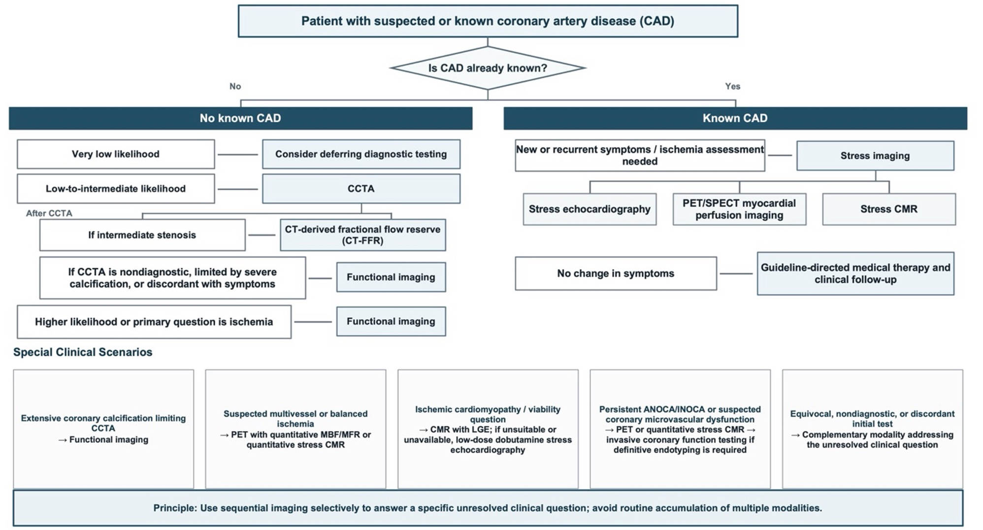
Figure 3. Question-Directed Clinical Decision Framework for Noninvasive Imaging in CAD. The flowchart summarizes a question-directed approach to noninvasive imaging according to the presence of known CAD, clinical likelihood of obstructive disease, the primary diagnostic question, and patient-specific factors affecting test selection. Sequential testing should be used selectively when the initial examination leaves a clinically relevant anatomic or physiologic question unresolved. ANOCA/INOCA: Angina with no obstructive coronary arteries/ischemia with no obstructive coronary arteries; CAD: Coronary artery disease; CCTA: coronary computed tomography angiography; CMD: coronary microvascular dysfunction; CMR: cardiac magnetic resonance; CT-FFR: computed tomography-derived fractional flow reserve; ICA: invasive coronary angiography; LGE: late gadolinium enhancement; MBF: myocardial blood flow; MFR: myocardial flow reserve; PET: positron emission tomography; SPECT: single-photon emission computed tomography.
Question-directed sequential imaging framework for CAD
| Clinical scenario | Suggested approach | Why/Supporting references |
| No known CAD, very low likelihood | Consider deferring diagnostic testing; reassess if symptoms or clinical likelihood change | Avoids low-yield testing in very-low-likelihood patients; consistent with the 2024 ESC risk-based approach[2] |
| No known CAD, low-to-intermediate likelihood | CCTA as the initial test; if an intermediate stenosis is identified, consider CT-FFR; if CCTA is nondiagnostic or discordant with symptoms, consider functional imaging | CCTA is effective for excluding obstructive CAD and characterizing coronary atherosclerosis; CT-FFR adds lesion-specific physiologic information when anatomic significance is uncertain[1,2,7,20] |
| Known CAD with new or recurrent symptoms | Stress imaging with stress echocardiography, PET/SPECT, or stress CMR according to patient characteristics and local expertise; consider ICA when high-risk findings or persistent symptoms warrant invasive assessment | The principal question commonly shifts from detection of atherosclerosis to assessment of inducible ischemia and its potential management implications[16,41] |
| Extensive coronary calcification limiting CCTA interpretation | Prefer functional imaging when the principal question is inducible ischemia | Extensive calcification may reduce CCTA interpretability and overestimate stenosis severity[2,16,52,53] |
| Intermediate stenosis on CCTA | CT-FFR for lesion-specific physiologic assessment; consider stress imaging or ICA if CT-FFR is unavailable, nondiagnostic, borderline, or discordant with the clinical presentation | Provides physiologic information without an additional image acquisition and may reduce low-yield invasive angiography[1,7,20] |
| Suspected multivessel or balanced ischemia | PET with quantitative MBF/MFR or quantitative stress CMR | Quantitative perfusion can identify globally or diffusely impaired perfusion that may be underestimated by relative perfusion imaging[9,37,47] |
| Ischemic cardiomyopathy or viability question | CMR with LGE; consider low-dose dobutamine stress echocardiography when CMR is unsuitable or unavailable | CMR provides integrated assessment of ventricular function, scar, and viability; dobutamine stress echocardiography can assess contractile reserve[8,10,28,29,46] |
| Persistent ANOCA/INOCA or suspected CMD | PET or quantitative stress CMR to assess myocardial flow/perfusion reserve; proceed to invasive coronary function testing when definitive endotyping or assessment for vasospasm is required | PET/CMR assess impaired vasodilator reserve but do not directly distinguish structural CMD from microvascular or epicardial vasospasm; invasive testing provides mechanistic endotyping[32-36] |
| Patient able to exercise and functional assessment is indicated | Prefer exercise-based stress imaging when feasible | Adds information on exercise capacity, symptom reproduction, heart-rate response, and hemodynamics[8,28,29] |
| Patient unable to exercise adequately | Use pharmacologic stress imaging, selected according to the clinical question, contraindications, and local expertise | Avoids a submaximal exercise study and permits functional assessment when exercise is not feasible[8,28,29] |
| Equivocal, nondiagnostic, or discordant initial test | Select a complementary modality that addresses the specific unresolved question; avoid additional testing when the result is unlikely to alter management | Sequential testing should resolve a clinically relevant anatomic or physiologic uncertainty rather than routinely accumulate multiple modalities[1-3,16] |
LIMITATIONS AND CHALLENGES
The limitations of noninvasive imaging extend beyond modality-specific technical constraints, which are summarized in Table 2, and include patient suitability, radiation or contrast exposure, cost, accessibility, and local expertise[16,52,53]. These factors should be considered when selecting the initial test and when determining whether additional imaging is likely to provide clinically meaningful information.
Discordance between anatomic stenosis severity and physiologic significance remains an important challenge, particularly for intermediate lesions, and imaging findings should therefore be interpreted within the broader clinical context[1]. Radiation exposure, contrast use, cost, and accessibility also influence test selection. Although contemporary acquisition protocols have reduced radiation dose for CCTA and nuclear imaging, cumulative exposure remains relevant when patients undergo repeated or sequential testing. In addition, iodinated contrast for CCTA and gadolinium-based contrast for CMR may be problematic in selected patients. Sequential multimodality testing can improve diagnostic confidence but may also increase patient burden, health-care costs, contrast exposure, and logistical complexity. Therefore, imaging pathways should aim to maximize diagnostic yield while avoiding unnecessary downstream testing[55,56]. Access to advanced imaging remains uneven across practice settings. PET, stress CMR, CT-FFR, and hybrid imaging require specialized infrastructure, trained personnel, and standardized protocols, which may limit their use in community, rural, or lower-resource environments. These differences in availability can contribute to variability in CAD diagnosis, risk stratification, and management, even when guideline recommendations support a given modality.
Environmental impact is also an emerging consideration, supporting more judicious and sustainable imaging strategies that avoid unnecessary testing while preserving diagnostic accuracy and patient safety[57].
FUTURE PERSPECTIVES
Emerging cardiovascular imaging technologies vary substantially in their level of validation and clinical readiness. Technical feasibility, association with prognosis, and incremental diagnostic information should not be interpreted as equivalent to demonstrated clinical utility or evidence that use of a technique improves patient management or outcomes. The technologies discussed below are therefore considered according to their current level of clinical implementation and supporting evidence. The future of imaging-guided decision-making in complex CAD is moving beyond stenosis-based diagnosis toward integrated, patient-specific atherosclerotic phenotyping. Artificial intelligence is likely to be a key enabling technology, with applications across patient selection, image acquisition, reconstruction, automated segmentation, stenosis quantification, plaque characterization, CT-FFR estimation, and prognostic modeling[23,58,59]. In CCTA, artificial intelligence-based tools may improve reproducibility and efficiency by automating plaque burden assessment, plaque composition analysis, calcium scoring, and CAD-RADS reporting. Machine learning and other automated CT-FFR approaches may enable faster on-site physiologic assessment compared with workflows requiring external processing. Representative clinical studies illustrate these applications. In the TARGET randomized trial, an on-site machine-learning CT-FFR strategy in 1,216 patients with stable CAD and intermediate stenosis on CCTA reduced invasive coronary angiography that demonstrated no obstructive disease or did not lead to intervention, although major adverse cardiovascular events and symptoms were similar at 1 year[21]. In a separate multicenter validation study, a fully automated deep-learning system evaluated coronary stenosis and high-risk plaque on CCTA, achieving 93.5% per-vessel agreement within one CAD-RADS category for stenosis and demonstrating good discrimination for low-attenuation plaque, spotty calcification, and positive remodeling[60]. However, these technologies use different computational methods and should be evaluated according to their platform-specific validation, generalizability, workflow characteristics, and regulatory status rather than assumed to be interchangeable with established computational fluid dynamics-based methods[23,58]. However, current artificial intelligence applications require broader external validation, standardization across vendors and scanners, assessment of bias in diverse populations, and prospective evidence that artificial intelligence-guided imaging improves clinical outcomes rather than simply increasing diagnostic output[23,58,59]. Recent AHA and society of cardiovascular computed tomography statements similarly emphasize that artificial intelligence is advancing rapidly but must be implemented with attention to validation, workflow integration, regulation, and clinical utility.
Several emerging CT-based technologies may expand cardiovascular imaging capabilities, although their levels of clinical readiness differ. Photon-counting detector CT is already available at selected centers and provides improved spatial and spectral resolution, reduced electronic noise, and reduced blooming from densely calcified structures, with potential advantages for heavily calcified vessels, coronary stents, plaque assessment, and myocardial characterization[61]. Early clinical studies have begun to demonstrate these advantages in practice. In a single-center observational cohort of 7,833 patients undergoing clinically indicated CCTA, photon-counting detector CT was associated with fewer subsequent referrals for invasive coronary angiography than conventional energy-integrating detector CT (9.9% vs. 13.1%) and demonstrated higher specificity and diagnostic accuracy for obstructive CAD among patients who underwent invasive angiography[62]. In another study of 100 patients undergoing both photon-counting CCTA and invasive coronary angiography, ultra-high-resolution photon-counting CCTA demonstrated excellent diagnostic performance for ≥ 50% coronary stenosis (Area under the curve 0.99) and improved luminal assessment compared with standard-resolution photon-counting imaging, particularly in severely calcified vessels[63]. These data provide early clinical support for photon-counting CT beyond technical feasibility, although multicenter outcome studies, protocol standardization, and cost-effectiveness analyses remain necessary before broader routine adoption. CT-derived extracellular volume fraction has demonstrated technical feasibility for quantifying myocardial extracellular volume and fibrosis and may be useful when CMR is unavailable or when CT is already being performed; however, its role in routine CAD pathways remains investigational and requires further protocol standardization, validation, and outcome-based evidence[64].
Other emerging approaches remain predominantly investigational. 18F-sodium fluoride PET can identify active coronary microcalcification and has demonstrated associations with high-risk plaque biology and subsequent cardiovascular events, but its ability to guide treatment decisions or improve clinical outcomes has not been established[65,66]. Coronary radiomics can extract high-dimensional plaque features not captured by conventional visual assessment, and radiomic phenotypes have shown prognostic associations, including in SCOT-HEART; however, reproducibility, external validation, and evidence of incremental clinical utility remain important barriers to routine implementation[67,68]. Hybrid PET/MR can integrate molecular, perfusion, ventricular, and tissue-characterization information, but its use in CAD remains restricted to specialized centers and lacks evidence supporting routine management decisions[40]. Digital twins and patient-specific computational models remain at an earlier conceptual or proof-of-concept stage, aiming to integrate imaging, clinical data, hemodynamics, and longitudinal information to simulate disease progression and treatment response[69].
Overall, the field is shifting toward precision imaging, integrating plaque burden, plaque composition, ischemia, inflammation, myocardial fibrosis, and patient-level risk to guide preventive therapy, revascularization decisions, and future drug-development trials[3,58,70].
Clinical-readiness categories: Clinically available indicates established incorporation into routine clinical pathways; selectively available indicates technology that can be implemented in specialized centers but is not broadly adopted or guideline-standard; investigational indicates human validation studies without an established routine clinical role; and conceptual indicates predominantly proof-of-concept or early translational development. Established modalities already summarized in Table 2 are not repeated here.
CONCLUSION
Noninvasive imaging in CAD should be selected according to the specific diagnostic or management question rather than through routine accumulation of multiple modalities. The available randomized evidence supports the use of specific imaging-guided strategies to improve diagnostic efficiency, better characterize or exclude obstructive CAD, reduce unnecessary invasive angiography and procedure-related complications, and, in some settings, influence preventive therapy and revascularization decisions. However, effects on symptoms and hard cardiovascular outcomes have been heterogeneous, and the evidence does not support routine use of multiple imaging modalities in every patient.
Accordingly, multimodality imaging should be applied selectively and, when necessary, sequentially, with each test chosen to answer a specific unresolved clinical question while avoiding redundant testing. The question-directed framework and visual flowchart proposed in this review are intended to support practical test selection by linking specific clinical scenarios to the modality most likely to provide clinically meaningful information. Future research should determine whether emerging imaging biomarkers and technologies provide incremental clinical utility, improve patient-important outcomes, and can be incorporated into efficient and cost-effective diagnostic pathways.
DECLARATIONS
Authors’ contributions
Conceptualization, investigation, writing the original draft, and review and editing: Hoyek K
Conceptualization, investigation, writing the original draft: Bedran A, Haroun E, Hajj J, Zalaquett Z, Agrawal A, Arockiam DA
conceptualization, supervision, writing - review and editing: Tan C, Wang TKM
All authors reviewed and approved the final manuscript.
Availability of data and materials
Not applicable.
AI and AI-assisted tools statement
During the preparation and revision of this manuscript, the authors used OpenAI ChatGPT (GPT-5.6, released on 07/09/2026) for language editing, improving clarity and organization, and assisting with editorial revision. Anthropic Claude (Opus-5.5, released on 09/22/2026) was used to generate the graphical abstract based on author-provided prompts and scientific content. The authors reviewed and revised all AI-assisted content and take full responsibility for the accuracy, integrity, interpretation, and final content of the manuscript. Generative AI was not used to generate original study data or perform primary data analysis.
Conflicts of interest
All authors declared that there are no conflicts of interest.
Financial support and sponsorship
None.
Ethical approval and consent to participate
Not applicable.
Consent for publication
Not applicable.
Copyright
© The Author(s) 2026.
REFERENCES
1. Gulati M, Levy PD, Mukherjee D, et al. 2021 AHA/ACC/ASE/CHEST/SAEM/SCCT/SCMR guideline for the evaluation and diagnosis of chest pain: a report of the american college of Cardiology/American Heart Association Joint Committee on clinical practice guidelines. Circulation. 2021;144:E368-454.
2. Vrints C, Andreotti F, Koskinas KC, et al. 2024 ESC Guidelines for the management of chronic coronary syndromes: Developed by the task force for the management of chronic coronary syndromes of the European Society of Cardiology (ESC) Endorsed by the European Association for Cardio-Thoracic Surgery (EACTS). Eur Heart J. 2024;45:3415-537.
3. Edvardsen T, Asch FM, Davidson B, et al. Non-invasive imaging in coronary syndromes: recommendations of The European Association of Cardiovascular Imaging and the American Society of Echocardiography, in collaboration with The American Society of nuclear cardiology, society of cardiovascular computed tomography, and society for cardiovascular magnetic resonance. J Am Soc Echocardiogr. 2022;35:329-54.
4. Serruys PW, Hara H, Garg S, et al. Coronary computed tomographic angiography for complete assessment of coronary artery disease: JACC state-of-the-art review. J Am Coll Cardiol. 2021;78:713-36.
5. Cury RC, Leipsic J, Abbara S, et al. CAD-RADS™ 2.0 - 2022 coronary artery disease-reporting and data system: an expert consensus document of the Society of Cardiovascular Computed Tomography (SCCT), the American College of Cardiology (ACC), the American College of Radiology (ACR), and the North America Society of Cardiovascular Imaging (NASCI). JACC Cardiovasc Imaging. 2022;15:1974-2001.
6. Williams MC, Kwiecinski J, Doris M, et al. Low-attenuation noncalcified plaque on coronary computed tomography angiography predicts myocardial infarction: results from the multicenter SCOT-HEART trial (scottish computed tomography of the HEART). Circulation. 2020;141:1452-62.
7. Tang CX, Leipsic JA, Nørgaard BL, et al. Clinical use of coronary computed tomography angiography-derived fractional flow reserve: expert consensus by an International Working Group. Eur Radiol. 2026;36:5303-16.
8. Picano E, Pierard L, Peteiro J, et al. The clinical use of stress echocardiography in chronic coronary syndromes and beyond coronary artery disease: a clinical consensus statement from the European Association of Cardiovascular Imaging of the ESC. Eur Heart J Cardiovasc Imaging. 2024;25:e65-90.
9. Vohra S, Sakul S, Agarwal V, Patel KK. Quantification of myocardial blood flow using PET: Current status, clinical applications, and future directions. Prog Cardiovasc Dis. 2025;93:10-20.
10. Patel AR, Salerno M, Kwong RY, Singh A, Heydari B, Kramer CM. Stress cardiac magnetic resonance myocardial perfusion imaging : JACC review topic of the week. J Am Coll Cardiol. 2021;78:1655-68.
11. Nagel E, Greenwood JP, Mccann GP, et al. Magnetic resonance perfusion or fractional flow reserve in coronary disease. N Engl J Med. 2019;380:2418-28.
12. Douglas PS, Hoffmann U, Patel MR, et al. Outcomes of anatomical versus functional testing for coronary artery disease. N Engl J Med. 2015;372:1291-300.
13. The DISCHARGE Trial Group. CT or invasive coronary angiography in stable chest pain. N Engl J Med. 2022;386:1591-602.
14. Douglas PS, Nanna MG, Kelsey MD, et al. Comparison of an initial risk-based testing strategy vs usual testing in stable symptomatic patients with suspected coronary artery disease: the PRECISE randomized clinical trial. JAMA Cardiol. 2023;8:904.
15. Williams MC, Wereski R, Tuck C, et al. Coronary CT angiography-guided management of patients with stable chest pain: 10-year outcomes from the SCOT-HEART randomised controlled trial in Scotland. Lancet. 2025;405:329-37.
16. Winchester DE, Maron DJ, Blankstein R, et al. ACC/AHA/ASE/ASNC/ASPC/HFSA/HRS/SCAI/SCCT/SCMR/STS 2023 multimodality appropriate use criteria for the detection and risk assessment of chronic coronary disease. J Am Coll Cardiol. 2023;81:2445-67.
17. Ferencik M, Mayrhofer T, Bittner DO, et al. Use of high-risk coronary atherosclerotic plaque detection for risk stratification of patients with stable chest pain: a secondary analysis of the PROMISE randomized clinical trial. JAMA Cardiol. 2018;3:144.
18. Osawa K, Nakanishi R, Budoff M. Coronary artery calcification; Report from the multi-ethnic study of atherosclerosis. Glob Heart. 2016;11:287.
19. Otaki Y, Arsanjani R, Gransar H, et al. What have we learned from CONFIRM? Prognostic implications from a prospective multicenter international observational cohort study of consecutive patients undergoing coronary computed tomographic angiography. J Nucl Cardiol. 2012;19:787-95.
20. Faulder TI, Prematunga K, Moloi SB, Faulder LE, Jones R, Moxon JV. Agreement of fractional flow reserve estimated by computed tomography with invasively measured fractional flow reserve: a systematic review and meta-analysis. J Am Heart Assoc. 2024;13:e034552.
21. Yang J, Shan D, Wang X, et al. On-site computed tomography-derived fractional flow reserve to guide management of patients with stable coronary artery disease: the TARGET randomized trial. Circulation. 2023;147:1369-81.
22. Guo B, Xing W, Hu C, et al. Clinical effectiveness of automated coronary CT-derived fractional flow reserve: a Chinese randomized controlled trial. Radiology. 2024;313:e233354.
23. Williams MC, Weir-Mccall JR, Baldassarre LA, et al. Artificial intelligence and machine learning for cardiovascular computed tomography (CCT): a white paper of the society of cardiovascular computed tomography (SCCT). J Cardiovasc Comput Tomogr. 2024;18:519-32.
24. Sagoo NS, Sagoo R, Legate J, Sathyamoorthy M. Pericoronary adipose tissue attenuation on CCTA as a marker of cardiovascular risk: a systematic review and meta-analysis. Eur J Radiol. 2026;195:112607.
25. Kato Y, Chehab O, Ambale-Venkatesh B, Lima JAC. Quantitative myocardial perfusion imaging across PET, SPECT, CMR, and CT. Front Radiol. 2026;6:1760241.
26. Sliwicka O, Sechopoulos I, Baggiano A, Pontone G, Nijveldt R, Habets J. Dynamic myocardial CT perfusion imaging—state of the art. Eur Radiol. 2023;33:5509-25.
27. Woodward W, Johnson CL, Krasner S, et al. Long-term outcomes after stress echocardiography in real-world practice: a 5-year follow-up of the UK EVAREST study. Eur Heart J Cardiovasc Imaging. 2025;26:187-96.
28. Zhang P, Tian J, Zhu T, et al. Stress echocardiography for chronic coronary syndrome: clinical practice guidelines (2023). J Geriatr Cardiol. 2024;21:475-505.
29. Haddad K, Ionescu AL, Pilehvar R, et al. Improving stress echocardiography for enhanced detection of left main and multivessel coronary artery disease. CJC Open. 2026;8:93-102.
30. Zivanic A, Stankovic I, Vranic Jovanovic I, et al. The role of post-systolic strain and electrocardiographic changes during dobutamine stress echocardiography in enhancing detection of symptomatic coronary artery disease. Front Cardiovasc Med. 2025;12:1641044.
31. Tjellaug Bråten A, Holte E, Wiseth R, Aakhus S. Dobutamine stress echocardiography after positive CCTA: diagnostic performance using fractional flow reserve and instantaneous wave-free ratio as reference standards. Open Heart. 2024;11:e002899.
32. Kunadian V, Chieffo A, Camici PG, et al. An EAPCI expert consensus document on ischaemia with non-obstructive coronary arteries in collaboration with european society of cardiology working group on coronary pathophysiology & microcirculation endorsed by coronary vasomotor disorders international study group. Eur Heart J. 2020;41:3504-20.
33. Murthy VL, Bateman TM, Beanlands RS, et al. Clinical quantification of myocardial blood flow using PET: joint position paper of the SNMMI cardiovascular council and the ASNC. J Nucl Med. 2018;59:273-93.
34. Taqueti VR, Hachamovitch R, Murthy VL, et al. Global coronary flow reserve is associated with adverse cardiovascular events independently of luminal angiographic severity and modifies the effect of early revascularization. Circulation. 2015;131:19-27.
35. Zhou W, Lee JCY, Leung ST, et al. Long-term prognosis of patients with coronary microvascular disease using stress perfusion cardiac magnetic resonance. JACC Cardiovasc Imaging. 2021;14:602-11.
36. Ford TJ, Stanley B, Good R, et al. Stratified medical therapy using invasive coronary function testing in angina. J Am Coll Cardiol. 2018;72:2841-55.
37. Alwan M, El Ghazawi A, El Yaman A, Al Rifai M, Al-Mallah MH. SPECT myocardial perfusion imaging in the era of PET and multimodality imaging: challenges and opportunities. Prog Cardiovasc Dis. 2025;93:3-9.
38. Dahdal J, Jukema RA, Harms HJ, et al. PET myocardial perfusion imaging: trends, challenges, and opportunities. J Nucl Cardiol. 2024;40:102011.
39. Veneziano FA, Gioia FA, Gentile F. Hybrid PET/CT and PET/MR in coronary artery disease: an update for clinicians, with insights into AI-guided integration. J Cardiovasc Dev Dis. 2025;12:338.
40. El Yaman A, Alwan M, El Ghazawi A, Al-Mallah MH, Al Rifai M. Advances in nuclear hybrid imaging: the role of SPECT/CT, PET/CT, and PET/MRI in cardiovascular diseases. Prog Cardiovasc Dis. 2025;93:43-50.
41. Virani SS, Newby LK, Arnold SV, et al. 2023 AHA/ACC/ACCP/ASPC/NLA/PCNA guideline for the management of patients with chronic coronary disease : a report of the American Heart Association/American College of Cardiology Joint Committee on clinical practice guidelines. J Am Coll Cardiol. 2023;82:833-955.
42. Litmanovich D, Hurwitz Kowee LM, Ghoshhajra BB, et al. ACR appropriateness criteria® chronic chest pain-high probability of coronary artery disease: 2021 update. J Am Coll Radiol. 2022;19:S1-18.
43. Ricci F, Khanji MY, Bisaccia G, et al. Diagnostic and prognostic value of stress cardiovascular magnetic resonance imaging in patients with known or suspected coronary artery disease: a systematic review and meta-analysis. JAMA Cardiol. 2023;8:662.
44. Greenwood JP, Maredia N, Younger JF, et al. Cardiovascular magnetic resonance and single-photon emission computed tomography for diagnosis of coronary heart disease (CE-MARC): a prospective trial. Lancet. 2012;379:453-60.
45. Greenwood JP, Ripley DP, Berry C, et al. Effect of care guided by cardiovascular magnetic resonance, myocardial perfusion scintigraphy, or NICE guidelines on subsequent unnecessary angiography rates: the CE-MARC 2 randomized clinical trial. JAMA. 2016;316:1051.
46. Garcia MJ, Kwong RY, Scherrer-Crosbie M, et al. State of the art: imaging for myocardial viability: a scientific statement from the American Heart Association. Circ Cardiovasc Imaging. 2020;13:e000053.
47. Wang S, Kim P, Wang H, et al. Myocardial blood flow quantification using stress cardiac magnetic resonance improves detection of coronary artery disease. JACC Cardiovasc Imaging. 2024;17:1428-41.
48. Narula J, Chandrashekhar Y, Ahmadi A, et al. SCCT 2021 expert consensus document on coronary computed tomographic angiography: a report of the society of cardiovascular computed tomography. J Cardiovasc Comput Tomogr. 2021;15:192-217.
49. Curzen N, Nicholas Z, Stuart B, et al. Fractional flow reserve derived from computed tomography coronary angiography in the assessment and management of stable chest pain: the FORECAST randomized trial. Eur Heart J. 2021;42:3844-52.
50. Knuuti J, Wijns W, Saraste A, et al. 2019 ESC guidelines for the diagnosis and management of chronic coronary syndromes. Eur Heart J. 2020;41:407-77.
51. National Institute for Health and Care Excellence (NICE). Recent-onset chest pain of suspected cardiac origin: assessment and diagnosis. Available from: https://www.nice.org.uk/guidance/cg95 [Last accessed on 6 Nov 2026].
52. Bax JJ, Di Carli M, Narula J, Delgado V. Multimodality imaging in ischaemic heart failure. Lancet. 2019;393:1056-70.
53. Baessato F, Guglielmo M, Muscogiuri G, et al. Stress CMR in known or suspected CAD: diagnostic and prognostic role. BioMed Res Int. 2021;2021:6678029.
54. Neglia D, Liga R, Gimelli A, et al. Use of cardiac imaging in chronic coronary syndromes: the EURECA Imaging registry. Eur Heart J. 2023;44:142-58.
55. Hirshfeld JW, Ferrari VA, Bengel FM, et al. 2018 ACC/HRS/NASCI/SCAI/SCCT expert consensus document on optimal use of ionizing radiation in cardiovascular imaging—best practices for safety and effectiveness, Part 1: radiation physics and radiation biology: A report of the American College of Cardiology Task Force on expert consensus decision pathways. J Am Coll Cardiol. 2018;71:2811-28.
56. Einstein AJ, Williams MC, Weir-Mccall JR, et al. Worldwide radiation dose in coronary artery disease diagnostic imaging. JAMA. 2026;335:1148.
57. Picano E. Economic, ethical, and environmental sustainability of cardiac imaging. Eur Heart J. 2023;44:4748-51.
58. Van Der Bijl P, Gulati M, Saraste A, et al. Contemporary, non-invasive imaging diagnosis of chronic coronary artery disease. Lancet. 2025;406:2577-87.
59. Hanneman K, Playford D, Dey D, et al. Value creation through artificial intelligence and cardiovascular imaging: a scientific statement from the American Heart Association. Circulation. 2024;149:E296-311.
60. Ihdayhid AR, Sehly A, He A, et al. Coronary artery stenosis and high-risk plaque assessed with an unsupervised fully automated deep learning technique. JACC Adv. 2024;3:100861.
61. Shiyovich A, Singh A, Blair CV, et al. Photon-counting computed tomography in cardiac imaging. JACC Cardiovasc Imaging. 2026;19:94-117.
62. Sakai K, Shin D, Singh M, et al. Diagnostic performance and clinical impact of photon-counting detector computed tomography in coronary artery disease. JACC. 2025;85:339-48.
63. Kotronias RA, De Maria GL, Xie C, et al. Benchmarking photon-counting computed tomography angiography against invasive assessment of coronary stenosis: implications for severely calcified coronaries. JACC Cardiovasc Imaging. 2025;18:572-85.
64. Han D, Lin A, Kuronuma K, et al. Cardiac Computed tomography for quantification of myocardial extracellular volume fraction: a systematic review and meta-analysis. JACC Cardiovasc Imaging. 2023;16:1306-17.
65. Joshi NV, Vesey AT, Williams MC, et al. 18F-fluoride positron emission tomography for identification of ruptured and high-risk coronary atherosclerotic plaques: a prospective clinical trial. Lancet. 2014;383:705-13.
66. Kwiecinski J, Tzolos E, Adamson PD, et al. Coronary 18F-sodium fluoride uptake predicts outcomes in patients with coronary artery disease. J Am Coll Cardiol. 2020;75:3061-74.
67. Lin A, Kolossváry M, Cadet S, et al. Radiomics-based precision phenotyping identifies unstable coronary plaques from computed tomography angiography. JACC Cardiovasc Imaging. 2022;15:859-71.
68. Kolossváry M, Lin A, Kwiecinski J, et al. Coronary plaque radiomic phenotypes predict fatal or nonfatal myocardial infarction. JACC Cardiovasc Imaging. 2025;18:308-19.
69. Thangaraj PM, Benson SH, Oikonomou EK, Asselbergs FW, Khera R. Cardiovascular care with digital twin technology in the era of generative artificial intelligence. Eur Heart J. 2024;45:4808-21.
Cite This Article
 Open Access
How to Cite
Hoyek K, Bedran A, Haroun E, Hajj J, Zalaquett Z, Agrawal A, Arockiam DA, Tan C, Wang TKM. Multimodality noninvasive imaging in the evaluation of coronary artery disease: a narrative review. Vessel Plus. 2026;10:58. https://dx.doi.org/10.20517/2574-1209.2026.55
Download Citation
If you have the appropriate software installed, you can download article citation data to the citation manager of your choice. Simply select your manager software from the list below and click on download.
Export Citation File
Type of Import
Tips on Downloading Citation
Citation Manager File Format
Type of Import
Direct Import: When the Direct Import option is selected (the default state), a dialogue box will give you the option to Save or Open the downloaded citation data. Choosing Open will either launch your citation manager or give you a choice of applications with which to use the metadata. The Save option saves the file locally for later use.
Indirect Import: When the Indirect Import option is selected, the metadata is displayed and may be copied and pasted as needed.
About This Article
Copyright

Data & Comments
Data
0









Comments
Comments must be written in English. Spam, offensive content, impersonation, and private information will not be permitted. If any comment is reported and identified as inappropriate content by OAE staff, the comment will be removed without notice. If you have any queries or need any help, please contact us at [email protected].