
fig1
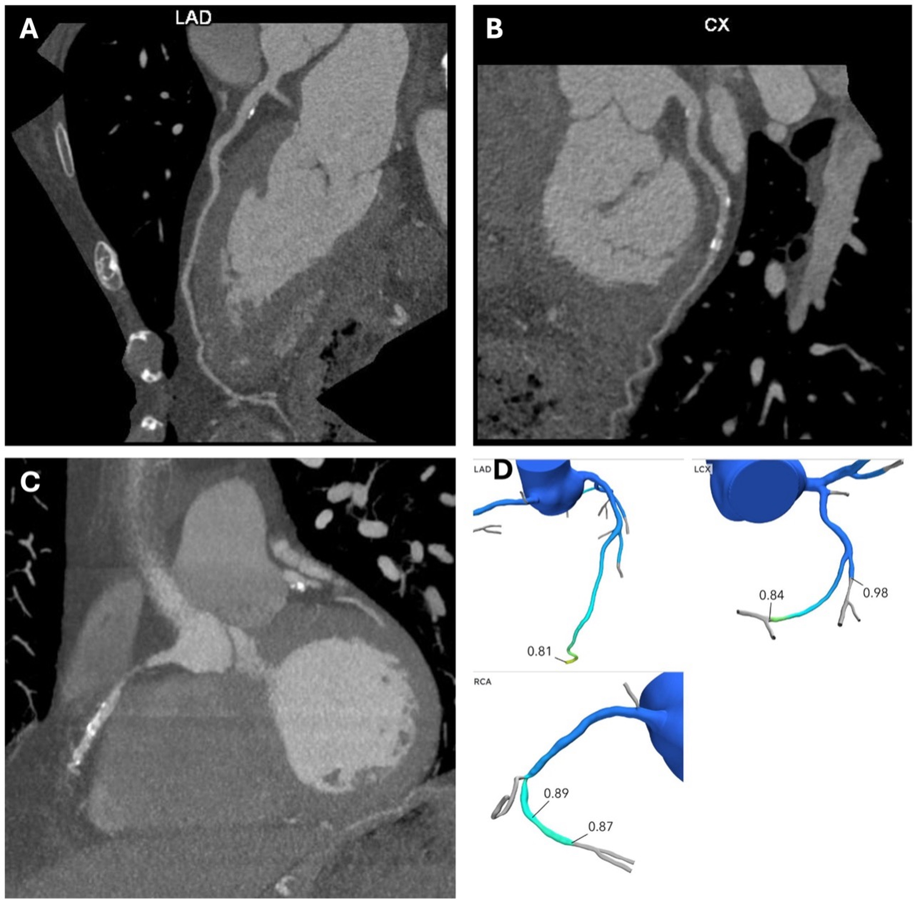
Figure 1. Coronary computed tomography angiography (CCTA) in a 72-year-old man presenting with exertional dyspnea and intermittent chest pain. (A) shows a curved planar reconstruction of the left anterior descending artery (LAD), demonstrating calcified plaque in the proximal LAD with minimal stenosis. (B) shows a curved planar reconstruction of the left circumflex artery (LCx), demonstrating calcified plaque in the proximal and mid LCx segments with mild stenosis. (C) shows a curved planar reconstruction of the right coronary artery (RCA), demonstrating calcified plaque in the mid RCA with moderate stenosis. (D) shows the CT-FFR analysis, with lesion-specific values > 0.80 measured distal to the corresponding stenoses, indicating no lesion-specific physiologic evidence of ischemia according to the CT-FFR assessment. The anatomic stenosis severity remains defined by the CCTA findings shown in (A-C).