
fig2
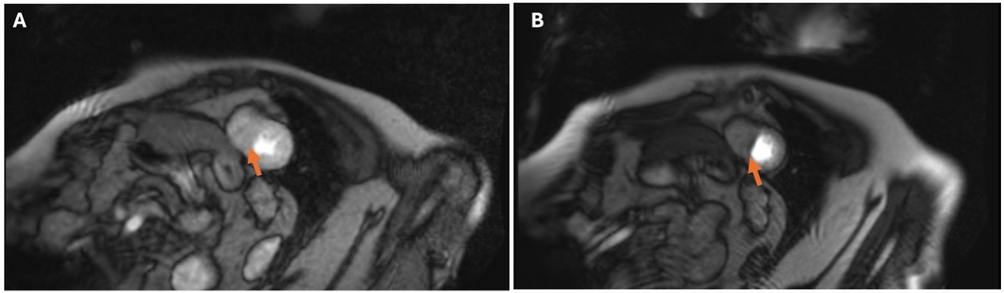
Figure 2. Stress CMR of a 64-year-old woman with exertional anginal symptoms despite no angiographic evidence of obstructive epicardial coronary artery disease. First-pass myocardial perfusion imaging was performed following intravenous administration of gadolinium-based contrast during pharmacologic stress, with resting perfusion images obtained for comparison. Adenosine was administered at 140 μg/kg/min for 2 min, with stress perfusion acquisition performed according to a protocol requiring an increase in heart rate of ≥ 15 beats/min. Perfusion assessment included evaluation of inducible and reversible versus persistent perfusion defects, the ratio of stress to resting myocardial blood flow (MBF), and the ratio of subendocardial to subepicardial stress MBF. (A) shows the resting perfusion image, with the orange arrow identifying the septal myocardial region that demonstrates no perfusion defect at rest. (B) shows the corresponding stress perfusion image, with the orange arrow indicating an inducible septal perfusion defect. In the absence of obstructive epicardial coronary artery disease, this finding may suggest impaired microvascular perfusion but does not independently establish coronary microvascular dysfunction; imaging artifacts and other potential causes should also be considered.