


Research progress on electrochemical immunosensors for the detection of cardiovascular biomarkers
 0
0 Abstract
Cardiovascular diseases (CVDs) are among the leading causes of global morbidity and mortality, placing a substantial burden on public health and socioeconomic development. Early and accurate diagnosis is essential for reducing CVDs mortality and optimizing individualized treatment strategies. Although conventional detection methods, such as enzymatic analysis and immunoassays, have reached a relative level of maturity in clinical applications, they are still constrained by high costs, complex procedures, and limited real-time capabilities. In recent years, electrochemical immunosensors have shown significant potential for detecting CVD-related biomarkers, owing to their advantages of high sensitivity, excellent selectivity, rapid response, and portability. In this review, we explore the latest advancements in electrochemical immunosensors for CVDs research, analyze innovative designs and fabrication techniques for various sensor types, and summarize their applications in detecting CVD-related biomarkers.
Keywords
INTRODUCTION
Cardiovascular diseases (CVDs) encompass a range of chronic conditions that impact the heart and vascular system, primarily including hypertension, atherosclerosis, and myocardial infarction. These diseases remain among the leading causes of death globally[1,2]. According to statistics from the World Heart Federation, CVDs are responsible for approximately 17.3 million deaths each year, accounting for about 32% of total global mortality. Projections indicate that by 2030, the number of deaths related to CVDs will rise to 23.6 million, thereby exacerbating the burden on public health systems[3]. Biomarkers play a crucial role in the early screening, auxiliary diagnosis, and risk assessment of CVDs[4]. These molecules, including proteins, nucleic acids, and various other bioactive substances[5], are primarily found in biological samples such as blood[6] and saliva[7]. Although traditional rapid analysis methods, such as Raman spectroscopy[8] and fluorescence detection[9], demonstrate commendable analytical performance under laboratory conditions, they still exhibit significant limitations in practical applications. Raman spectroscopy typically relies on high-precision optical equipment, requires prolonged acquisition times, and is susceptible to interference from complex biological matrices, thereby adversely affecting detection sensitivity and accuracy[10,11]. Fluorescence detection often depends on exogenous dyes or probes, increasing operational complexity and potentially compromising the reliability of detection results due to background noise and insufficient probe stability[12]. While traditional techniques such as liquid chromatography-selected reaction monitoring (LC-SRM)[13], enzyme-linked immunosorbent assay (ELISA)[14], and Western blot[15] can quantitatively analyze CVD-related biomarkers, they often involve cumbersome procedures and lack adequate real-time detection capabilities. Consequently, their use in rapid screening for critical and emergency care scenarios is restricted. Currently, assistive diagnostic technologies for CVDs have significant potential for improvement in sensitivity, timeliness, and clinical accessibility. Innovative research aimed at the early detection of CVD-related biomarkers will play a crucial role in advancing clinical diagnosis and treatment[16,17].
In recent years, with the continuous development of precision medicine, novel detection technologies targeting specific biomarkers have emerged[18,19]. Electrochemical immunosensors utilize the specific recognition between antigens and antibodies to convert biological recognition events into measurable electrochemical signals, thereby enabling the highly sensitive detection of biomarkers[20]. Compared with traditional methods, electrochemical immunosensors generally exhibit higher sensitivity and faster response times. They show promising potential for applications, particularly in trace analysis and the detection of complex biological samples[21], while addressing limitations associated with prolonged measurement times and insufficient dye stability. Additionally, electrochemical immunosensors provide significant advantages, including ease of operation, reduced costs, and the elimination of the need for expensive optical equipment. These features make them particularly valuable for clinical diagnostics and primary healthcare, especially in rapid screening situations during emergencies. Furthermore, these sensors can minimize the preprocessing steps necessary for complex biological sample testing, thereby enhancing detection efficiency and clinical usability. Electrochemical immunosensors can be categorized into two primary types based on their labeling strategies[22]: label-free and labeled immunosensors. Label-free electrochemical immunosensors do not require the introduction of exogenous labels, allowing for in situ dynamic monitoring of antigen-antibody binding processes. These sensors offer several advantages, including straightforward operation, rapid response times, and suitability for portable detection[23,24]. However, their performance may be constrained in high-sensitivity detection scenarios due to limited signal amplification, posing challenges for trace analysis and detection in complex matrices. In contrast, labeled electrochemical immunosensors significantly enhance detection sensitivity and reduce detection limits by incorporating labeled secondary antibodies or other signal amplification elements[25-27]. Overall, both types of electrochemical immunosensors have been extensively utilized in the detection of CVD-related biomarkers.
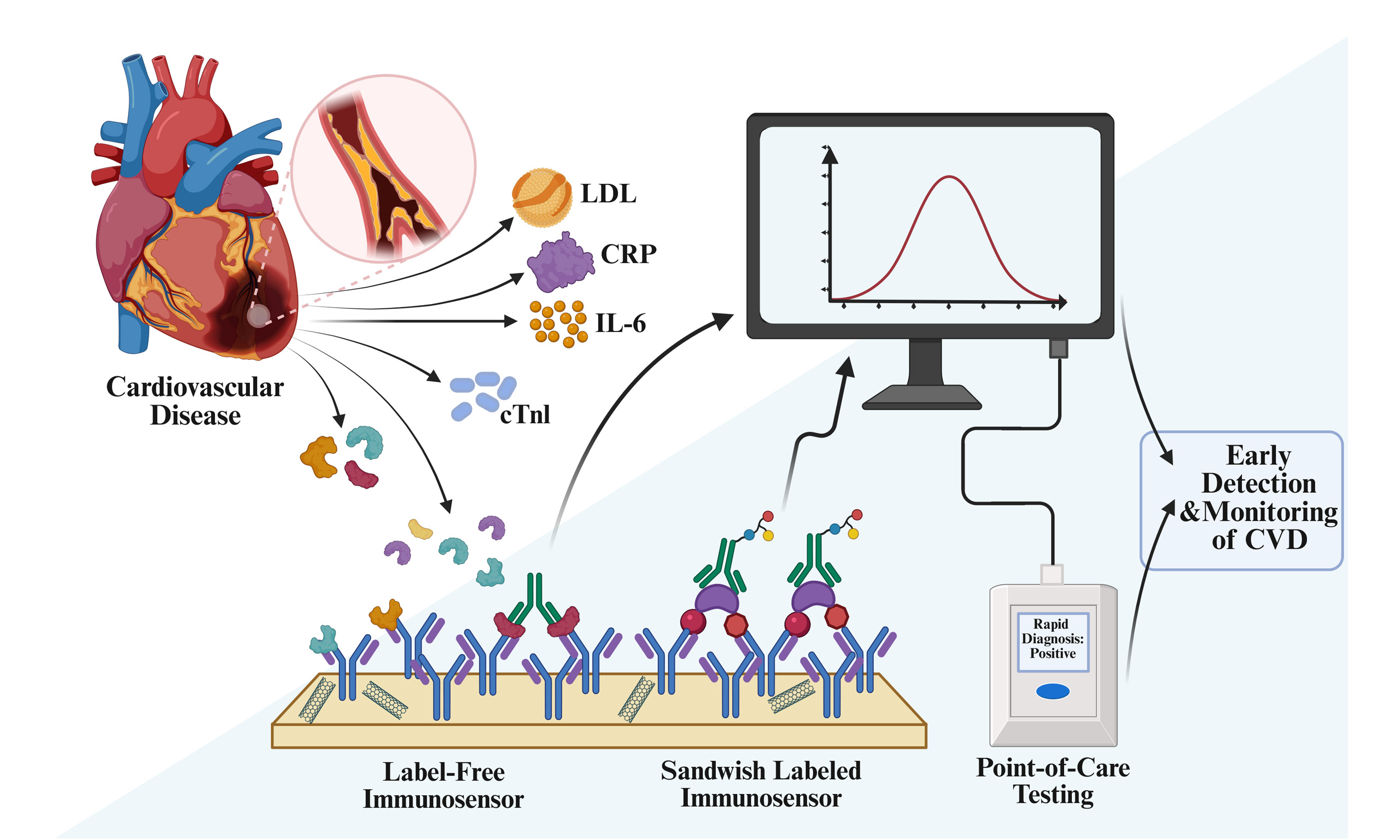
In this review, we systematically summarize the latest advances in electrochemical immunosensors for the detection of CVD-related biomarkers. The article outlines core strategies for nanomaterial modification and signal amplification, while summarizing their application characteristics and clinical value across various CVDs [Figure 1]. In particular, it provides an in-depth analysis of the current bottlenecks hindering clinical translation and explores future development directions. Finally, this review provides a solid theoretical foundation and practical guidance for subsequent research and clinical applications in this field.
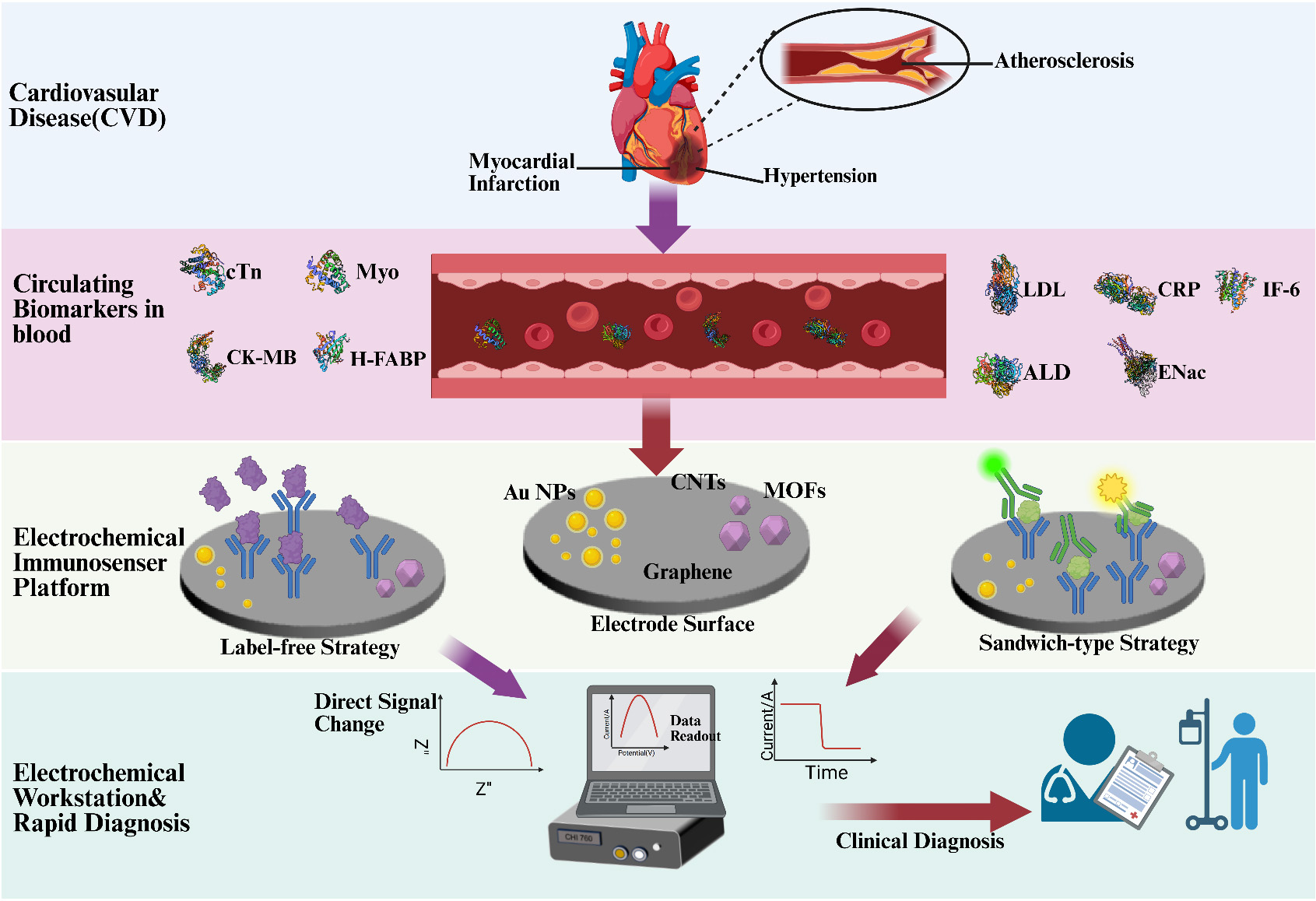
Figure 1. Electrochemical Immunosensors for Detecting CVD-related biomarkers. Created with BioRender. Wang S (2026) https://BioRender.com/mofcq6e. CVD: Cardiovascular disease; cTn: cardiac troponin; Myo: myoglobin; H-FABP: heart-type fatty acid-binding protein; LDL: low-density lipoprotein; CRP: C-reactive protein; IL-6: interleukin-6; ALD: aldosterone; ENac: epithelial sodium channel; CNTs: carbon nanotubes; MOFs: metal-organic frameworks; CK-MB: creatine Kinase-MB; AuNPs: gold nanoparticles.
ELECTROCHEMICAL IMMUNOSENSORS
An electrochemical immunosensor is a type of biosensor that integrates electrochemical detection technology with immunological reactions. Its principle is based on the specific recognition between antigens and antibodies, which converts the concentration of the analytes into detectable electrochemical signals, thereby facilitating the qualitative or quantitative detection of the target analyte[24,28]. Compared with other electrochemical biosensors, electrochemical immunosensors demonstrate higher selectivity and sensitivity towards target biomarkers. Consequently, these sensors can achieve efficient and accurate detection even in complex matrices such as blood[29]. Capitalizing on these advantages, electrochemical immunosensors have found widespread application in the detection of CVD-related biomarkers. Based on the use of labels to assist signal detection, electrochemical immunosensors can be classified into label-free and labeled types.
Label-free electrochemical immunosensors
Label-free electrochemical immunosensors (LFEIs) detect target analytes through electrochemical signal changes induced by specific antigen-antibody binding, thus eliminating the need for auxiliary labels such as enzymes, fluorescent probes, or nanomarkers[30,31]. This “label-free” approach significantly simplifies sensor preparation and operational procedures while enhancing detection efficiency and reducing costs. Additionally, it mitigates potential instability introduced by labeling substances. In recent years, this technology has made remarkable progress in laboratory diagnostics and point-of-care testing (POCT) applications[32-34].
In the realm of materials, metallic nanomaterials and carbon nanotubes are widely used to enhance the conductivity at electrode interfaces and the loading capacity for biomolecules. This enhancement leads to lower detection limits and improved sensitivity in various applications[35-37]. Oliveira et al. developed a LFEI that employed gold nanoparticle modification for the detection of low-density lipoprotein (LDL)[38]. By incorporating gold nanoparticles, the research team significantly increased the specific surface area of the electrode substrate, thereby enhancing the loading capacity of anti-LDL monoclonal antibodies and providing more antigen-antibody binding sites. In terms of applications, LFEIs are widely used for the detection of clinical biomarkers, such as LDL and C-reactive protein (CRP), which aid in the diagnosis of CVDs. Guillem et al. developed a LFEI utilizing screen-printed electrodes for the quantitative detection of CRP[34]. This label-free approach simplifies the detection process into three steps: sample addition, incubation, and electrochemical detection, significantly reducing the time required for a single test. Furthermore, the absence of labels considerably lowers the fabrication costs of the sensor. These advantages render LFEIs highly applicable in clinical biomarker detection, particularly in the realm of POCT.
Labeled electrochemical immunosensors
Labeled electrochemical immunosensors (LEIs) enhance the electrochemical signals produced by antigen-antibody interactions through the introduction of specific labels, such as enzymes, conjugated polymers, and noble metal nanoparticles. When applied to CVD-related biomarker detection, these sensors typically utilize a sandwich configuration[39,40]. In comparison to label-free sensors, the inclusion of specific labels significantly amplifies the signals, thereby improving sensitivity and selectivity. This approach provides substantial advantages for accurate detection in laboratory environments.
Enzyme-LEIs represent the most prevalent category of LEIs. Martins et al. developed a LEI utilizing an Au-Reduced Graphene Oxide (rGO) composite material[41]. During the immunorecognition process, the target CA15-3 first binds specifically to Ab1, which is immobilized on the electrode surface. Subsequently, it combines with horseradish peroxidase (HRP)-labeled Ab2 to form a stable Ab1-Ag-Ab2-HRP sandwich complex. In the detection phase, the enzyme-catalyzed reaction generates a stable current signal, enabling precise detection of the biomarker. Noble metal nanoparticles are important signal amplification materials that are widely employed in the construction of LEIs. Awan et al. utilized silver nanoparticles (AgNPs) conjugated with Ab2 as signal labels. Following the formation of the Ab1-Ag-Ab2 complex, quantitative analysis conducted through the oxidation current response of Ag0[42]. The incorporation of AgNPs significantly improved the sensor’s detection performance. Conjugated polymers exhibit promising application prospects due to their advantages, including facile preparation processes and high electrical conductivity. Song et al. developed a CPS@PANI@Au-BA (Carboxylated Polystyrene Spheres@ polyaniline@Au-BA) probe that utilizes PANI as the carrier for labeling secondary antibodies and constructed a LEI based on conductive conjugated polymer-supported gold nanoparticles[43]. This sensor demonstrated an ultra-low detection limit of 1.56 pg/mL, along with excellent selectivity, reproducibility, and long-term stability.
ELECTROCHEMICAL IMMUNOSENSORS FOR CARDIOVASCULAR DISEASES
CVDs represent a significant global public health challenge, contributing to both mortality and disability. These diseases encompass a range of pathological conditions that affect the heart and vascular system[44]. CVDs account for over one-third of total global deaths, and their morbidity and mortality risks continue to rise, particularly in aging populations and those with a high prevalence of metabolic syndrome[3]. Among the various types of CVDs, hypertension[45], atherosclerosis[46], and myocardial infarction[47] are the most prevalent and life-threatening conditions.
Atherosclerosis
Atherosclerosis (AS) is the primary pathological basis for various CVDs[48,49]. Biomarkers such as LDL[50], CRP[51,52], and Interleukin-6 (IL-6)[53] reflect different disease stages of AS, and their levels are closely correlated with lesion progression. Therefore, the accurate detection of these markers facilitates the early identification of AS at the subclinical stage and provides crucial evidence for personalized prevention, diagnosis, and intervention[54]. Novel electrochemical immunosensor-based detection technologies can offer technical support for the highly sensitive and rapid monitoring of these biomarkers, potentially advancing the development of early screening, diagnosis, and comprehensive health management for AS.
LDL/ox-LDL
LDL and its oxidized form (ox-LDL) are critical in the initiation and progression of AS. LDL tends to accumulate beneath the vascular endothelium, where it undergoes further oxidation to form ox-LDL. The latter induces endothelial dysfunction, inflammatory responses, and immune activation. Additionally, ox-LDL can be internalized by macrophages via scavenger receptors, such as CD36, which promotes foam cell formation and further contributes to the development of atherosclerotic plaques[50,55]. Therefore, monitoring and effectively controlling serum LDL levels is of paramount importance for the prevention and treatment of AS.
Traditional LFEIs often exhibit insufficient signal sensitivity, which restricts their application in LDL detection. To address this limitation, Luo et al. developed a LFEI utilizing a dual-enzyme-catalyzed silver deposition reaction for the detection of LDL levels in human serum[56]. The sensor captures LDL on a gold nanoparticle and poly-o-aminothiophenol film-modified electrode surface. Subsequently, cholesterol esterase and cholesterol oxidase catalyze the reaction to produce H2O2, which facilitates the reduction and deposition of silver ions, thereby enabling electrochemical detection of LDL. The incorporation of the enzyme-catalyzed signal amplification strategy significantly enhances the analytical performance of the sensor. However, the stability and activity of enzymes are influenced by environmental factors, and enzyme activity may diminish with prolonged use, potentially impacting the long-term stability and reproducibility of the sensor. In contrast, labeled electrochemical immunosensing strategies typically achieve higher sensitivity in LDL detection. Rudewicz-Kowalczyk et al. constructed a LEI utilizing antibody-ferrocene (Fc) conjugates[57][Figure 2A]. They successfully immobilized the covalent conjugate of the Apolipoprotein B (ApoB) antibody and Fc onto the surface of a gold electrode, enabling it to serve as both the recognition element for LDL and the signal transduction element. The incorporation of the Fc label facilitated highly sensitive detection of LDL, achieving a detection limit as low as 0.53 ng/mL, which surpasses the performance of most reported electrochemical immunosensors. Furthermore, Fc exhibits highly reversible redox properties, allowing it to maintain stable electrochemical signals over extended periods, thereby enhancing the operational stability of the sensor. Overall, this strategy demonstrates significant potential for clinical translation. Table 1 lists other electrochemical immunosensors for detecting LDL.
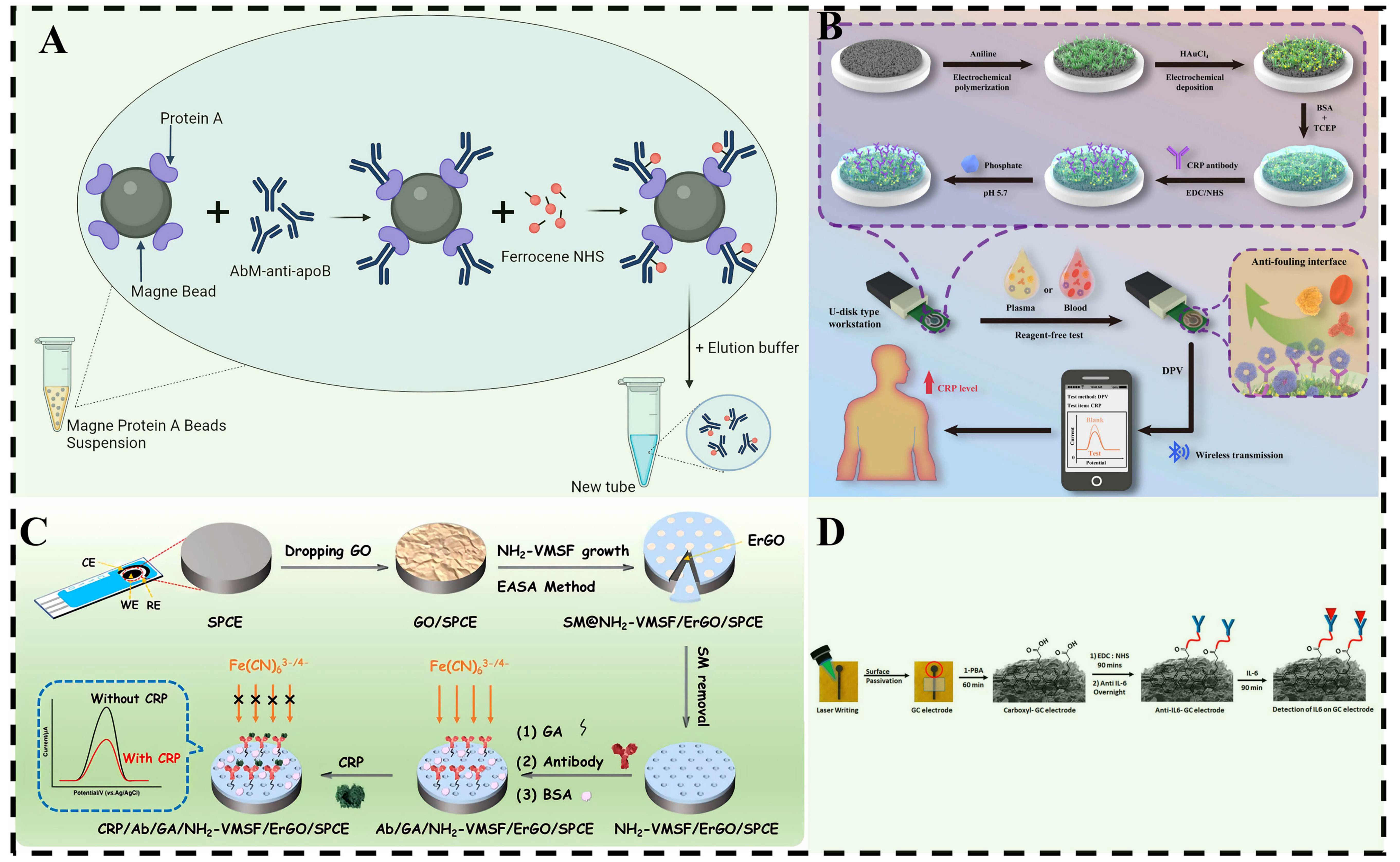
Figure 2. (A) Schematic illustration of the conjugation procedure between the AbM-anti-apoB and ferrocene carboxylic N-hydroxysuccinimide ester (ferrocene-NHS). Reproduced from Ref.[57]; (B) Preparation and detection principle of reagent-free anti-fouling electrochemical immunosensor. Reproduced from Ref.[68] with permission from Copyright Clearance Center; (C) Schematic illustration for fabrication of electrochemical sensor on Ni/Fe-WS2 modified SPCE and the detection of CRP based on gated electrochemical signal. Reproduced from Ref.[69]; (D) Schematic of graphitic carbon electrode fabrication, surface modification and IL-6 capture. Reproduced from Ref.[83]. NHS: N-hydroxysuccinimide; AbM: antibody monoclonal; BSA: bovine serum albumin; TCEP: tris(2-carboxyethyl)phosphine; EDC: 1-ethyl-3-(3-dimethylaminopropyl) carbodiimide hydrochloride; CE: counter electrode; WE: working electrode; RE: reference electrode; SPCE: screen-printed carbon electrode; GO: graphene oxide; ErGO: electrochemically reduced graphene oxide; NH2-VMSF: amino-functionalized vertically-ordered mesoporous silica film; GA: glutaraldehyde; 1-PBA: 1-pyrenebutyric acid; IL-6: interleukin-6; CRP: C-reactive protein; SM: surfactant micelle; GC: glassy carbon.
Electrochemical Immunosensor for LDL detection
| Marker | Electrode | Method | LOD | Linear | Ref. |
| LDL | AAB/NiO/ITO | EIS | 0.015 μM | 0.018-0.5 μM | [58] |
| LDL- | PVF/AuNPs/MAb/BSA | EIS | 3.50 μg/mL | 3.50-8.75 μg/mL | [38] |
| LDL | AAB/AuNPs-AgCl@PANI/GC | EIS | 0.34 pg/mL | 0.34-13.4 ng/mL | [59] |
| LDL | Au/4-ATP/AbM/BSA | EIS | 0.31 ng/mL | - | [60] |
| LDL | Au/Cys/mAbs/BSA | EIS SWV | 0.22 μg/mL | 0.5-18.0 μg/mL | [61] |
| LDL | A: DB/LDL/ApoB-Fc B: DB/MDA/LDL/ApoB-AQ | DPV | A: 0.2 ng/mL B: 0.1 ng/mL | A: 0.001-1.0 ng/mL B: 0.01-10.0 ng/mL | [62] |
| LDL | Au-IDEs/anti-LDL/BSA | EIS | 120 pg/mL | 50 pg/mL-500 ng/mL | [63] |
| LDL | PVF/AuNPs/MAb/BSA | EIS | 20 mg/dL | 30-135 mg/dL | [64] |
| LDL | GCE/poly(oATP)/AuNPs/AAB/LDL/Ag | LSV | 3.25 ng/mL | 10-1000 ng/mL | [56] |
| LDL | Au/4-ATP/AbM-Fc/BSA | SWV | 0.53 ng/mL | 0.01-1.0 ng/mL | [57] |
CRP
CRP serves not only as a sensitive acute-phase marker of systemic inflammation in the development and progression of AS, but also as a potential pro-inflammatory agent. Clinically, CRP has been widely used for risk stratification of AS, with elevated levels closely associated with acute coronary syndrome, plaque rupture, and recurrent cardiovascular events[65-67]. Therefore, CRP is an important biomarker for AS diagnosis and risk assessment, making its highly sensitive and accurate monitoring of significant importance.
Nonspecific adsorption and biofouling in complex biological fluids, such as serum, plasma, and whole blood, represent significant bottlenecks that limit the practical application of electrochemical immunosensors. Constructing sensing platforms with excellent antifouling properties to achieve highly sensitive detection of biomarkers remains a critical technical challenge that requires urgent attention. Lu et al. developed a LFEI exhibiting outstanding antifouling performance for the sensitive detection of CRP[68][Figure 2B]. By constructing an antifouling coating [Amyloid- Bovine Serum Albumin-Gold Nanoparticles-PANI (AL-BSA/AuNPs/PANI)], this study effectively reduced nonspecific adsorption in plasma and whole blood, thereby enhancing the stability and reliability of the sensor in complex biological matrices. Additionally, the authors proposed a reagent-free operational mode: by pre-loading sodium dihydrogen phosphate on the electrode surface, the local pH can be automatically adjusted upon sample addition, eliminating the need for additional reagents and simplifying the detection process. This method is particularly suitable for POCT and rapid on-site analysis. The integrated application of novel nanomaterials can significantly enhance the detection performance of electrochemical immunosensors. Ma et al. constructed a LFEI for the sensitive detection of CRP[69][Figure 2C]. The team innovatively combined electrochemically reduced graphene oxide (ErGO) with amino-functionalized vertically-ordered mesoporous silica nanochannel film (NH2-VMSF). In this system, ErGO serves as a “conductive adhesive bridge” that performs dual functions: it forms stable binding with the screen-printed carbon electrode substrate through hydrophobic interactions and π-π stacking effects, while simultaneously anchoring the mesoporous silica film firmly via condensation reactions between its surface oxygen-containing groups and the silanol groups of NH2-VMSF. This strategy effectively addresses the limitations of traditional VMSF, including easy detachment and insufficient conductivity when directly assembled on carbon electrode surfaces. Meanwhile, the excellent electron transfer capability of ErGO facilitates the rapid transmission of detection signals. The synergistic integration of these two nanomaterials significantly enhances the detection performance of the electrochemical immunosensor, offering a novel technical approach for the rapid detection of low-concentration CRP in complex biological matrices. Table 2 lists other electrochemical immunosensors for detecting CRP.
Table of electrochemical immunosensors for detecting CRP
| Marker | Electrode | Method | LOD | Linear | Ref. |
| CRP | Au/f-MWCNTs/Ab/BSA | DPV | PBS: 0.745 µg/mL Whole blood: 0.177 µg/mL | PBS: 1.25-80 µg/mL Whole blood: 0.005-10 µg/mL | [70] |
| CRP | GE/ErGO/PTyr | DPV | 1.25 μg/L | 1.09-100 μg/L | [71] |
| CRP | Ab/GA/NH2-VMSF/ErGO/SPCE | DPV | 8 pg/mL | 10 pg/mL-100 ng/mL | [69] |
| CRP | BSA/CRP-Ab/OLC-PAN/GCE | DPV | 0.9 fg/mL | 0.94 pg/mL-30 μg/mL | [72] |
| CRP | PET/Ag/CeO2-NRs/Anti-CRP/BSA | DPV | 0.18 ng/mL | 0.3-7.0 ng/mL | [73] |
| CRP | CSPE-COOH-AuNPs/Anti-CRP/BSA | DPV | 0.058 μg/mL | 1-100 μg/mL | [34] |
| CRP | SPCE/COF-PtRuC/Anti-CRP/BSA | AMP | 0.1 ng/mL | 0.2-20 ng/mL | [74] |
| CRP | Au-N-CNSs/AuPtRh NBCs/Anti-CRP/BSA | DPV | 7.70 pg/mL | 0.01 ng/mL-100 μg/mL | [75] |
| CRP | GCE/MB-Ab₁/CRP/Ab2-IrNPs/GO | DPV | 3.3 pg/mL | 0.01-100 ng/mL | [76] |
| CRP | LIG/Mx-AuNPs/Ab/BSA | DPV | 1.45 pg/mL | 0.01-10,000 ng/mL | [77] |
| CRP | Ab/AL-BSA/AuNPs/PANI/SPCE | DPV | 0.09 μg/mL | 0.1-25 μg/mL | [68] |
| CRP | Cu-MWCNT-Gra@rGO/AuNPs/Anti-CRP/BSA/CRP/QC-Ab2 | DPV | 81 pg/mL | 0.20-100 ng/mL | [78] |
| CRP | anti-CRP/CRP/BSA/anti-CRP/AuNPs@CoFe/N-GCT/GCE | DPV | 166.7 pg/mL | 0.5-200 ng/mL | [79] |
IL-6
In the initiation and progression of AS, IL-6 plays a pivotal role in triggering and amplifying inflammatory responses. The persistently activated IL-6 signaling pathway not only promotes lipid deposition and foam cell formation but also drives plaque destabilization, thereby accelerating the progression of AS lesions[80,81]. Therefore, the precise detection of IL-6 holds significant value for the early warning of AS.
The cost of electrode substrates has become a critical bottleneck that constrains the large-scale fabrication of electrochemical immunosensors. Cancelliere et al. reported a LFEI based on biochar-modified screen-printed electrodes (Bio-SPE) for the sensitive detection of IL-6 in human serum[82]. This study employed EDC/NHS [N-(3-dimethylaminopropyl)-N’ethyl carbodiimide/N-Hydroxysuccinimide] to activate carboxyl groups on the Bio-SPE surface for the immobilization of anti-mouse Immunoglobulin G (IgG), followed by immobilization of two different clones of anti-IL-6 monoclonal antibodies. The authors modified the SPE using biochar derived from brewers' spent grains, a widely available material that provides both environmental sustainability and cost-effectiveness. Furthermore, the biochar’s surface is abundant in functional groups, particularly carboxyl groups, which enhance the electrochemical activity at the electrode interface and improve charge transfer efficiency. In comparison to gold- or graphene-modified SPEs, this biochar modification strategy allows the sensor to maintain detection performance while significantly lowering material costs, thereby achieving a balance between economic viability and performance. Graphite electrodes prepared using traditional methods often face challenges such as high costs and low preparation efficiency, which hinder the large-scale and cost-effective production of sensors. Tan et al. developed an electrochemical immunosensor utilizing laser-induced graphene electrodes, successfully achieving efficient detection of IL-6[83][Figure 2D]. This study employed a visible laser diode with a power output of 3 W and a wavelength of 450 nm to directly write on commercial polyimide tape through raster scanning under ambient temperature and pressure conditions. The method achieves both graphitization and electrode patterning in a single step, enabling the direct fabrication of three-dimensional porous conductive Glassy carbon electrodes without the need for precursors or subsequent annealing modifications. This technique introduces a low-power laser direct writing strategy into the preparation of electrochemical immunosensor electrodes for the first time, providing a novel technical pathway for large-scale manufacturing. In the future, this technology has the potential to further optimize the electrode fabrication process, thereby reducing the manufacturing costs of sensors. Table 3 lists other electrochemical immunosensors for detecting IL-6.
Electrochemical immunosensor for IL-6 detection
| Marker | Electrode | Method | LOD | Linear | Ref. |
| IL-6 | Anti-IL-6-AuNPs/rGO/Au electrode | IT | 0.41 pg/mL | 0.97-250 pg/mL | [84] |
| IL-6 | Anti-IL-6-AuNPs/thionine-CMWCNTs/GCE | SWV | 2.97 pg/mL | 10-8.0 × 10⁵ pg/mL | [85] |
| IL-6 | Anti-IL-6-PPy/AuNPs/SPCE | EIS | 0.33 pg/mL | 1-100,000 pg/mL | [86] |
| IL-6 | Anti-IL-6-p-ABA/p-ATP/AuNPs/GCE | EIS | 1.66 pg/mL | 5-100,000 pg/mL | [87] |
| IL-6 | mAb-IL-6 clone-5/SPCE | SWV | 4.8 pg/mL | 26-125 pg/mL | [82] |
| IL-6 | PBCaNP-mAb2-target-mAb1-IL-6-GCE | SWV | 0.078 pg/mL | 0.1-1,000 pg/mL | [88] |
| IL-6 | Thi-Ab2-target-mAb1/SPGE | SWV | 8.14 fg/mL | 20 fg/mL-2 ng/mL | [89] |
| IL-6 | mAb-IL-6/GCE | DPV | 5.1 pg/mL | 10-500 pg/mL | [83] |
| IL-6 | PVA-TEGO-PANI/mAb | CV | 15.4 pg/mL | 15.4-400 pg/mL | [90] |
| IL-6 | MB-mAb-IL-6/PPC/GC rod | SWV | 5 pg/mL | 5-150 pg/mL | [91] |
| IL-6 | mMb-IL-6/Co3O4/SNF/ITO | ECL | 0.64 fg/mL | 1 fg/mL-10 ng/mL | [92] |
| IL-6 | mMb-IL-6/NAE | DPV | 0.45 pg/mL | / | [93] |
| IL-6 | mMb-IL-6/PANI/CdS@ ZIF-8-NH | SWV | 5.761 pg/mL | 5.761 × 10⁻⁵-20.00 ng/mL | [94] |
| IL-6 | mMb-IL-6/ZIF-8@Ag NWs/SPCE | DPV | 10 pg/mL | 10 pg/mL | [95] |
| IL-6 | mMb-IL-6/NiCoO2@CeO2 NBs | IT | 7 fg/mL | 2.5 × 10⁻⁵-10 ng/mL | [96] |
Hypertension
Hypertension is a major risk factor for the onset and progression of CVDs, with its pathophysiological processes closely linked to vascular dysfunction and damage to target organs[97,98]. Renin, aldosterone, and the epithelial sodium channel (ENaC) are recognized as crucial biomarkers for the development and progression of hypertension, and their abnormal variations are strongly correlated with the advancement of the disease[99,100]. Therefore, the precise detection of these biomarkers can assist in identifying hypertension risks at early stages and provide essential evidence for personalized prevention, treatment, and efficacy evaluation.
Renin-angiotensin-aldosterone system
The renin-angiotensin-aldosterone system (RAAS) is a vital neuroendocrine regulatory system responsible for the regulation of blood pressure, sodium-water balance, and cardiovascular homeostasis. Excessive activation of this system is a primary pathological factor contributing to hypertension and target organ damage[101-103]. In this context, renin and aldosterone (ALD) serve as important biomarkers, and their accurate detection is crucial for the early identification of hypertension.
Electrochemical immunosensors generally utilize high-surface-area nanomaterials to increase the number of antibody immobilization sites, thereby enhancing detection sensitivity. However, achieving stable dispersion of these nanomaterials and constructing biocompatible interfaces remain critical challenges that limit performance improvements. Schuck et al. constructed a LFEI utilizing a carbon electrode modified with a bilayer AuNS-P(Arg) nanostructure[104][Figure 3A]. This study employed polyarginine [P(Arg)] to coat gold nanostars (AuNS), which enhanced their dispersibility and biocompatibility while maintaining the electrical properties of AuNS. The sensor developed through this approach exhibited excellent analytical performance for detecting renin in undiluted human plasma and demonstrated high consistency with ELISA results. In the RAAS, ALD serves as a crucial hormone in regulating water-salt balance; however, its concentration in biological fluids is typically extremely low. In clinical testing, the aldosterone-to-renin ratio (ARR) is frequently utilized to aid in the evaluation of related pathological conditions. Nonetheless, the direct and precise quantification of ALD itself continues to pose challenges. To address this issue, Dong et al. developed a LFEI utilizing BSA/Anti-ALD/Mn@Hollow Nitrogen-Doped Carbon Nanotubes (H-NCNTs), which enabled the quantitative detection of ALD in human blood and urine samples[105][Figure 3B]. The team innovatively constructed tunable helical N-doped mesoporous carbon nanotubes encapsulating manganese nanoparticles, which served as ALD biosensing probes. This material exhibits a large active specific surface area, a unique helical structure, and synergistic catalytic effects due to the encapsulation of Mn nanoparticles. These features significantly enhance the detection sensitivity of the sensor and facilitate electron transport. Consequently, the sensing platform enables the accurate determination of trace ALD in blood and urine using chronoamperometry. However, the tendency of Mn nanoparticles to aggregate presents challenges for the large-scale fabrication of this innovative electrochemical immunosensor. Therefore, the development of sensing platforms that demonstrate high reproducibility, excellent stability, and strong clinical applicability is essential to advance their practical use.
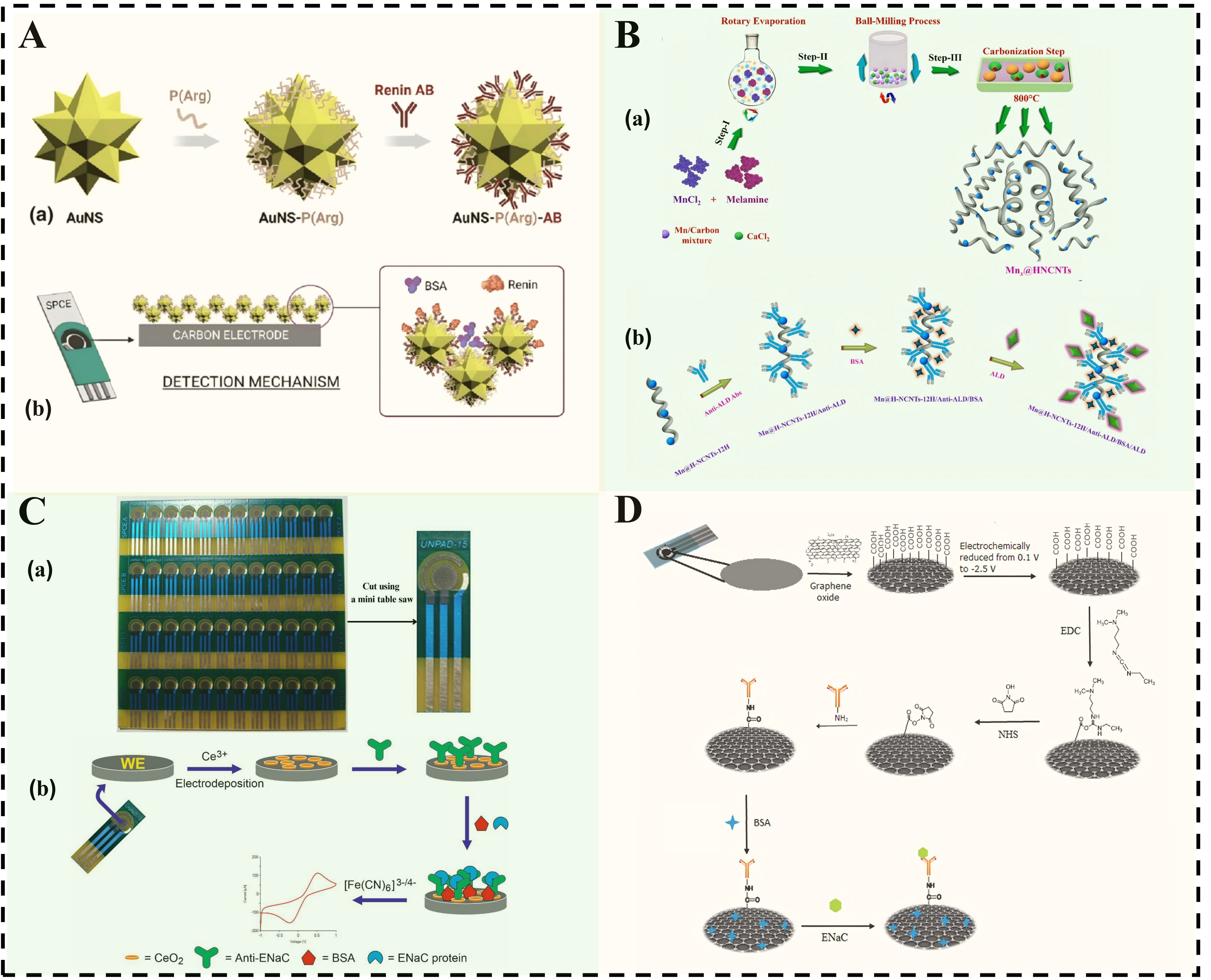
Figure 3. (A) Schematic diagram of renin detection sensing mechanism: (a) After the AuNS surface is coated with P (Arg), renin antibodies are immobilized on its surface; (b) When using a double-layer AuNSs-P (Arg) structure for detection, after immobilizing renin antibodies on the top layer, the active layer is then processed. Reproduced from Ref.[104] with permission from Copyright Clearance Center; (B) Synthesis and fabrication process of ALD immunosensor: (a) Schematic diagram showing the synthesis process of Mnx@H-NCNTs nanostructures; (b) Schematic diagram showing the construction of the BSA/anti-ALD/Mn@H-NCNTs-12H biosensor electrode used for ALD detection. Reproduced from Ref.[105] with permission from Copyright Clearance Center; (C) Schematic diagram of batch preparation of ENaC immunosensors: (a) PCB-SPCE printed circuit board-screen-printed carbon electrode sheets; (b) Schematic diagram of the electrochemical immunosensor used for the detection of ENaC protein. Reproduced from Ref.[109]; (D) Immunosensing scheme based on anti-ENaC/SPCE-rGO. Reproduced from Ref.[109]. AuNS: Gold nanostars; P(Arg): poly(arginine); HNCNTs: hollow nitrogen-doped carbon nanotubes; PCB: printed circuit board; BSA: bovine serum albumin; ENaC: epithelial sodium channel; NHS: N-hydroxysuccinimide; EDC: 1-ethyl-3-(3-dimethylaminopropyl) carbodiimide hydrochloride; ALD: aldosterone; H-NCNTs: hollow nitrogen-doped carbon nanotubes.
Epithelial sodium channel
The epithelial sodium channel (ENaC) is a transmembrane channel protein located on the apical membrane of epithelial cells, where it plays a crucial role in mediating sodium reabsorption and maintaining sodium transport[106,107]. Additionally, ENaC serves as a significant biomarker for hypertension. It is essential for regulating extracellular fluid volume, electrolyte balance, and long-term blood pressure homeostasis. Consequently, the specific recognition and precise detection of ENaC are of great significance[108].
Cost control and large-scale manufacturing capabilities have emerged as critical bottlenecks that restrict the practical applications of sensors. The key to addressing this issue lies in the development of sensing methods that integrate operational simplicity, low cost, and high detection performance. Setiyono et al. developed a LFEI for the detection of ENaC[109]. The research team constructed a composite electrode using self-made printed circuit board-screen-printed carbon electrodes (PCB-SPCE) combined with cerium oxide (CeO2), followed by the immobilization of anti-ENaC antibodies on its surface [Figure 3C]. Notably, a single PCB can simultaneously fabricate 48 independent electrodes, demonstrating low cost and significant potential for scalable production. Furthermore, the team integrated this sensor with their self-developed portable potentiostat, UnpadStat, to achieve real-time data display, facilitating rapid reading of test results and enhancing its suitability for POCT scenarios. However, this potentiostat continues to serve as an external instrument for data visualization and analysis. The physical separation between the sensor and the detection terminal may increase operational complexity and the risk of human error. Consequently, the platform requires further development towards integrated design in the future. The team led by Hartati et al. modified the surface of SPCE using rGO and immobilized anti-ENaC antibodies to construct a LFEI for ENaC detection[110][Figure 3D]. This method achieves a low detection limit while eliminating the need for expensive chemical labels, thereby significantly reducing both reagent and consumable costs, as well as overall testing expenses. This study establishes a significant foundation for the development of portable, efficient, and cost-effective early diagnostic tools, showcasing promising applications and transformative potential, particularly in POCT. Table 4 lists other electrochemical immunosensors for detecting ENaC and Renin.
Electrochemical immunosensors for detecting hypertension biomarkers
| Marker | Electrode | Method | LOD | Linear | Ref. |
| ENaC | Anti-ENaC-rGO-SPCE | DPV | 0.198 ng/mL | 0.01-1.5 ng/mL | [110] |
| Renin | CNT-PEG-AuNS-anti-Renin | DPV | 0.7412 pg/mL | 31.3-1,000 pg/mL | [111] |
| ENaC | Anti-ENaC-SPCE-Au | DPV | 0.037 ng/mL | 0.1-1.5 ng/mL | [112] |
| ENaC | AuNP/HS-PEG-COOH/anti-ENaC | DPV | 8.4 × 10⁻² ng/mL | 9.375 × 10⁻²-1.0 ng/mL | [113] |
| ENaC | SPCE/Au/Cysteamine/Glutaraldehyde/Anti-ENaC | DPV | 0.0372 ng/mL | 0.09375-1.0 ng/mL | [114] |
Myocardial infarction
Myocardial infarction (MI) is a clinical condition characterized by the necrosis of myocardial tissue due to persistent severe ischemia[115], which results from a sudden reduction or interruption of blood flow in the coronary arteries. Following MI onset, myocardial necrosis progresses to pump failure and life-threatening arrhythmias, with severe cases culminating in sudden cardiac death[116]. Therefore, the accurate detection of relevant biomarkers in the early stages of MI is of significant importance[117].
Cardiac troponin
Cardiac troponin (cTn) is a critical protein complex that regulates myocardial contraction and consists of three isoforms: cTnI, cTnT, and TnC. Among these isoforms, cTnI and cTnT are primarily expressed in cardiomyocytes, with minimal presence in non-cardiac tissues such as skeletal muscle[118]. When myocardial cells sustain damage, cTnI and cTnT are released from the affected cells into the bloodstream. Due to their exceptional cardiac specificity and release characteristics following cellular injury, both cTnI and cTnT demonstrate high sensitivity and specificity, establishing them as the widely accepted “gold standard” for diagnosing MI[115,119].
Traditional electrochemical immunosensors for cTnI detection predominantly utilize sandwich-type detection strategies, which typically necessitate the labeling of secondary antibodies. This approach involves complex operational procedures and incurs higher detection costs, thereby limiting its clinical translation and large-scale application. To address these limitations, Liu et al. developed a LFEI based on AuNPs/CuTA@Cu[120]. This strategy leverages the specific binding between cTnI and immobilized antibodies to form insulated immune complexes, which directly inhibit the electrochemical signal of CuTA@Cu, enabling one-step label-free detection. This approach offers a novel technical pathway for the miniaturization and cost-effective production of electrochemical immunosensors [Figure 4A]. However, the glassy carbon electrode (GCE) utilized in this study still encounters challenges, such as stringent pretreatment requirements and inadequate reproducibility in batch preparation, which hinder its ability to fully satisfy the application needs of POCT. The construction of the AuNPs/CuTA@Cu interface on the surface of a screen-printed electrode is anticipated to enhance the manufacturability and scalability potential of such sensors. Non-invasive detection is a vital developmental direction in wearable health monitoring. In contrast to traditional invasive methods, this approach avoids disrupting the skin barrier, minimizes infection risks, and shows potential for enabling real-time, continuous monitoring during treatment. Yengin et al. achieved non-invasive detection of target biomarkers (cTnT and cTnI) in artificial body fluids by combining reverse iontophoresis for extraction with LFEIs[121]. This method employs low-intensity direct current to facilitate the transdermal transport of cTnT and cTnI across simulated skin (cellulose ester dialysis membrane) into a collection gel, thereby mitigating complications associated with blood sampling, such as pain, infection risks, and challenges in continuous monitoring [Figure 4B]. The study offers novel technical insights for the development of wearable platforms aimed at monitoring myocardial injury biomarkers. Table 5 lists other electrochemical immunosensors for detecting cTn.
Figure 4. (A) Schematic illustration of the preparation and detection of the CuTA@Cu-based cTnI LFEI. Reproduced from Ref.[120] with permission from Copyright Clearance Center; (B) Schematic illustration of the preparation of the electrochemical immunochemical sensor and the non-invasive detection of cTnT in artificial body fluids. Reproduced from Ref.[121] with permission from Copyright Clearance Center. GCE: Glassy carbon electrode; CuTA: copper(II)-annic acid; EDC: 1-ethyl-3-(3-dimethylaminopropyl)carbodiimide; NHS: N-hydroxysuccinimide; BSA: bovine serum albumin; TA: tannic acid; CPE: carbon paste electrode.
Electrochemical Immunosensors for cTnI and cTnT Detection
| Marker | Electrode | Method | Linear | LOD | Ref. |
| cTnI | Au/MHDA/Ab | EIS | - | 10 ag/mL | [122] |
| cTnI | GCE-Au-Ab1/CuFe2O4-Pd-Ab2 | CA | 0.001-100 ng/mL | 1.9 1fg/mL | [123] |
| cTnI | GCE-β-CD@3DG-AgNCs-Ab1/β-CD@3DG-Fc-COOH-Ab2 | DPV | 100 fg/mL-100 ng/mL | 45.5 fg/mL | [124] |
| cTnI | MoS2@Cu2O-Ag-Ab1/Ce:ZnO-NGQDs-Ab2 | ECL | 0.01-100 ng/mL | 2.9 fg/mL | [125] |
| cTnI | HRP-Ab2-Au-COF | DPV | 5 pg/mL-10 ng/mL | 1.7 pg/mL | [126] |
| cTnI | GE/rGO/pTyr/anti-cTnI | DPV | 4 pg/mL-4 ng/mL | 4 pg/mL | [127] |
| cTnI | GCE/Pt/Au-B,S,N-Rgo/Ab | CA | 0.1 pg/mL-50 ng/mL | 0.082 pg/mL | [128] |
| cTnI | GCE/Cu2O-CuO@CeO2-Pd/Ab | CA | 100 fg/mL-100 ng/mL | 15.85 fg/mL | [129] |
| cTnT | caf-TCQDs@AuNPs-HO-BNNS-Ab1/Fc-COOH-Ab2 | DPV | 0.0001-100 ng/mL | 0.0013 ng/mL | [130] |
| cTnT | SPE/GNPs/Ab- cTnT-Ag | ECL | 5 fg/mL-100 pg/mL | 0.05 fg/mL | [131] |
| cTnT | GCE/ZnSnO3/NHS-EDC/Ab | EIS | 0.187 fg/mL | [132] | |
| cTnT | PCB/Biochar/ glutaraldehyde/Ab | CV | 0.01-5.00 ng/mL | 0.003 ng/mL | [133] |
| cTnT | GP-HCL-EDC/NHS-Ab | SWV | 0.5-1,000 fg/mL | 1.28 fg/mL | [134] |
| cTnT | GCE/COOH-PPy-EDC/NHS-NH2 /NCY-GA-Ab1-gly | SWV | - | 0.35 pg/mL | [135] |
Myoglobin
Myoglobin (Myo) is a small oxygen-binding protein that is predominantly found in cardiac and skeletal muscles, where it primarily functions in oxygen storage and transport[136]. Following myocardial injury, Myo can be released into the peripheral circulation within 1 h to 3 h, peaking at 6 h to 9 h, and exhibits an earlier elevation compared to cTnT and cTnI. Due to this early-release characteristic, Myo is recognized as a crucial early biomarker for the diagnosis of acute myocardial infarction (AMI) in the hyperacute phase, making it particularly suitable for rapid emergency screening[137,138].
Nanomaterials can be conjugated with antibody probes to enhance detection signals through efficient signal amplification, thereby achieving highly sensitive and accurate detection of Myo. Zhang et al. developed a LEI based on mesoporous SiO2@PDA@PtPd nanocrystals (m-SiO2@PDA@PtPd NCs) and PtNi nanodendrites (NDs) for detecting Myo[139]. In this system, m-SiO2@PDA@PtPd NCs effectively accelerate interfacial electron transfer and increase the loading capacity of the primary antibody, while PtNi NDs-labeled secondary antibodies enhance signal amplification, thus improving detection sensitivity [Figure 5A]. However, the sandwich-structured detection method involves multiple incubation and washing steps, rendering the procedure relatively cumbersome and challenging to meet the rapid detection requirements for POCT during the acute phase of myocardial infarction. Traditional molybdenum disulfide (MoS2)-based sensors often encounter significant technical challenges, including high background signals and inadequate conductivity. These issues limit advancements in detection sensitivity and signal transduction efficiency. In response, Yang et al. developed a LFEI utilizing Au/Co-BDC@MoS2, which successfully achieved quantitative detection of Myo[140]. The team leveraged the low catalytic activity of Co-BDC (a 2D cobalt-based metal-organic framework) for the reduction of H2O2. By compositing it with MoS2 nanosheets to selectively cover certain active sites, they effectively mitigated the non-specific background current attributed to the inherent high catalytic performance of MoS2. This strategy significantly addressed challenges such as strong background signals and low signal-to-noise ratios commonly encountered in traditional MoS2-based sensors. Overall, this synergistic functional design presents a novel approach to overcoming the inherent limitations of conventional MoS2-based sensors and provides a viable technical pathway for the highly sensitive detection of Myo. Table 6 lists other electrochemical immunosensors for detecting Myo.
Figure 5. (A) Schematic diagram of the stepwise fabrication of an electrochemical immunosensor for detecting Myo using m-SiO2@PDA@PtPd NC-modified electrodes and PtNi NDs-labeled secondary antibodies. Reproduced from Ref.[139] with permission from Copyright Clearance Center; (B) Schematic illustration of the fabrication and detection of CK-MB using an electrochemical immunosensor based on AuPdCu NWNs. Reproduced from Ref.[150] with permission from Copyright Clearance Center; (C) Schematic illustration of the construction of a LFEI based on mf-Pd@PtCu for H-FABP detection. Reproduced from Ref.[159] with permission from Copyright Clearance Center; (D) Schematic illustration of the preparation of RuMn MOFs and FePtRh MOFs-Ab2 and the fabrication and detection of H-FABP using labeled electrochemical sensors. Reproduced from Ref.[160] with permission from Copyright Clearance Center. AuPdCu NWs: Gold-palladium-copper nanowires; Ru(bpy)32+: Tris (2,2’ bipyridine) ruthenium (II) ion; mf-Pd@PtCu: metal-organic framework-derived palladium@platinum-CopperMOF; CTAC: cetyltrimethylammonium chloride; PDA: polydopamine; H-FABP: heart-type fatty acid-binding protein; DPV: differential pulse voltammetry; BSA: bovine serum albumin; Myo: myoglobin; GCE: glassy carbon electrode; EDC: 1-ethyl-3-(3-dimethylaminopropyl) carbodiimide hydrochloride; NHS: N-hydroxysuccinimide; HCL: hydrochloric acid; DMA: dimethylacetamide; DA: dopamine; CK-MB: creatine kinase-MB isoenzyme.
Electrochemical immunosensor for Myo detection table
| Marker | Electrode | Method | Linear | LOD | Ref. |
| Myo | Myo-Ab/MnO2@CNTs/GCE | DPV | 0-15 μg/mL | 3 ng/mL | [141] |
| Myo | Ab-Au/Co-BDC@MD/GCE | DPV | 10 fg/mL-10 ng/mL | 10.03 fg/mL | [140] |
| Myo | Ab-AuPtAg PHNR/GCE | DPV | 0.0001-1000 ng/mL | 0.46 pg/mL | [142] |
| Myo | Ab/CNTs@CS-FET | CA | 1-4000 ng/mL | 4.2 ng/mL | [143] |
| Myo | Ab/CdSeS/ZnS QDs/BTO/FTO | - | 0.01-1000 ng/mL | - | [144] |
| Myo | PtNi NDs/Ab2-Ab1/SiO2@PDA@PtPd NCs/GCE | DPV | 0.001-1000 ng/mL | 0.21 pg/mL | [145] |
| Myo | DDCE/Ru(bpy)32+@RuSi NPs/BSA/Myo/-Au@Ag2S | ECL | 0.1 pg/mL-100 ng/mL | 0.05 pg/mL | [146] |
Creatine kinase-MB isoenzyme
Creatine kinase (CK) is a dimeric enzyme that primarily catalyzes the reversible phosphorylation reaction between creatine and phosphocreatine. Among its important isoenzymes, creatine kinase MB (CK-MB) is predominantly found in myocardial cells. When these cells are damaged or undergo necrosis due to ischemia and hypoxia, the integrity of the cell membrane is compromised, resulting in the release of intracellular CK-MB into the bloodstream and consequently leading to elevated serum CK-MB levels. Therefore, monitoring the dynamic changes in blood CK-MB levels serves as a crucial indicator for assessing the occurrence and severity of myocardial injury[147,148].
Traditional LEIs designed for the detection of CK-MB predominantly depend on a singular signale amplification mechanism. This typically involves either enhancing the conductivity of nanomaterials or utilizing a single probe, which constrains their ultra-sensitive detection capabilities. To overcome this limitation, Wang et al. developed a LEI that utilizes PdPtCoNi@Pt-skin nano-polyhedrons for the detection of CK-MB[149]. This sensor utilizes a sandwich immunoassay approach by labeling signal probes on the secondary antibody. The Pt-skin shell of the signal probe, PdPtCoNi@Pt-skin nano-polyhedron, exhibits exceptional catalytic activity, protects the internal alloy core from oxidation, and enhances binding stability with the secondary antibody, thereby extending the sensor’s operational lifespan. However, the sandwich method necessitates multiple incubation and washing steps, making it difficult to meet rapid response requirements for POCT during AMI episodes. Additionally, the synthesis process of traditional polymetallic nanomaterials involves cumbersome procedures and relies heavily on environmentally harmful chemical reagents, which not only escalate production costs but also pose a risk of environmental pollution. To address this issue, Cen et al. adopted a green synthesis strategy to overcome the bottleneck in nanomaterial preparation and constructed a LFEI for the highly sensitive detection of CK-MB[150]. The team prepared ultrathin gold-palladium-copper nanowire networks (AuPdCu NWNs) using a seedless, template-free, and surfactant-free aqueous one-pot method, and immobilized anti-CK-MB antibodies onto the modified electrode surface via physical adsorption [Figure 5B]. The AuPdCu NWNs exhibit a large specific surface area, abundant active sites, excellent biocompatibility, and high conductivity, significantly enhancing the detection sensitivity for CK-MB. Although this sensor has completed spiked serum recovery experiments, it still lacks large-scale validation with clinical case samples, indicating a gap that must be addressed before practical clinical application can be achieved. Table 7 lists other electrochemical immunosensors for detecting CK-MB.
Electrochemical immunosensor for CK-MB detection
| Marker | Electrode | Method | Linear | LOD | Ref. |
| CK-MB | GCE/AuNP/Ab | EIS | 0.05-1 ng/mL | 0.018 ng/mL | [151] |
| CK-MB | ITO-PET/AuNPs/11-MuA/EDC/NHS/Ab | SWV | 0.1-100 pg/mL | 0.0118 pg/mL | [152] |
| CK-MB | SWCNT-SPE/CNOs/Fe3O4/AuNP/CS/Ab | ECL | 10 ng/mL-50 fg/mL | 5 fg/mL | [153] |
| CK-MB | GP/AuNP/6-MH/3-GOPE | EIS | 1-50 pg/mL | 0.045 pg/mL | [154] |
Heart-type fatty acid-binding protein
Heart-type fatty acid-binding protein (H-FABP) is a small molecular protein found in the cytoplasm of cardiomyocytes, where it plays a crucial role in fatty acid transport and energy metabolism[155]. Following myocardial injury, H-FABP can be rapidly released into the bloodstream within one hour, with its elevation occurring earlier than that of cTnI and CK-MB. Therefore, it is regarded as an important biomarker for the early diagnosis of AMI[156,157].
Due to the extremely low concentration of H-FABP in blood and its rapid fluctuations during the early stages of injury, the relevant detection technology must exhibit exceptionally high sensitivity and rapid response capabilities[158]. Ai et al. designed a LFEI for the quantitative detection of H-FABP. By modifying the surface of the GCE with mesoporous flower-like Pd@PtCu core-shell nanocrystals (MF-Pd@PtCu), this sensor significantly enhanced the electrochemically active surface area and facilitated rapid interfacial electron transfer[159]. The interface facilitates rapid electron transfer. Meanwhile, MF-Pd@PtCu can incorporate a greater number of biomolecular recognition elements, significantly enhancing the sensor’s detection performance for H-FABP [Figure 5C]. However, this study encounters industrial challenges concerning material cost control and scalable production. Although the introduction of Cu reduces the usage of Pt and Pd to some extent, Pt remains the primary catalytic component; therefore, its economic feasibility for large-scale clinical applications necessitates further evaluation. Future research should focus on developing low-Pt-content alloys or carbon-based composite carriers to further minimize noble metal consumption while preserving sensing performance. In contrast, electrochemical luminescence immunosensors detect optical signals, effectively minimizing background interference from electroactive substances present in complex samples. These sensors typically demonstrate higher signal-to-noise ratios and enhanced sensitivity, thereby providing robust support for the efficient and accurate detection of biomarkers. Li et al. constructed a labeled electrochemical luminescence immunosensor utilizing bimetallic RuMn- Metal-Organic Framework (MOF) as the electrochemiluminescence emitter and trimetallic FePtRh-MOF as the quencher-labeled secondary antibody[160]. Under the synergistic effect of MOF and Ru(bpy)32+ [Tris(2,2’-bipyridine)ruthenium(II) ion], RuMn-MOF demonstrates enhanced and stable electrochemiluminescence performance [Figure 5D]. Meanwhile, FePtRh-MOF improves electron transfer efficiency, which in turn enhances the quenching effect. However, the processes of antibody incubation and surface modification for this sensor are time-consuming, typically requiring several hours. This limitation confines its application primarily to static laboratory detection scenarios and hinders its potential for further transformation and application in POCT. Table 8 lists other electrochemical immunosensors for detecting H-FABP.
Electrochemical immunosensors for H-FABP detection
| Marker | Electrode | Method | Linear | LOD | Ref. |
| H-FABP | GE/PTh-CNT-MB-EDA/Ab | - | 3-25ng/mL | 1.47ng/mL | [161] |
| H-FABP | ITO/Cu2+-Cys-ABEI-GNPs-CS/Ab-GNPs | ECL | 0.1-1000pg/mL | 0.09pg/mL | [162] |
| H-FABP | AuNDs/Chit-g-Fc/Ab1- Ab2/Thi/AuPt/PDA/OHCSs | DPV | 0.001-200ng/mL | 0.53pg/mL | [163] |
| H-FABP | GCE/Ni-TCPP(Fe)@PSS-Gr/Ab1-Ab2-Ag@Au/Pt | CA | 10fg/mL-100ng/mL | 5.75fg/mL | [164] |
| H-FABP | Ru-g-C3N4-Ab1-Ab2/NH2-MIL-101(Fe)/Luminol | ECL | 5fg/mL-50ng/mL | 2.43fg/mL | [165] |
| H-FABP | GCE/PICA-Ab1-Ab2-Ni-TCPP (Fe)-PEI-Lum | ECL | 100fg/mL-100ng/mL | 44.5fg/mL | [166] |
| H-FABP | GCE/hc-g-C3N4@CDsE /Ab1-Ab2/Cd0.5Zn0.5S/d-Ti3C2Tx MXene | DPV | - | 3.3fg/mL | [167] |
| H-FABP | ITO/ Luminol@Ag/Cu2O/Ti3C2-Ab1-Ab2-AgS QDs@NH2-MIL-101(Fe) | ECL | 1fg/mL-100ng/mL | 0.36fg/mL | [168] |
Multiplex immunoelectrochemical sensor for detecting cardiovascular disease biomarkers
The occurrence and progression of CVDs involve complex pathological mechanisms. Changes in the concentration of a single biomarker often fail to provide accurate diagnostic evidence. Consequently, multiplex electrochemical immunosensors can simultaneously identify and quantify multiple CVD-related biomarkers on the same detection platform. This approach significantly enhances testing efficiency and diagnostic accuracy, while also providing a more comprehensive characterization of the dynamic evolution of myocardial injury[169,170].
Multiplex detection encounters significant challenges in complex testing environments, making the development of high-performance antifouling coatings essential for achieving precise detection. Timilsina et al. constructed a nanocomposite coating through the cross-linking of BSA, partially reduced Graphene Oxide, and Glutaraldehyde (GA), which was successfully modified on the surface of a gold electrode[171]. This innovative coating effectively suppresses nonspecific adsorption in complex biological fluids while preserving the conductivity and stability of the electrode. Utilizing this modified electrode, researchers were able to simultaneously detect multiple disease markers, including cTnI, in whole blood and plasma, demonstrating remarkable sensitivity and selectivity. However, the current system is limited by insufficient clinical validation and a lack of long-term stability evaluation. Furthermore, avoiding signal crosstalk and achieving synchronous multi-index analysis continue to pose significant challenges in current multiplex detection. Boonkaew et al. constructed sample inlets by stacking wax-printed paper with transparent film and incorporating laser-cut double-sided adhesive tape, which formed three independent detection zones and microfluidic channels[172]. A LFEI was developed by modifying SPCE with Graphene Oxide (GO). Carboxyl groups were activated through anodic oxidation, followed by antibody conjugation via EDC-NHS coupling, and the blocking of nonspecific binding sites with BSA. Antibodies were covalently immobilized through amide bonds to enhance stability. Square wave voltammetry (SWV) was employed for detection, where the formation of immunocomplexes impeded electron transfer of the redox probe, resulting in a decreased current response as biomarker concentration increased. The target concentration was ultimately quantified by measuring the changes in current. This sensor can simultaneously detect three biomarkers [CRP, cTnI, and Procalcitonin (PCT)] in a single sample, demonstrating high selectivity and sensitivity. The integration of electrochemical immunosensors with microfluidic technology represents a significant research direction. Zhou et al. developed a microfluidic chip-based electrochemical immunosensor for the simultaneous detection of cTnI and CRP[173]. This sensor utilizes the high specific surface area of gold nanoparticles, providing an excellent substrate for antibody immobilization. Additionally, the miniaturized design of the microfluidic channels significantly reduces the required sample volume to only 30 μL, making it suitable for POCT. Furthermore, to address signal interference issues in dual-marker synchronous detection, a differential quantum dot labeling strategy was employed to overcome the limitations of traditional signal superposition. Although the team has completed preliminary validation with 20 serum samples, a gap remains for clinical application. Future work should focus on expanding clinical sample validation to enhance the practical applicability.
THE INTEGRATION OF ELECTROCHEMICAL IMMUNOSENSORS WITH ADVANCED TECHNOLOGIES
As highly sensitive and efficient detection platforms, electrochemical immunosensors are increasingly being integrated with cutting-edge fields such as microfluidic technology[174,175], artificial intelligence[176], and portable devices[177]. This integration is driving the continuous advancement of biomarker detection technologies. The synergy of these multiple technologies not only enhances the analytical performance of the sensors but also broadens their application potential in the early diagnosis, dynamic monitoring, and personalized treatment of CVDs.
Microfluidic technology enables the precise manipulation of liquids at the micrometer scale, making it particularly suitable for the efficient analysis of minute samples. This technology offers significant advantages, including reduced sample consumption, improved reaction efficiency, and enhanced controllability in detection[178,179]. Singh et al. developed a LFEI based on a microfluidic chip for the ultra-low concentration simultaneous detection of cTnI and CK-MB[180]. The microfluidic chip automates the detection process, minimizes manual intervention, and enhances experimental repeatability and stability. However, this platform has not yet fully integrated machine learning methods with electrochemical sensing data, which somewhat limits its potential for complex data analysis and disease prediction. In the future, the introduction of artificial intelligence algorithms to integrate and analyze multi-parameter sensing data may optimize the testing process and enhance clinical diagnostic decision support capabilities. Ganguly et al. developed a LFEI utilizing non-Faradaic electrochemical impedance spectroscopy, which enables the detection of ultra-low concentrations of IL-6 and IL-8[181]. Furthermore, the study employed a random forest model to analyze the concentration data of IL-6 and IL-8 obtained from the sensor, facilitating disease classification and grading discrimination. By optimizing the number and depth of decision trees within the model, the final classification accuracy reached 98.437%. This strategy aids in the early identification of diseases and may help mitigate diagnostic and treatment delays. Additionally, the sensor platform demonstrates excellent modularity and flexibility, making it applicable not only for the detection of IL-6 and IL-8 but also extensible to other inflammatory markers and various disease models. In the future, by integrating more CVD-related biomarkers such as cTnI and CRP, this platform is anticipated to further enhance its application value in the precision diagnosis of CVDs. With the growing demand for rapid diagnosis of critical illnesses, traditional laboratory testing models have struggled to meet clinical requirements for timeliness, particularly in emergency scenarios such as myocardial infarction and trauma care, where obtaining test results promptly can be challenging. To address this issue, Boonkaew et al. developed a LFEI suitable for POCT applications, enabling rapid and accurate detection of three key cardiovascular biomarkers: CRP, cTnI, and PCT[172]. Utilizing paper-based materials as the substrate, the sensor employs a label-free immunoassay strategy to immobilize antibodies on the electrode surface, achieving controlled liquid transport on the chip and simultaneous multi-region, multi-marker detection. This analytical platform is compatible with various sample types, such as serum and whole blood, and features advantages including a simple structure, low cost, and user-friendly operation, making it particularly well-suited for rapid screening in resource-limited settings. Especially in emergency situations like myocardial infarction and trauma care, this platform can provide timely test results. With its portability and high sensitivity, this technology offers a promising solution for POCT and has the potential to facilitate early identification and timely intervention in CVDs, thereby improving patient prognosis.
CONCLUSION
Currently, electrochemical immunosensors have made significant advances in the innovation of functional materials, optimization of devices, and enhancement of performance. However, they continue to encounter challenges in scaling for clinical applications and in complex scenarios. Firstly, biofouling issues arising from complex biological samples limit both detection accuracy and long-term stability. Patients and clinicians alike wish to detect abnormalities in vivo using complex biological fluids, such as a drop of plasma or serum. However, interference from non-target electroactive substances can result in electrode contamination. Although antifouling coating solutions are available, they frequently compromise the electron transfer efficiency of the electrode, making it challenging to meet the requirements for continuous clinical monitoring. Secondly, insufficient manufacturability and reproducibility in mass production impede the translation of laboratory performance into industrial products. Variations in the production of printed electrodes, challenges in substrate cost control, and difficulties in scaling up modification processes contribute to fluctuations in sensor performance, increased production costs, and compromised manufacturing consistency, which pose significant obstacles to commercialization. Furthermore, insufficient system integration and dependence on sample pretreatment impede the effective implementation of POCT scenarios. Ideally, POCT should facilitate fully integrated workflows. However, most current sensors still necessitate manual preprocessing or coordination among multiple devices. Additionally, the immature built-in algorithms often struggle to calibrate complex interference signals in real time, ultimately undermining diagnostic accuracy and timeliness. Lastly, the absence of clinical validation protocols remains a critical barrier to the clinical translation of electrochemical immunosensors. In the medical field, any novel diagnostic technology must undergo stringent clinical validation before achieving widespread application. However, current electrochemical immunosensors exhibit significant deficiencies in this area: on one hand, the limited sample sizes do not adequately represent the complexities of clinical scenarios; on the other hand, the lack of comparative studies with established gold-standard diagnostic methods impedes their clinical adoption and promotion.
In the future, with the continuous integration of multidisciplinary fields such as materials science, nanotechnology, molecular biology, and artificial intelligence, electrochemical immunosensors are anticipated to achieve multidimensional improvements. Firstly, non-invasive detection represents a pivotal development direction, broadening their application scope from traditional laboratory testing to areas such as home self-monitoring and sports health management. To realize this goal, two significant technical bottlenecks must be addressed: First, innovative sample extraction and enrichment technologies need to be developed, such as reverse iontophoresis, to efficiently extract target biomarkers from body fluids without damaging the skin. This approach aims to tackle the challenges posed by low concentrations and complex matrices in non-invasive samples. Secondly, the sensor interface must be optimized by integrating antifouling nano-coatings to prevent the non-specific adsorption of proteins and lipids in samples, while also leveraging the signal amplification effects of nanomaterials to enhance detection sensitivity for low-concentration biomarkers. Additionally, efforts should be directed towards advancing the intelligent development of POCT platforms based on electrochemical immunosensors. Previous studies have developed a smartphone-controlled NFC electrochemical immunosensor for the detection of CRP, establishing a foundational reference for the advancement of intelligent POCT. With the rapid progress in artificial intelligence technology, future POCT platforms will not only depend on hardware innovations but also utilize sophisticated data analysis and diagnostic systems. By integrating artificial intelligence and machine learning algorithms, these platforms can process multiplex signals in real time, automatically identify disease patterns, and deliver personalized treatment plans based on patient data. For example, by analyzing multi-marker profiles in blood samples alongside historical physiological data such as electrocardiograms, POCT can forecast patients’ risks of CVDs and provide customized intervention strategies. This intelligent data processing will significantly enhance the application value of electrochemical immunosensors in clinical settings, particularly in practical scenarios such as home testing. Furthermore, it is essential to strengthen the methodological validation of electrochemical immunosensors in clinical practice. Future efforts should focus on conducting multicenter, large-sample clinical studies to systematically compare these sensors with gold-standard diagnostic methods, establish standardized quality control systems, and improve awareness and participation among clinicians. These initiatives will further advance the clinical utility of electrochemical immunosensors, providing reliable technical support for precise disease diagnosis.
In summary, electrochemical immunosensors exhibit unique advantages and hold significant promise for early screening, risk assessment, and real-time monitoring of CVDs. This review summarizes the research advancements in electrochemical biosensors for detecting CVD-related biomarkers, emphasizing recent breakthroughs in this field. Fueled by the deep integration of multidisciplinary approaches and clinical needs, this technology is anticipated to play an increasingly vital and irreplaceable role in the precise diagnosis and intelligent healthcare of CVDs.
DECLARATIONS
Acknowledgments
The Graphical Abstract was created with BioRender.com (Created with BioRender. Wang S (2026) https://BioRender.com/0ucecul).
Authors’ contributions
Contributed to conceptualization, original draft writing, data integration, overall manuscript coordination, final polishing, and manuscript review and revision: Zhang J
Contributed to figure and table preparation, literature investigation, original draft writing, reference management, and manuscript formatting: Wang S, Zeng Y
Contributed to conceptualization, supervision, funding acquisition, project administration, manuscript review and revision, and final approval of the manuscript: Lan H, Zang G
Availability of data and materials
Not applicable.
AI and AI-assisted tools statement
Not applicable.
Conflict of interest
Lan H is from the Chongqing Quality Testing & Inspection Center for Medical Devices. The other authors declared that there are no conflicts of interest.
Financial support and sponsorship
This work was supported by the Natural Science Foundation of Chongqing (CSTB2023NSCQ-MSX0232, CSTB2024NSCQ-MSX0105), the Pharmaceutical Regulatory Science Research Project of Chongqing Medical Products Administration (Project No. CQYJKJ2025-17), the Project of Tutorial System of Medical Undergraduate in the Lab Teaching & Management Center at Chongqing Medical University (LTMCEMTS202627, LTMCEMTSDC202617, LTMCEMTSDC202624), and the National Training Program of Innovation and Entrepreneurship for Undergraduates (202410631001, 202510631020, 202510631039).
Ethical approval and consent to participate
Not applicable.
Consent for publication
Not applicable.
Copyright
© The Author(s) 2026.
REFERENCES
1. Roth GA, Mensah GA, Johnson CO, et al. Global burden of cardiovascular diseases and risk factors, 1990-2019. J Am Coll Cardiol. 2020;76:2982-3021.
2. Yang L, Zang G, Li J, Li X, Li Y, Zhao Y. Cell-derived biomimetic nanoparticles as a novel drug delivery system for atherosclerosis: predecessors and perspectives. Regen Biomater. 2020;7:349-58.
3. Yang F, Xue J, Wang G, Diao Q. Nanoparticle-based drug delivery systems for the treatment of cardiovascular diseases. Front Pharmacol. 2022;13:999404.
4. Kogularasu S, Lin W, Lee Y, et al. Advancements in electrochemical biosensing of cardiovascular disease biomarkers. J Mater Chem. B. 2024;12:6305-27.
5. Yousefi F, Movahedpour A, Shabaninejad Z, et al. Electrochemical-based biosensors: new diagnosis platforms for cardiovascular disease. Curr Med Chem. 2020;27:2550-75.
6. Bryan T, Luo X, Bueno PR, Davis JJ. An optimised electrochemical biosensor for the label-free detection of C-reactive protein in blood. Biosens Bioelectron. 2013;39:94-8.
7. Diao W, Zhou C, Zhang Z, et al. EGaIn-modified ePADs for simultaneous detection of homocysteine and C-reactive protein in saliva toward early diagnosis of cardiovascular disease. ACS Sens. 2024;9:4265-76.
8. Li C, Hsu SH, Chang C, Wang G. Direct bilirubin detection using surface-enhanced raman spectroscopy. IEEE Sensors J. 2021;21:21458-64.
9. Wang K, Wang S, Lin L, Zhao Z, Li G. Detection of total bilirubin based on multi-wavelength excitation fluorescence spectroscopy. Measurement. 2025;253:117807.
10. Oshima Y, Haruki T, Koizumi K, et al. Practices, potential, and perspectives for detecting predisease using raman spectroscopy. Int J Mol Sci. 2023;24:12170.
11. Delrue C, Speeckaert MM. The potential applications of raman spectroscopy in kidney diseases. J Pers Med. 2022;12:1644.
12. Yao B, Giel M, Hong Y. Detection of kidney disease biomarkers based on fluorescence technology. Mater. Chem Front. 2021;5:2124-42.
13. Huillet C, Adrait A, Lebert D, et al. Accurate quantification of cardiovascular biomarkers in serum using protein standard absolute quantification (PSAQTM) and selected reaction monitoring. Mol Cell Proteomics. 2012;11:M111.008235.
14. Osorio LA, Lozano M, Soto P, et al. Levels of small extracellular vesicles containing hERG-1 and Hsp47 as potential biomarkers for cardiovascular diseases. Int J Mol Sci. 2024;25:4913.
15. Längst N, Adler J, Kuret A, et al. Cysteine-rich LIM-Only protein 4 (CRP4) promotes atherogenesis in the ApoE-/- mouse model. Cells. 2022;11:1364.
16. Greco A, Occhipinti G, Giacoppo D, et al. Antithrombotic therapy for primary and secondary prevention of ischemic stroke. J Am Coll Cardiol. 2023;82:1538-57.
17. Poznyak AV, Sukhorukov VN, Eremin II, Nadelyaeva II, Orekhov AN. Diagnostics of atherosclerosis: overview of the existing methods. Front. Cardiovasc. Med. 2023;10:1134097.
18. Debnath S, Paramasivam SS, Pradhan D, Manickam P, Chatterjee PB. A redox‐active copper complex for orthogonal detection of homocysteine involving fluorescence and electrochemical techniques. Small. 2025;21:2409982.
19. Stewart AJ, O’reilly EJ, Moriarty RD, et al. A cholesterol biosensor based on the NIR electrogenerated-chemiluminescence (ECL) of water-soluble CdSeTe/ZnS quantum dots. Electrochim Acta. 2015;157:8-14.
20. Özçelikay-akyıldız G, Ünal MA, Atakan Ş, et al. Ultrasensitive electrochemical immunosensor system for determination of autologous SOX2 antibody. J Pharm Biomed Anal. 2024;241:115992.
21. Alsefri S, Balbaied T, Moore E. Electrochemical development of an immunosensor for detection polychlorinated biphenyls (PCBs) for environmental analysis. Chemosensors. 2021;9:307.
22. Filik H, Avan AA. Nanostructures for nonlabeled and labeled electrochemical immunosensors: Simultaneous electrochemical detection of cancer markers: a review. Talanta. 2019;205:120153.
23. Sun J, Tian D, Guo Q, Zhang L, Jiang W, Yang M. A label-free electrochemical immunosensor for the detection of cancer biomarker α-fetoprotein (AFP) based on hydroxyapatite induced redox current. Anal Methods. 2016;8:7319-23.
24. De Castro ACH, Alves LM, Siquieroli ACS, Madurro JM, Brito-madurro AG. Label-free electrochemical immunosensor for detection of oncomarker CA125 in serum. Microchem J. 2020;155:104746.
25. Regiart M, Gimenez AM, Lopes AT, Carreño MN, Bertotti M. Ultrasensitive microfluidic electrochemical immunosensor based on electrodeposited nanoporous gold for SOX-2 determination. Anal Chim Acta. 2020;1127:122-30.
26. Lah ZMANH, Ahmad SAA, Zaini MS, Kamarudin MA. An electrochemical sandwich immunosensor for the detection of HER2 using antibody-conjugated PbS quantum dot as a label. J Pharm Biomed Anal. 2019;174:608-17.
27. Liu Y, He G, Liu H, et al. Electrochemical immunosensor based on AuBP@Pt nanostructure and AuPd-PDA nanozyme for ultrasensitive detection of APOE4. RSC Adv. 2020;10:7912-7.
28. Pollap A, Kochana J. Electrochemical immunosensors for antibiotic detection. Biosensors. 2019;9:61.
29. Zhang B, Li H, Li W, et al. Gold/vertical-graphene-based dual-channel electrochemical immunosensor for point-of-care testing of tetanus toxin in blood. Biosens Bioelectron. 2026;302:118521.
30. Robinson C, Juska VB, O’riordan A. Electrochemical impedance spectroscopy (EIS) based label-free immunosensors. ChemRxiv 2022.
31. Rahn KL, Peramune U, Zhang T, Anand RK. Label-free electrochemical methods for disease detection. Annu Rev Anal Chem. 2023;16:49-69.
32. Mradula, Kumar P, Raj R, Mishra S. Development of a label-free electrochemical immunosensor for thyroxine detection with CdS@MOF-5 nanocomposite. Microchem J. 2025;216:114656.
33. Junlapak N, Khumngern S, Nontipichet N, et al. A label-free electrochemical immunosensor for bladder tumor marker NMP22 using AuNPs@OMC and Thi@Gr-COOH nanocomposites. Bioelectrochemistry. 2026;167:109074.
34. Guillem P, Bustos R, Garzon V, Munoz A, Juez G. A low-cost electrochemical biosensor platform for C-reactive protein detection. Sens Bio Sens Res. 2021;31:100402.
35. Police Patil AV, Chuang Y, Li C, Wu C. Recent advances in electrochemical immunosensors with nanomaterial assistance for signal amplification. Biosensors. 2023;13:125.
36. Guo L, Zhao Y, Huang Q, et al. Electrochemical protein biosensors for disease marker detection: progress and opportunities. Microsyst Nanoeng. 2024;10:65.
37. Cancelliere R, Paialunga E, Grattagliano A, Micheli L. Label-free electrochemical immunosensors: a practical guide. Trends Anal Chem. 2024;180:117949.
38. Oliveira MD, Abdalla DS, Guilherme DF, Faulin TE, Andrade CA. Impedimetric immunosensor for electronegative low density lipoprotein (LDL-) based on monoclonal antibody adsorbed on (polyvinyl formal)-gold nanoparticles matrix. Sens Actuators B Chem. 2011;155:775-81.
39. Wang H, Wang M, Chi H, et al. Sandwich-type photoelectrochemical immunosensor for procalcitonin detection based on Mn2+ doped CdS sensitized Bi2WO6 and signal amplification of NaYF4:Yb, Tm upconversion nanomaterial. Anal Chim Acta. 2021;1188:339190.
40. Li X, Han D, Li X, et al. PdPtCu mesoporous nanocube-based electrochemical sandwich immunosensor for detection of HIV-p24. Bioelectrochemistry. 2025;161:108819.
41. Martins TS, Bott-neto JL, Oliveira ON, Machado SAS. A sandwich-type electrochemical immunosensor based on Au-rGO composite for CA15-3 tumor marker detection. Microchim Acta. 2021;189:38.
42. Awan M, Rauf S, Abbas A, et al. A sandwich electrochemical immunosensor based on antibody functionalized-silver nanoparticles (Ab-Ag NPs) for the detection of dengue biomarker protein NS1. J Mol Liq. 2020;317:114014.
43. Song D, Zheng J, Myung NV, Xu J, Zhang M. Sandwich-type electrochemical immunosensor for CEA detection using magnetic hollow Ni/C@SiO2 nanomatrix and boronic acid functionalized CPS@PANI@Au probe. Talanta. 2021;225:122006.
44. Bułdak Ł. Cardiovascular diseases-a focus on atherosclerosis, its prophylaxis, complications and recent advancements in therapies. Int J Mol Sci. 2022;23:4695.
46. Zhu Y, Xian X, Wang Z, et al. Research progress on the relationship between atherosclerosis and inflammation. Biomolecules. 2018;8:80.
48. Roger VL, Sidney S, Fairchild AL, et al. ; On behalf of the American Heart Association Advocacy Coordinating Committee. Recommendations for cardiovascular health and disease surveillance for 2030 and beyond: a policy statement from the American Heart Association. Circulation. 2020;141:e104-19.
49. Yu X, Zhang D, Zheng X, Tang C. Cholesterol transport system: an integrated cholesterol transport model involved in atherosclerosis. Prog Lipid Res. 2019;73:65-91.
50. Pirillo A, Norata GD, Catapano AL. LOX-1, OxLDL, and atherosclerosis. Mediators Inflamm. 2013;2013:152786.
52. Song X, Zhang J, Li S. Recent advances in biomarkers for cardiovascular diseases and biosensing precisely via modification method of electrochemical biosensor: a review. Electrochem Commun. 2025;178:107968.
53. Tyrrell DJ, Goldstein DR. Ageing and atherosclerosis: vascular intrinsic and extrinsic factors and potential role of IL-6. Nat Rev Cardiol. 2020;18:58-68.
55. Khatana C, Saini NK, Chakrabarti S, et al. Mechanistic Insights into the oxidized low-density lipoprotein-induced atherosclerosis. Oxid Med Cell Longev. 2020;2020:5245308.
56. Luo R, Li J, Huang G, Li G, Guo S, Yuan Y. Electrochemical biosensor for the detection of low density lipoprotein based on gold nanoparticles mediated bi-enzymes catalytic silver deposition reaction. Microchem J. 2024;199:109927.
57. Rudewicz-Kowalczyk D, Grabowska I. Antibody-ferrocene conjugates as a platform for electro-chemical detection of low-density lipoprotein. Molecules. 2022;27:5492.
58. Kaur G, Tomar M, Gupta V. Realization of a label-free electrochemical immunosensor for detection of low density lipoprotein using NiO thin film. Biosens Bioelectron. 2016;80:294-9.
59. Yan W, Chen X, Li X, Feng X, Zhu J. Fabrication of a label-free electrochemical immunosensor of low-density lipoprotein. J. Phys. Chem. B. 2008;112:1275-81.
60. Rudewicz-kowalczyk D, Grabowska I. Detection of low density lipoprotein-comparison of electrochemical immuno- and aptasensor. Sensors. 2021;21:7733.
61. Cabral-miranda G, Yamashiro-kanashiro EHG, Gidlund M, Sales MGF. Specific label-free and real-time detection of oxidized low density lipoprotein (oxLDL) using an immunosensor with three monoclonal antibodies. J Mater Chem B. 2014;2:477-84.
62. Rudewicz-kowalczyk D, Grabowska I. Simultaneous electrochemical detection of LDL and MDA-LDL using antibody-ferrocene or anthraquinone conjugates coated magnetic beads. Int J Mol Sci. 2023;24:6005.
63. Assaifan AK, Alqahtani FA, Alnamlah S, Almutairi R, Alkhammash HI. Detection and real-time monitoring of LDL-cholesterol by redox-free impedimetric biosensors. BioChip J. 2022;16:197-206.
64. Egito EMND, Frias IAM, Oliveira MDL, Andrade CASD. Electrochemical immunosensing of low-density lipoprotein based on sol-gel encapsulation. Braz J Pharm Sci. 2023;59:e22430.
65. Badimon L, Peña E, Arderiu G, et al. C-reactive protein in atherothrombosis and angiogenesis. Front Immunol. 2018;9:430.
66. Singh SK, Suresh MV, Voleti B, Agrawal A. The connection between C‐reactive protein and atherosclerosis. Ann Med. 2009;40:110-20.
67. Calabrò P, Golia E, Yeh ETH. CRP and the risk of atherosclerotic events. Semin Immunopathol. 2009;31:79-94.
68. Lu T, Lin Y, Xiao W, et al. Reagent-free anti-fouling electrochemical immunosensor based on AL-BSA/AuNPs/PANI coating for the point-of-care detection of C-reactive protein in plasma and whole blood. Biosens Bioelectron. 2024;264:116667.
69. Ma N, Luo X, Wu W, Liu J. Fabrication of a disposable electrochemical immunosensor based on nanochannel array modified electrodes and gated electrochemical signals for sensitive determination of C-reactive protein. Nanomaterials. 2022;12:3981.
70. Zambry NS, Prathaban SN, Ibrahim F, et al. A label-free electrochemical immunosensor on gold-printed electrode for the electrochemical detection of C-reactive protein from blood samples. Microchim Acta. 2025;192:437.
71. Ribeiro SHD, Alves LM, Flauzino JMR, et al. Reusable immunosensor for detection of C‐reactive protein in human serum. Electroanalysis. 2020;32:2316-22.
72. Ozoemena OC, Boateng E, Chen A. Ultrasensitive electrochemical immunosensor for the detection of C-reactive protein antigen. Analyst. 2024;149:3773-82.
73. Amoresi RAC, Roza NAV, Mazon T. Applying CeO2 nanorods in flexible electrochemical immunosensor to detect C-reactive protein. J Electroanal Chem. 2023;935:117353.
74. Liu T, Hu R, Liu Y, Zhang K, Bai R, Yang Y. Amperometric immunosensor based on covalent organic frameworks and Pt/Ru/C nanoparticles for the quantification of C-reactive protein. Microchim Acta. 2020;187:320.
75. Feng Y, He J, Jiang L, Chen D, Wang A, Feng J. Novel sandwich-typed electrochemical immunosensing of C-reactive protein using multiply twinned AuPtRh nanobead chains and nitrogen-rich porous carbon nanospheres decorated with Au nanoparticles. Sens Actuators B Chem. 2022;358:131518.
76. Ma Y, Yang J, Yang T, et al. Electrochemical detection of C-reactive protein using functionalized iridium nanoparticles/graphene oxide as a tag. RSC Adv. 2020;10:9723-9.
77. Torati SR, Slaughter G. Advanced laser-induced graphene-based electrochemical immunosensor for the detection of C-reactive protein. Bioelectrochemistry. 2025;161:108842.
78. Du J, Ma Y, Wang Y. An electrochemical immunosensor based on a graphene/multi-wall carbon nanotube composite platform for the detection of cardiovascular marker C-reactive protein. Carbon Lett. 2023;34:519-28.
79. Li M, Xia X, Meng S, et al. An electrochemical immunosensor coupling a bamboo-like carbon nanostructure substrate with toluidine blue-functionalized Cu(ii)-MOFs as signal probes for a C-reactive protein assay. RSC Adv. 2021;11:6699-708.
80. Papastamos C, Antonopoulos AS, Simantiris S, et al. Interleukin-6 signaling in atherosclerosis: from molecular mechanisms to clinical outcomes. Curr Top Med Chem. 2023;23:2172-83.
81. Hartman J, Frishman WH. Inflammation and atherosclerosis: a review of the role of interleukin-6 in the development of atherosclerosis and the potential for targeted drug therapy. Cardiol Rev. 2014;22:147-51.
82. Cancelliere R, Di Tinno A, Di Lellis AM, Contini G, Micheli L, Signori E. Cost-effective and disposable label-free voltammetric immunosensor for sensitive detection of interleukin-6. Biosens Bioelectron. 2022;213:114467.
83. Tan PS, Vaughan E, Islam J, Burke N, Iacopino D, Tierney JB. Laser scribing fabrication of graphitic carbon biosensors for label-free detection of interleukin-6. Nanomaterials. 2021;11:2110.
84. Zhang C, Shi D, Li X, Yuan J. Microfluidic electrochemical magnetoimmunosensor for ultrasensitive detection of interleukin-6 based on hybrid of AuNPs and graphene. Talanta. 2022;240:123173.
85. Wang Z, Yang S, Wang Y, et al. A novel oriented immunosensor based on AuNPs-thionine-CMWCNTs and staphylococcal protein a for interleukin-6 analysis in complicated biological samples. Anal Chim Acta. 2020;1140:145-52.
86. Tertiş M, Melinte G, Ciui B, et al. A novel label free electrochemical magnetoimmunosensor for human interleukin‐6 quantification in serum. Electroanalysis. 2018;31:282-92.
87. Tertis M, Leva PI, Bogdan D, Suciu M, Graur F, Cristea C. Impedimetric aptasensor for the label-free and selective detection of Interleukin-6 for colorectal cancer screening. Biosens Bioelectron. 2019;137:123-32.
88. Chen Z, He W, Lin R, Wu D, Jiang X, Cheng Y. Prussian blue-doped CaCO3 nanoparticle-labeled secondary antibodies for electrochemical immunoassay of interleukin-6 with migraine patients. Analyst. 2025;150:94-102.
89. Liu L, Sheng X, Xue Y, et al. Ultrasensitive electrochemical immunosensor for multiplex sandwich bioassaying based on the functional antibodies. ACS Omega. 2024;9:14249-54.
90. Rani S, Bandyopadhyay-ghosh S, Ghosh SB. Personalized assessment and monitoring of bone health from sweat: unveiling TEGO doped wearable, non-invasive hydrogel nanocomposite biosensor empowered by IL-6 detection. Biomed Mater. 2025;20:035010.
91. Shen Z, Huang J, Wei H, et al. Validation of an in vivo electrochemical immunosensing platform for simultaneous detection of multiple cytokines in Parkinson’s disease mice model. Bioelectrochemistry. 2020;134:107532.
92. Zhu C, Zhao Y, Liu J. Sensitive Detection of biomarker in gingival crevicular fluid based on enhanced electrochemiluminescence by nanochannel-confined Co3O4 nanocatalyst. Biosensors. 2025;15:63.
93. Sung S, Choi M, Kim J, Nam U, Kim S, Lee J. Effect of nanoarray density on enhanced electron transfer efficiency and analytical sensitivity for electrochemical immunosensors. Talanta. 2024;279:126637.
94. Li T, Kosgei BK, Soko GF, et al. An immunosensor for the near real-time and site of inflammation detections of multiple proinflammatory cytokines. Biosens Bioelectron. 2024;263:116618.
95. Lv Y, Deng M, Wang X, et al. In-situ formation of “electron conductive wires” threaded ZIF-8 membrane for multiplexed immunoassay of human interleukins. Nano Res. 2022;16:2866-74.
96. Cao L, Cai J, Deng W, Tan Y, Xie Q. NiCoO2@CeO2 nanoboxes for ultrasensitive electrochemical immunosensing based on the oxygen evolution reaction in a neutral medium: application for interleukin-6 detection. Anal Chem. 2020;92:16267-73.
97. Williams B, Mancia G, Spiering W, et al. ; ESC Scientific Document Group. 2018 ESC/ESH Guidelines for the management of arterial hypertension. Eur Heart J. 2018;39:3021-104.
98. Joint Committee for Guideline Revision. 2018 Chinese guidelines for prevention and treatment of hypertension-a report of the revision committee of Chinese guidelines for prevention and treatment of hypertension. J Geriatr Cardiol. 2019;16:182-241.
99. Deng Y, Huang C, Su J, Pan C, Ke C. Identification of biomarkers for essential hypertension based on metabolomics. Nutr Metab Cardiovasc Dis. 2021;31:382-95.
100. Wang J, Qian J, Wang Y, et al. Serological biomarkers as risk factors of SLE-associated pulmonary arterial hypertension: a systematic review and meta-analysis. Lupus. 2017;26:1390-400.
101. Takeda Y, Demura M, Yoneda T, Takeda Y. Epigenetic regulation of the renin-angiotensin-aldosterone system in hypertension. Int J Mol Sci. 2024;25:8099.
102. Ames MK, Atkins CE, Pitt B. The renin-angiotensin-aldosterone system and its suppression. J Vet Intern Med. 2019;33:363-82.
103. Patel S, Rauf A, Khan H, Abu-izneid T. Renin-angiotensin-aldosterone (RAAS): the ubiquitous system for homeostasis and pathologies. Biomed Pharmacother. 2017;94:317-25.
104. Schuck A, Kim HE, Kang M, Kim Y. Gold nanostar-modified electrochemical sensor for highly sensitive renin quantification as a marker of tissue-perfusion. MRS Communications. 2023;13:1150-5.
105. Dong Q, Lei L, Wang H, Kannan P, Zhou Q, Ji S. Manganese nanoparticles catalyzed the formation of mesoporous helical nitrogen-doped carbon nanotubes enabled aldosterone biosensing. Chem Eng J. 2024;500:156700.
106. Hanukoglu I, Hanukoglu A. Epithelial sodium channel (ENaC) family: phylogeny, structure-function, tissue distribution, and associated inherited diseases. Gene. 2016;579:95-132.
107. Loffing J, Schild L. Functional domains of the epithelial sodium channel. J Am Soc Nephrol. 2005;16:3175-81.
108. Jones E, Rayner B. The importance of the epithelial sodium channel in determining salt sensitivity in people of African origin. Pediatr Nephrol. 2020;36:237-43.
109. Setiyono R, Zein MIH, Daud P, Anggraeni A, Hartati YW, Bahti HH. A novel in-house built printed circuit board-ceria based electrochemical device for rapid detection of Epithelial Sodium Channel (ENaC), a hypertension biomarker. Sens Actuators Rep. 2024;7:100200.
110. Hartati YW, Yusup SF, Wyantuti FS, Sofiatin Y, Gaffar S. A voltammetric epithelial sodium channels immunosensor using screen printed carbon electrode modified with reduced graphene oxide. Curr Chem Lett. 2020:151-60.
111. Schuck A, Kang M, Kim Y. Electrochemical detection of biomarkers via affinity binding with functionalized nanocomposite for assessment of tissue-perfusion. J Electr Eng Technol. 2024;19:3309-16.
112. Hartati Y, Satriana N, Gaffar S, Mulyana J, Wyantuti S, Sofiatin Y. Electrochemical label-free immunosensor for the detection of epithelial sodium channels using gold modified screen-printed carbon electrode. In: Widiana A, Wulan ER, Hidayat C, Fuadi RS, Rahim R, Editors. Proceedings of the 1st International Conference on Islam, Science and Technology, ICONISTECH 2019; 2019 July 11-12; Bandung, Indonesia. Available from: https://eudl.eu/doi/10.4108/eai.11-7-2019.2298070. [Last accessed on 25 May 2026].
113. Hartati YW, Gaffar S, Alfiani D, Pratomo U, Sofiatin Y, Subroto T. A voltammetric immunosensor based on gold nanoparticle - Anti-ENaC bioconjugate for the detection of epithelial sodium channel (ENaC) protein as a biomarker of hypertension. Sens Bio Sens Res. 2020;29:100343.
114. Hanifa Lestari TF, Setiyono R, Tristina N, Sofiatin Y, Hartati YW. The optimization of electrochemical immunosensors to detect epithelial sodium channel as a biomarker of hypertension. ADMET DMPK. 2023;11:211-26.
115. Thygesen K, Alpert JS, Jaffe AS, et al. ; The Executive Group on behalf of the Joint European Society of Cardiology (ESC)/American College of Cardiology (ACC)/American Heart Association (AHA)/World Heart Federation (WHF) Task Force for the Universal Definition of Myocardial Infarction. Fourth universal definition of myocardial infarction (2018). Circulation. 2018;138:e618-51.
117. Yufera-sanchez A, Lopez-ayala P, Nestelberger T, et al. ; The APACE Investigators. Combining glucose and high-sensitivity cardiac troponin in the early diagnosis of acute myocardial infarction. Sci Rep. 2023;13:14598.
118. Arifulin EA, Sheval EV. Non-canonical localization of cardiac troponins: expanding functions or causing pathologies? Int J Mol Sci. 2024;25:3117.
119. Collet J, Thiele H, Barbato E, et al. ; ESC Scientific Document Group. 2020 ESC guidelines for the management of acute coronary syndromes in patients presenting without persistent ST-segment elevation. Eur Heart J. 2021;42:1289-367.
120. Liu X, Yang H, Ni J, et al. Copper(II)-tannic acid@Cu with in situ grown gold nanoparticles as a bifunctional matrix for facile construction of label-free and ultrasensitive electrochemical cTnI immunosensor. ACS Appl. Bio Mater. 2024;7:5258-67.
121. Yengin C, Der FG, Alcin I, Cihan B, Kilinc E. Non-invasive electrochemical immunosensor for reverse iontophoretic determination of cardiac troponins (cTnT & cTnI) in a simulated artificial skin model. Significance of raw DPV and CV data for chemometric discrimination. Talanta. 2023;256:124276.
122. Spain E, Carrara S, Adamson K, et al. Cardiac troponin I: ultrasensitive detection using faradaic electrochemical impedance. ACS Omega. 2018;3:17116-24.
123. Meng Y, Li Y, Liu S, et al. Sandwich-type electrochemical immunosensor based on CuFe2O4-Pd for cardiac troponin I detection. Microchim Acta. 2023;190:249.
124. Liu F, Gao Y, Guo Y, Li J, Huo W, Dai W. A sandwich-type electrochemical immunosensor for cardiac troponin I based on β-cyclodextrin functionalized 3D graphene incorporated with Ag nanoclusters. Electrochem Commun. 2025;177:107978.
125. Wang B, Wang C, Li Y, Liu X, Wu D, Wei Q. Electrochemiluminescence biosensor for cardiac troponin I with signal amplification based on a MoS2@Cu2O-Ag-modified electrode and Ce:ZnO-NGQDs. Analyst. 2022;147:4768-76.
126. Feng S, Yan M, Xue Y, Huang J, Yang X. Electrochemical immunosensor for cardiac troponin I detection based on covalent organic framework and enzyme-catalyzed signal amplification. Anal Chem. 2021;93:13572-9.
127. Brussasco JG, Guedes PHG, Moço ACR, et al. Electro‐immunosensor for ultra‐sensitive determination of cardiac troponin I based on reduced graphene oxide and polytyramine. J Mol Recognit. 2022;36:e2995.
128. Li M, Wu Y, Ke C, et al. An ultrasensitive unlabeled electrochemical immunosensor for the detection of cardiac troponin I based on Pt/Au-B,S,N-rGO as the signal amplification platform. Talanta. 2024;270:125546.
129. Jiang F, Meng Y, Mo M, et al. A sensitive electrochemical immunosensor based on high-efficiency catalytic cycle amplification strategy for detection of cardiac troponin I. Bioelectrochemistry. 2024;159:108730.
130. Kim SE, Yoon JC, Muthurasu A, Kim HY. Functionalized triangular carbon quantum dot stabilized gold nanoparticles decorated boron nitride nanosheets interfaced for electrochemical detection of cardiac troponin T. Langmuir. 2024;40:25051-60.
131. Noor Azam NF, Mohammad NA, Lim SA, Ahmed MU. A label-free cardiac troponin T electrochemiluminescence immunosensor enhanced by graphene nanoplatelets. Anal Sci. 2019;35:973-8.
132. Supraja P, Sudarshan V, Tripathy S, Agrawal A, Singh SG. Label free electrochemical detection of cardiac biomarker troponin T using ZnSnO3 perovskite nanomaterials. Anal Methods. 2019;11:744-51.
133. Faria AM, Amoresi RAC, Bach-toledo L, Andrés J, Mazon T. Surface modification of biochar to prepare environmentally friendly electrochemical biosensors for detection of cardiac troponin T. ACS Omega. 2025;10:25842-54.
134. Demirbakan B, Kemal Sezgintürk M. A novel ultrasensitive immunosensor based on disposable graphite paper electrodes for troponin T detection in cardiovascular disease. Talanta. 2020;213:120779.
135. Landim VPA, Foguel MV, Prado CM, et al. A polypyrrole/nanoclay hybrid film for ultra-sensitive cardiac troponin T electrochemical immunosensor. Biosensors. 2022;12:545.
136. Li Q, Liu H, Jiang S, et al. The effects of high pressure treatment on the structural and digestive properties of myoglobin. Food Res Int. 2022;156:111193.
137. Grenadier E, Keidar S, Kahana L, Alpan G, Marmur A, Palant A. The roles of serum myoglobin, total CPK, and CK-MB isoenzyme in the acute phase of myocardial infarction. Am Heart J. 1983;105:408-16.
138. Al-Hadi HA, Fox KA. Cardiac markers in the early diagnosis and management of patients with acute coronary syndrome. Sultan Qaboos Univ Med J. 2009;9:231-46.
139. Zhang J, Tang C, Chen D, Jiang L, Wang A, Feng J. Ultrasensitive label-free sandwich immunoassay of cardiac biomarker myoglobin using meso-SiO2@ploydapamine@PtPd nanocrystals and PtNi nanodendrites for effective signal amplification. Appl Surf Sci. 2023;608:155216.
140. Yang Q, Chen W, Shi J, et al. Highly sensitive and label-free electrochemical detection of cardiac myoglobin using an Au/Co-BDC@MD-based Immunosensor. Microchem J. 2025;217:114831.
141. Li X, Rouhi O. Electrochemical sensor based on antibody modified of MnO2@CNTs/GCE for cardiac myoglobin detection in human blood serum as a sensitive marker of muscle damage. Int J Electrochem Sci. 2022;17:221041.
142. Tang C, Wang A, Feng J, Cheang TY. Mulberry-like porous-hollow AuPtAg nanorods for electrochemical immunosensing of biomarker myoglobin. Microchim Acta. 2023;190:233.
143. Rabbani G, Ahmad E, Khan ME, et al. Synthesis of carbon nanotubes-chitosan nanocomposite and immunosensor fabrication for myoglobin detection: an acute myocardial infarction biomarker. Int J Biol Macromol. 2024;265:130616.
144. Lima FMR, De Menezes AS, Maciel AP, et al. Zero-biased photoelectrochemical detection of cardiac biomarker myoglobin based on CdSeS/ZnS quantum dots and barium titanate perovskite. Molecules. 2022;27:4778.
145. Liu Y, Sun B, Wu Y, et al. Fabricating electrochemical immunosensor based on magnetic multi-walled carbon nanotubes for rapid detection of early warning marker of acute myocardial infarction-Mb. Microchem J. 2025;215:114449.
146. Chen C, Ma J, Wang H, et al. A spatially resolved ratiometric electrochemiluminescence immunosensor for myoglobin detection using Au@Ag2S as signal amplification tags. New J Chem. 2022;46:17331-7.
147. Robinson DJ, Christenson RH. Creatine kinase and its CK-MB isoenzyme: the conventional marker for the diagnosis of acute myocardial infarction. J Emerg Med. 1999;17:95-104.
148. Ammirati E, Dobrev D. Conventional Troponin-I versus high-sensitivity troponin-T: performance and incremental prognostic value in non-ST-elevation acute myocardial infarction patients with negative CK-MB based on a real-world multicenter cohort. Int J Cardiol Heart Vasc. 2018;20:38-9.
149. Wang X, Chen Y, Mei L, Wang A, Yuan P, Feng J. Confining signal probe in porous PdPtCoNi@Pt-skin nanopolyhedra to construct a sandwich-type electrochemical immmunosensor for ultrasensitive detection of creatine kinase-MB. Sens Actuators B Chem. 2020;315:128088.
150. Cen S, Feng Y, Zhu J, et al. Eco-friendly one-pot aqueous synthesis of ultra-thin AuPdCu alloyed nanowire-like networks for highly sensitive immunoassay of creatine kinase-MB. Sens Actuators B Chem. 2021;333:129573.
151. Zhong T, Jiang Z, Xing Y, Jiang S, Wu Y, Yao S. Electrochemical immunosensor based on gold nanoparticles for the detection of creatine kinase as a cardiac marker. Int J Electrochem Sci. 2024;19:100821.
152. Demirbakan B. A highly sensitive creatine kinase detection in human serum using 11-mercaptoundecanoic acid modified ITO-PET electrodes. Anal Biochem. 2025;700:115768.
153. Adhikari J, Keasberry NA, Mahadi AH, Yoshikawa H, Tamiya E, Ahmed MU. An ultra-sensitive label-free electrochemiluminescence CKMB immunosensor using a novel nanocomposite-modified printed electrode. RSC Adv. 2019;9:34283-92.
154. Demirbakan B, Sezgintürk MK. An electrochemical immunosensor based on graphite paper electrodes for the sensitive detection of creatine kinase in actual samples. J Electroanal Chem. 2022;921:116656.
155. Van Der Vusse GJ, Glatz JF, Stam HC, Reneman RS. Fatty acid homeostasis in the normoxic and ischemic heart. Physiol Rev. 1992;72:881-940.
156. Pavel A, Kundnani NR, Morariu S, et al. Importance of H-FABP in early diagnosis of acute myocardial infarction. Int J Gen Med. 2024;17:4335-46.
157. Ye X, He Y, Wang S, Wong GT, Irwin MG, Xia Z. Heart-type fatty acid binding protein (H-FABP) as a biomarker for acute myocardial injury and long-term post-ischemic prognosis. Acta Pharmacol Sin. 2018;39:1155-63.
158. Okamoto F, Sohmiya K, Ohkaru Y, et al. Human heart-type cytoplasmic fatty acid-binding protein (H-FABP) for the diagnosis of acute myocardial infarction. clinical evaluation of H-FABP in comparison with myoglobin and creatine kinase isoenzyme MB. Clin Chem Lab Med. 2000;38:231-8.
159. Ai Q, Xu F, Wang A, Mei L, Song P, Feng J. Pd@PtCu core-shell nanocrystal-assisted signal amplification for label-free electrochemical immunoanalysis of the human fatty acid binding protein biomarker. ACS Appl. Nano Mater. 2024;7:26136-44.
160. Li Y, Zhao G, An B, et al. Multimetal-based metal-organic framework system for the sensitive detection of heart-type fatty acid binding protein in electrochemiluminescence immunoassay. Anal Chem. 2024;96:4067-75.
161. Prado CM, Burgos Ferreira PA, Alves De Lima L, Gomes Trindade EK, Fireman Dutra R. A methylene blue-enhanced nanostructured electrochemical immunosensor for H-FABP myocardial injury biomarker. Biosensors. 2023;13:873.
162. Guo M, Pan Y, Du D, Wang L, Nie W. Single-electrode electrochemiluminescence immunoarray combined with smartphone for high-throughput detection of heart-type fatty acid binding protein. Microchem J. 2025;212:113249.
163. Feng Y, Zhu J, Wang A, Mei L, Luo X, Feng J. AuPt nanocrystals/polydopamine supported on open-pored hollow carbon nanospheres for a dual-signaling electrochemical ratiometric immunosensor towards h-FABP detection. Sens Actuators B Chem. 2021;346:130501.
164. Zhang Z, Liu J, Li Y, Dong J, Qiu J, Li C. A novel electrochemical immunosensor based on rifter-like Ni-TCPP(Fe) nanosheets and PSS-functionalized graphene for ultrasensitive detection of H-FABP. J Solid State Electrochem. 2023;28:389-98.
165. Li X, Lu Y, Feng J, et al. Competitive electrochemiluminescence immunosensor based on self-deposited ultrasmall Ru nanoparticles on g-C3N4 and luminol for H-FABP detection. Microchim Acta. 2025;192:369.
166. Gan X, Han D, Wang J, et al. A highly sensitive electrochemiluminescence immunosensor for h-FABP determination based on self-enhanced luminophore coupled with ultrathin 2D nickel metal-organic framework nanosheets. Biosens Bioelectron. 2021;171:112735.
167. Karaman C, Karaman O, Atar N, Yola ML. Electrochemical immunosensor development based on core-shell high-crystalline graphitic carbon nitride@carbon dots and Cd0.5Zn0.5S/d-Ti3C2Tx MXene composite for heart-type fatty acid-binding protein detection. Microchim Acta. 2021;188:182.
168. Li X, Lu X, Wang Y, Zhang Z, Zhang Y. A signal-off electrochemiluminescence immunosensor based on AgS quantum dot quenching luminol modified Ag/Cu2O/Ti3C2 nanocomposites for h-FABP detection. New J Chem. 2025;49:12980-5.
169. Gupta RK, Pandya R, Sieffert T, Meyyappan M, Koehne JE. Multiplexed electrochemical immunosensor for label-free detection of cardiac markers using a carbon nanofiber array chip. J Electroanal Chem. 2016;773:53-62.
170. Wang N, Zhao X, Chen H, et al. Fabrication of novel electrochemical immunosensor by mussel-inspired chemistry and surface-initiated PET-ATRP for the simultaneous detection of CEA and AFP. React Funct Polym. 2020;154:104632.
171. Timilsina SS, Ramasamy M, Durr N, Ahmad R, Jolly P, Ingber DE. Biofabrication of multiplexed electrochemical immunosensors for simultaneous detection of clinical biomarkers in complex fluids. Adv Healthc Mater. 2022;11:2200589.
172. Boonkaew S, Jang I, Noviana E, Siangproh W, Chailapakul O, Henry CS. Electrochemical paper-based analytical device for multiplexed, point-of-care detection of cardiovascular disease biomarkers. Sens Actuators B Chem. 2021;330:129336.
173. Zhou F, Lu M, Wang W, Bian Z, Zhang J, Zhu J. Electrochemical immunosensor for simultaneous detection of dual cardiac markers based on a poly(dimethylsiloxane)-gold nanoparticles composite microfluidic chip: a proof of principle. Clin Chem. 2010;56:1701-7.
174. Kim S, Kim S, Ko C, Lee W, Kim HD. A microfluidic electrochemical immunosensor for detection of CEA and Ki67 in 3D tumor spheroids. Materials Today Bio. 2025;32:101768.
175. Sun J, Shi Z, Holman JB, et al. An electrochemical immunosensor chip for sensing c-reactive protein and fibrinogen in early atherosclerosis. J Electrochem Soc. 2026;173:037502.
176. Tumrani SH, Soomro RA, Thabet HK, et al. Au-decorated Ti3C2Tx/porous carbon immunoplatform for ECM1 breast cancer biomarker detection with machine learning computation for predictive accuracy. Talanta. 2024;278:126507.
177. Laochai T, Yukird J, Promphet N, Qin J, Chailapakul O, Rodthongkum N. Non-invasive electrochemical immunosensor for sweat cortisol based on L-cys/AuNPs/ MXene modified thread electrode. Biosens Bioelectron. 2022;203:114039.
178. Bhardwaj H, Hashmi Z, Singh AK, Kumar G, Lakshmi GBVS, Solanki PR. An electrochemical immunosensor based on a nano-ceria integrated microfluidic chip for interleukin-8 biomarker detection. Nanoscale Adv. 2025;7:196-208.
179. Schmidt-speicher LM, Länge K. Microfluidic integration for electrochemical biosensor applications. Curr Opin Electrochem. 2021;29:100755.
180. Singh N, Kaushik A, Ghori I, et al. Electrochemical and plasmonic detection of myocardial infarction using microfluidic biochip incorporated with mesoporous nanoscaffolds. ACS Appl Mater Interfaces. 2024;16:32794-811.
Cite This Article
How to Cite
Download Citation
Export Citation File:
Type of Import
Tips on Downloading Citation
Citation Manager File Format
Type of Import
Direct Import: When the Direct Import option is selected (the default state), a dialogue box will give you the option to Save or Open the downloaded citation data. Choosing Open will either launch your citation manager or give you a choice of applications with which to use the metadata. The Save option saves the file locally for later use.
Indirect Import: When the Indirect Import option is selected, the metadata is displayed and may be copied and pasted as needed.
About This Article
Special Topic
Copyright

Data & Comments
Data
0


Comments
Comments must be written in English. Spam, offensive content, impersonation, and private information will not be permitted. If any comment is reported and identified as inappropriate content by OAE staff, the comment will be removed without notice. If you have any queries or need any help, please contact us at [email protected].