

Mechanisms and therapeutic implications of interactions between the gut microbiome and host epigenetics in hepatocellular carcinoma
 0
Abstract
This narrative review summarizes current evidence on reciprocal interactions between the gut microbiome and host epigenetic regulation in hepatocellular carcinoma (HCC), with emphasis on mechanistic pathways and potential translational implications. Disruption of the intestinal barrier can increase hepatic exposure to microbial products and activate inflammatory signaling, including toll-like receptor 4 (TLR4)-nuclear factor kappa B (NF-κB) and interleukin 6 (IL-6)-Janus kinase (JAK)-signal transducer and activator of transcription 3 (STAT3) pathways, which may interact with epigenetic regulators. Microbiota-derived short-chain fatty acids can modulate histone deacetylase (HDAC) activity and acetylation-related processes, while microbiota-dependent bile acid remodeling influences hepatic metabolic and immune signaling through receptors such as farnesoid X receptor (FXR) and Takeda G protein-coupled receptor 5 (TGR5). Polyamine, one-carbon, and α-ketoglutarate-related metabolism provide plausible biochemical links between microbial metabolism and DNA or RNA modification, although direct causal evidence for these connections in HCC remains limited. HCC-specific studies also support roles for N6-methyladenosine (m6A) regulation in metabolic reprogramming and antitumor immunity, whereas evidence that host epigenetic regulation reshapes the gut microbiota derives mainly from non-HCC intestinal models. These findings highlight a heterogeneous evidence base in which selected microbiome-metabolite-host pathways are mechanistically supported, while others remain inferential. Future studies should integrate longitudinal microbiome and metabolite profiling with cell-resolved epigenomic, immune, and clinical data. These approaches may help identify reproducible biomarkers and mechanistically validated therapeutic targets. However, prospective and experimental validation remains necessary before microbiome- or epigenetics-based strategies can inform routine HCC management.
Keywords


 Download PDF
INTRODUCTION
Primary liver cancer remains a major global cause of cancer-related mortality, and hepatocellular carcinoma (HCC) represents its predominant histological subtype. Global burden models from 2020 project increases in the incidence and mortality rates of primary liver cancer over the subsequent decades[1]. Epidemiological analyses from 2022 quantify regional variations in the contribution of nine modifiable risk factors, categorizing them primarily into infectious factors [hepatitis B or C virus (HBV, HCV) infection or Clonorchis sinensis], metabolic factors [obesity, type 2 diabetes, and metabolic dysfunction-associated steatotic liver disease (MASLD)], and behavioral/toxic exposures (heavy alcohol consumption, smoking, and aflatoxin B1)[2]. HCC formation rarely results from an isolated somatic mutation; it develops progressively within the altered architectural framework of chronic liver disease. This process involves persistent hepatic inflammation, continuous cycles of hepatocyte necrosis and regeneration, the deposition of a cirrhotic extracellular matrix, systemic metabolic anomalies, and the progressive attenuation of localized immune surveillance[3].
The gut-liver axis provides a direct vascular connection through the portal circulation, allowing intestinal-derived molecules, including microbial metabolites and inflammatory mediators, to reach hepatic cells. The portal vein transports dietary nutrients, microbial metabolites, and microbe-associated molecular patterns (MAMPs) to the hepatic sinusoids, where the fenestrated endothelium and resident Kupffer cells process these inputs before systemic distribution. The enterohepatic circulation of bile acids represents an interconnected metabolic network that coordinates hepatic metabolism, intestinal epithelial function, and gut microbial activity. During chronic liver disease progression, disruption of intestinal barrier integrity increases permeability and facilitates the translocation of microbial components, including lipopolysaccharide (LPS), peptidoglycans, and unmethylated bacterial CpG DNA, into the hepatic immune microenvironment. Research correlates specific gut microbiota compositions, the enzymatic capacity for bile acid transformation, peripheral short-chain fatty acid concentrations, and subsequent clinical responses to immunotherapy[4-8]. These data indicate that the gut microbiome participates in the pathogenesis of HCC, necessitating the characterization of specific host transcriptional and epigenetic responses to these microbial stimuli.
Epigenetic dysregulation operates alongside genetic mutations to drive HCC pathogenesis. Documented abnormalities include global DNA hypomethylation coupled with specific promoter hypermethylation, the reduction of 5-hydroxymethylcytosine (5hmC) marks, the abnormal genomic deposition of histone H3 lysine 27 trimethylation (H3K27me3), imbalances in global histone acetylation, the dysregulation of N6-methyladenosine (m6A) RNA modifications, and the structural remodeling of non-coding RNA regulatory networks[9-13]. Aberrant promoter DNA methylation can transcriptionally silence tumor suppressor-related genes such as RASSF1A, CDKN2A/p16, SOCS1, and GSTP1[12,14]. In addition, abnormalities in DNA methylation, histone modifications, RNA methylation, and non-coding RNAs may regulate cell-cycle proteins, major histocompatibility complex (MHC)-associated antigen processing and immune-recognition pathways, and immune checkpoint molecules such as programmed death-ligand 1 (PD-L1), thereby promoting tumor-cell proliferation and immune escape.
Epigenetic enzymes require specific intracellular metabolites for their catalytic functions. DNA methyltransferases (DNMTs) and RNA methyltransferase complexes (METTL3/METTL14) utilize S-adenosylmethionine (SAM) as the methyl group donor. The ten-eleven translocation (TET) family of dioxygenases requires divalent iron (Fe2+) and alpha-ketoglutarate (α-KG) for catalytic activity. The sirtuin (SIRT) family of histone deacetylases depends on nicotinamide adenine dinucleotide (NAD+), whereas histone acetyltransferases (HATs) use acetyl-CoA as the acetyl donor. Microbiota-derived short-chain fatty acids (SCFAs) modulate histone acetylation through several complementary mechanisms[15-17]. These findings illustrate biochemical links between microbial metabolites and host epigenetic regulation.
This review focuses on intestine-derived inflammatory signals, SCFAs, bile acids, tryptophan metabolism, polyamine metabolism, and host epigenetic regulation, and further discusses their relationships with etiological heterogeneity, immunotherapy, and biomarker development in HCC [Figure 1]. The principal challenge in the field is not a lack of candidate pathways, but the scarcity of studies that simultaneously manipulate microbial composition, quantitatively measure key metabolites, and resolve epigenetic changes in specific intrahepatic cell populations within the same experimental system. The supporting evidence and biological context of each pathway are discussed in the relevant sections to distinguish well-established mechanisms from emerging or insufficiently validated models in HCC. Table 1 summarizes the experimental contexts and limitations of these pathways.
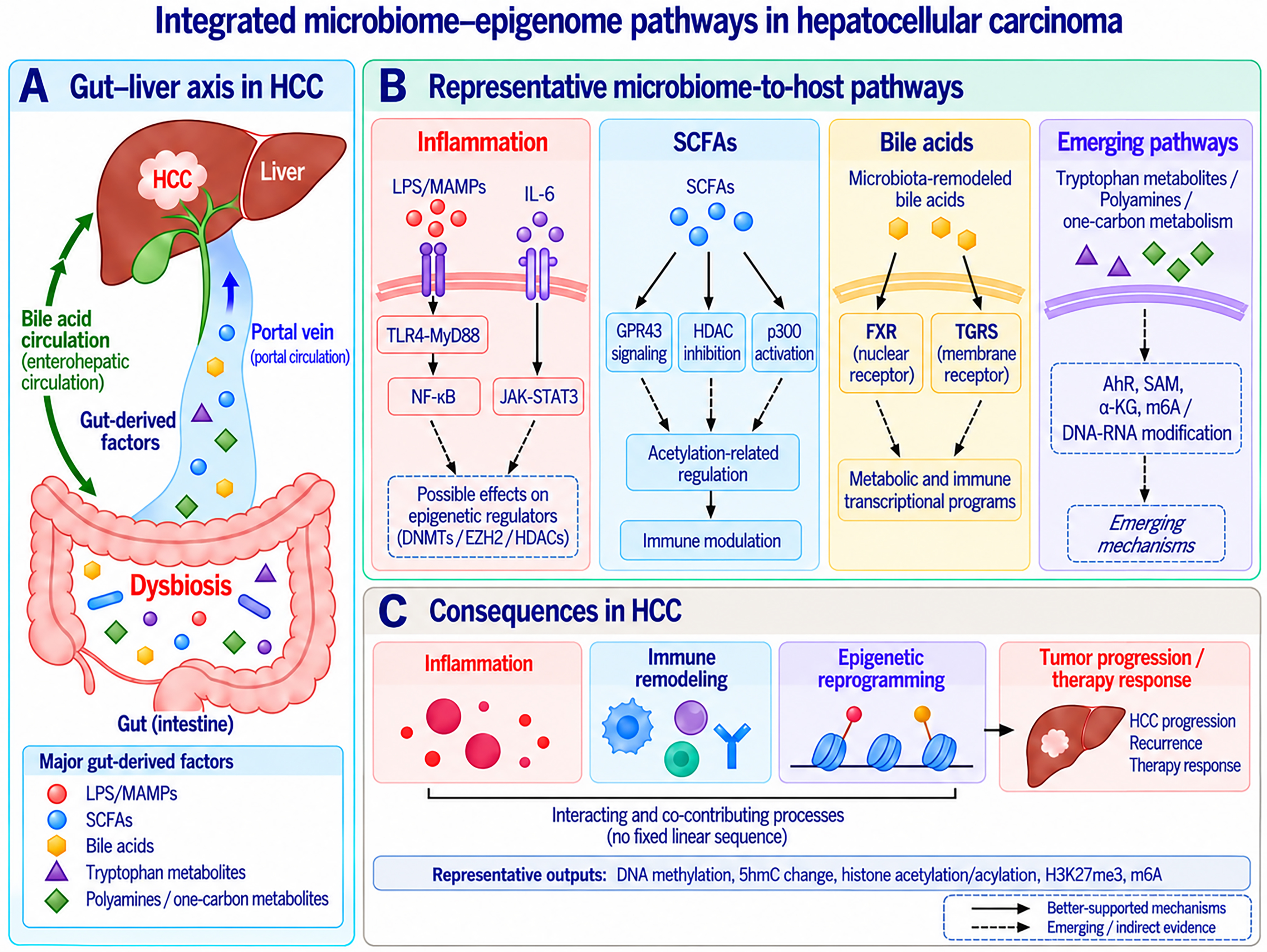
Figure 1. Integrated microbiome-epigenome pathways in HCC. (A) Gut dysbiosis and intestinal barrier dysfunction increase hepatic exposure to gut-derived factors through the portal circulation, including LPS/MAMPs, SCFAs, bile acids, tryptophan metabolites, and polyamine/one-carbon metabolites. Bile acids additionally participate in enterohepatic circulation; (B) Representative microbiome-to-host pathways include inflammatory signaling involving TLR4-MyD88-NF-κB and IL-6-JAK-STAT3, SCFA-mediated GPR43 signaling and acetylation-related regulation, microbiota-dependent bile acid signaling through FXR and TGR5, and emerging pathways involving tryptophan, polyamine, and one-carbon metabolism. Potential links between inflammatory signaling and epigenetic regulators, as well as between microbial metabolism and DNA/RNA modification, remain incompletely established in HCC; (C) These pathways may contribute to inflammatory responses, immune remodeling, epigenetic reprogramming, tumor progression, and therapeutic response. Representative epigenetic outputs include DNA methylation, 5hmC changes, histone acetylation/acylation, H3K27me3, and m6A RNA modification. Solid arrows denote relatively better-supported mechanisms, whereas dashed arrows indicate emerging or indirect evidence. The pathways are presented schematically and do not imply a fixed linear or temporal sequence. AhR: Aryl hydrocarbon receptor; α-KG: alpha-ketoglutarate; 5hmC: 5-hydroxymethylcytosine; DNA: deoxyribonucleic acid; DNMTs: DNA methyltransferases; EZH2: enhancer of zeste homolog 2; FXR: farnesoid X receptor; GPR43: G protein-coupled receptor 43; H3K27me3: histone H3 lysine 27 trimethylation; HCC: hepatocellular carcinoma; HDAC: histone deacetylase; HDACs: histone deacetylases; IL-6: interleukin 6; JAK: Janus kinase; LPS: lipopolysaccharide; m6A: N6-methyladenosine; MAMPs: microbe-associated molecular patterns; MyD88: myeloid differentiation primary response 88; NF-κB: nuclear factor kappa B; p300: histone acetyltransferase p300; RNA: ribonucleic acid; SAM: S-adenosylmethionine; SCFAs: short-chain fatty acids; TGR5: Takeda G protein-coupled receptor 5; TLR4: toll-like receptor 4; STAT3: signal transducer and activator of transcription 3.
Representative microbiome-associated and host epigenetic pathways relevant to hepatocellular carcinoma
| Principal pathway and epigenetic relevance | Cell type or experimental model | Evidence context and limitations | Potential therapeutic or translational relevance | References |
| TLR4-NF-κB signaling; proposed links to DNMTs, EZH2 and HDACs | Hepatic cells in HCC models; complementary non-HCC tumor studies | HCC inflammatory-pathway evidence; direct gut-derived epigenetic coupling remains unproven | Barrier restoration and inflammatory-pathway studies; epigenetic targets require validation | [28,31-33] |
| GPR43 activation; suppression of IL-6/JAK1/STAT3 signaling. No direct epigenetic endpoint | Hepatocytes in NAFLD-associated HCC mouse models | HCC preclinical microbiome-metabolite mechanism | Defined strain-metabolite intervention; clinical efficacy remains unestablished | [22] |
| HDAC-associated Sox13 acetylation and reduced IL-17A; a non-histone mechanism | HCC mouse models, isolated hepatic ILC3s, and complementary transfected 293T-cell assays | HCC-related functional evidence with histone H3 acetylation and complementary Sox13 assays; locus-specific chromatin mediation remains unproven | Test acetate-ILC3 immunomodulation with HDAC, Sox13 and IL-17A readouts | [38] |
| HDAC inhibition; p300 auto-acylation; histone acetylation, methylation and butyrylation | Cultured cells; non-tumor mouse liver, intestine and adipose tissue | Biochemical and mainly non-HCC evidence; HCC chromatin effects require validation | Measure metabolite exposure and cell-specific histone marks in HCC | [15-17,41,42] |
| CXCL16-CXCR6+ NKT-cell recruitment and antitumor immunity. No direct epigenetic endpoint | Liver sinusoidal endothelial cells and NKT cells in mouse liver-tumor models | Preclinical microbiome-bile acid-immune mechanism; human correlative support | Investigate bile acid composition and NKT-cell recruitment as linked intervention readouts | [45] |
| Microbial butyrate induces CXCL14 and DC/CD8+ T-cell responses. No direct epigenetic endpoint | Hepatocyte-specific Af6 deficiency in HCC models | HCC preclinical host-microbiota-metabolite-immune mechanism | Test butyrate-CXCL14 immunomodulation; distinguish it from direct bile acid effects | [46] |
| Nuclear FXR transcriptional coregulation; membrane TGR5-cAMP signaling | Hepatic and intestinal receptor studies, mainly outside HCC | Receptor mechanisms support plausibility; microbiota-driven HCC chromatin remodeling is unproven | Study receptor- and tissue-specific effects alongside quantitative bile acid profiles | [43,44] |
| TIE2-PI3K/AKT signaling and tumor promotion. No direct epigenetic endpoint | HCC cells; conventional and germ-free mouse models | HCC preclinical bacterial species-metabolite-receptor mechanism | Investigate bacterial, metabolite or receptor targeting in preclinical HCC | [56] |
| Ligand- and cell-dependent AhR immune regulation; proposed RNA-modification links | Intestinal immune and general tumor models; host kynurenine and microbial indoles | Mainly non-HCC mechanisms; a microbial metabolite-AhR-m6A axis in HCC is unproven | Separate host and microbial metabolite sources and test cell-specific effects | [54,55] |
| SAM/dcSAM partitioning; putative effects on methyl-donor availability | Biochemical and microbial metabolic studies; HCC ODC1 models | Biochemical rationale; microbial control of hepatic DNA/RNA methylation flux is unproven | Combine isotope tracing with metabolomic and epigenomic measurements | [50-53] |
| m6A-SCAP-cholesterol and m6A-TUG1-PD-L1/CD47 pathways | HCC cells and mouse models; downstream CD8+ T-cell and macrophage effects | HCC host mechanistic evidence; an upstream microbiome link was not established | Preclinical rationale for m6A-targeted treatment combined with checkpoint blockade | [48,49] |
| miR-122a/miR-200c-3p-occludin; PIM1-HDAC2; fecal miRNA-bacterial interactions | Intestinal epithelial, goblet-cell, bacterial and colitis models | Non-HCC experiments; barrier effects and direct bacterial regulation are distinct mechanisms | Test barrier/mucus regulation and host miRNA signaling in HCC-relevant models | [58-61] |
| Microbial, metabolite and inflammatory associations with outcome; no defined epigenetic target | Clinical HCC cohorts receiving ICI monotherapy or combination regimens | Observational HCC associations; findings and response definitions vary across cohorts | Candidate biomarkers require prospective external validation and control of clinical confounding | [67-72] |
FUNCTIONAL SIGNIFICANCE OF GUT MICROBIOTA ALTERATIONS IN HCC
Microbiome profiling studies in patients with HCC have revealed changes in microbial diversity, altered abundance of specific bacterial taxa, together with changes in predicted or inferred microbial functional potential[18,19]. However, these cross-sectional observations may be influenced by multiple confounding factors, including the underlying cause of liver disease, geographic differences, dietary patterns, cirrhosis severity, and recent medication exposure, including antibiotics. Characterizing specific functional pathways provides a stronger foundation for mechanistic biological linkage than taxonomy alone. Relevant functional pathways include the capacity for short-chain fatty acid biosynthesis, the enzymatic deconjugation and dehydroxylation of bile acids, LPS biosynthesis potential, and the metabolic processing of tryptophan and polyamines.
Several independent HCC cohorts have reported reductions in selected SCFA-producing or SCFA-associated bacterial taxa, although the affected taxa and the direction of these alterations vary according to disease etiology, stage, and comparator group[18-20]. For example, reduced butyrate-producing genera have been reported in early HCC, while reductions in Faecalibacterium and Bifidobacterium have been observed in selected etiological subgroups of HCC[21]. In mouse models of non-alcoholic fatty liver disease (NAFLD)-associated HCC, Bifidobacterium pseudolongum-derived acetate suppresses tumor development through G protein-coupled receptor 43 (GPR43) activation and subsequent inhibition of the interleukin 6 (IL-6)/Janus kinase 1 (JAK1)/signal transducer and activator of transcription 3 (STAT3) signaling pathway[22]. This model provides direct mechanistic evidence by linking a defined bacterial strain, its specific metabolite, the disease context, and the corresponding host signaling pathway.
Bile acid metabolism constitutes an additional enterohepatic pathway associated with HCC pathophysiology. The gut microbiota regulates the conversion of primary bile acids into secondary bile acids through specific enzymatic processes, particularly bile salt hydrolase (BSH)-mediated deconjugation and 7α-dehydroxylation. Microbiota-driven remodeling of bile acid composition subsequently influences host receptor signaling, including pathways mediated by farnesoid X receptor (FXR), Takeda G protein-coupled receptor 5 (TGR5), vitamin D receptor (VDR), pregnane X receptor (PXR), and constitutive androstane receptor (CAR)[23-27].
In parallel, enrichment of pro-inflammatory bacterial taxa and Gram-negative bacteria-associated functional pathways may increase systemic exposure to LPS. Preclinical HCC evidence further indicates that modulation of the gut microbiota and intestinal barrier can reduce LPS leakage and attenuate hepatic toll-like receptor 4 (TLR4)-nuclear factor kappa B (NF-κB) signaling[28]. These microbial alterations occur in the broader context of cirrhosis, bile acid dysregulation, intestinal barrier dysfunction, and immune disturbance, all of which may independently influence microbiome composition and HCC biology and therefore confound cross-sectional associations[4,5,29,30].
INTESTINE-DERIVED INFLAMMATORY SIGNALS AND EPIGENETIC REGULATION
Following the structural compromise of the intestinal epithelial barrier, MAMPs enter the portal venous system. Microbial components such as LPS can translocate to the liver, where they engage pattern recognition receptors on various hepatic cell types, including Kupffer cells, liver sinusoidal endothelial cells, hepatic stellate cells, and hepatocytes[4,5]. Activation of the TLR4-myeloid differentiation primary response 88 (MyD88)-NF-κB axis together with the IL-6/JAK/STAT3 signaling pathway reshapes the expression of inflammatory cytokines, chemokines, and anti-apoptotic mediators in these intrahepatic cells. Increasing evidence indicates that these inflammatory signaling pathways interact with epigenetic regulators to influence gene expression programs.
Mechanistic links between inflammatory transcription factors and epigenetic enzymes have been demonstrated in non-HCC tumor models. In particular, STAT3 acetylation at Lys685 promotes its interaction with DNMT1 and contributes to promoter methylation and transcriptional silencing of selected tumor-suppressor genes[31]. Independently, dysregulation of DNMTs, enhancer of zeste homolog 2 (EZH2), and histone deacetylases (HDACs) is well documented in HCC[9,32]. However, current evidence does not establish intestine-derived inflammatory signaling as the direct upstream cause of these epigenetic abnormalities in HCC[30,33].
The abnormal hypermethylation of promoter CpG islands regulating genes such as RASSF1A, CDKN2A/p16, SOCS1, and GSTP1 is extensively characterized in HCC tissue arrays[12,14]. EZH2 is the catalytic subunit of Polycomb Repressive Complex 2 (PRC2) and promotes local deposition of H3K27me3, thereby facilitating transcriptional silencing of selected differentiation-related genes, tumor-suppressor genes, and tumor-suppressive miRNA loci[32]. In chronic liver disease, persistent exposure to intestine-derived LPS and downstream inflammatory cytokines such as IL-6 and TNF may maintain intrahepatic cells in prolonged states of inflammatory stress and regeneration and may thereby influence the expression or activity of epigenetic regulators such as DNMTs, EZH2, and HDACs. Ohtani reviewed how persistent senescence-associated secretory phenotype (SASP) programs can connect cellular stress, chronic inflammation, and cancer, providing a conceptual framework for long-lasting inflammatory remodeling of the tissue microenvironment[34]. In murine models simulating obesity-driven HCC, Yoshimoto et al. demonstrate that specific gut microbial metabolites induce a SASP in hepatic stellate cells, which promotes hepatocarcinogenesis[35]. Collectively, these findings indicate that intestine-derived inflammation and microbial metabolic signals can contribute to long-term remodeling of the premalignant microenvironment.
Alterations in TET enzyme function and the genomic distribution of 5hmC connect cellular oxidative stress, epigenetic remodeling, and immunomodulation. The TET dioxygenase family initiates active DNA demethylation by sequentially oxidizing 5-methylcytosine to 5hmC, 5-formylcytosine (5fC), and 5-carboxylcytosine (5caC)[36]. Lu et al. showed that oxidative stress within the HCC microenvironment activates TET2 in B cells. Aryl hydrocarbon receptor (AhR) recruits TET2 to specific genomic sites, facilitating hydroxymethylation at the Il10 locus and promoting IL-10+ regulatory B-cell generation; conversely, B-cell-specific Tet2 inactivation reduces this immunosuppressive population and enhances antitumor immunity[37]. These studies identify distinct regulatory mechanisms of different evidentiary strength rather than a single established epigenetic cascade initiated by intestine-derived inflammation. Establishing relationships between intestinal-derived inflammatory signals, TET enzyme activity, 5hmC patterns, and the functional status of tissue-resident immune cells requires integrated quantitative analyses using intact liver specimens or comprehensive liquid biopsy approaches.
SCFAs AND HISTONE ACETYLATION/ACYLATION MODIFICATIONS
SCFAs, predominantly acetate, propionate, and butyrate, are synthesized through the anaerobic bacterial fermentation of dietary fiber in the intestinal lumen. The enzymatic mechanisms connecting SCFAs with host epigenetic regulation have been increasingly characterized. Waldecker et al. showed that butyrate inhibits HDAC activity in nuclear extracts from human colon carcinoma cells, whereas propionate exerts a weaker inhibitory effect[15]. Subsequently, Thomas and Denu reported that propionate and butyrate can be converted into their corresponding acyl-coenzyme A (acyl-CoA) species - activated metabolic intermediates formed from fatty acid-derived acyl groups and coenzyme A - and enhance the histone and protein acetyltransferase activity of p300 by promoting auto-acylation of its autoinhibitory loop[16]. Therefore, the effects of SCFAs on histone modifications are likely mediated through multiple mechanisms, including HDAC inhibition, p300 activation, and alterations in intracellular acyl-CoA availability. The relative contribution of each mechanism may depend on the specific metabolite, its absolute concentration, exposure duration, and the metabolic state of the target cells.
Extrapolating the enzymatic interactions of SCFAs to HCC requires accounting for absolute molecular concentrations and tissue-specific metabolic rates. SCFA availability varies substantially across different biological compartments, including the colonic lumen, fecal samples, peripheral blood, and portal circulation, resulting in distinct exposure levels within the liver. Furthermore, the capacity to transport and metabolize SCFAs differs among functional hepatocytes, glycolytic tumor cells, resident Kupffer cells, hepatic stellate cells, and specific subsets of infiltrating lymphocytes. Donohoe et al. showed in a colorectal cancer model that the effect of butyrate depends on its intracellular metabolic fate. Normal colonocytes efficiently oxidized butyrate as an energy substrate, whereas the Warburg phenotype of cancerous colonocytes limited butyrate oxidation, allowing intracellular accumulation and HDAC inhibition, with consequent changes in histone acetylation and cell proliferation[17]. The findings from colorectal cancer raise the testable possibility that SCFA effects in HCC may vary with the metabolic state of malignant and non-malignant intrahepatic cell populations.
In HCC-bearing mice, Hu et al. showed that microbiota-associated acetate inhibits HDAC activity, increases Sox13 K30 acetylation, reduces Sox13 mRNA expression and Sox13-dependent IL-17A transcription in hepatic group 3 innate lymphoid cell (ILC3s), and thereby enhances antitumor immunity[38]. In non-HCC immune models, Arpaia et al. found that butyrate and propionate promote peripheral regulatory T cell (Treg) differentiation[39]. In a non-tumor systemic bacterial infection model, Balmer et al. showed that stress-associated increases in circulating acetate expand the acetyl-CoA pool in memory CD8+ T cells and promote glyceraldehyde-3-phosphate dehydrogenase (GAPDH) acetylation, glycolysis, and rapid effector responses[40]. Krautkramer et al. further demonstrated that microbial colonization and dietary composition alter histone acetylation and methylation in mouse liver, colon, and adipose tissue, and that SCFA supplementation partially reproduces the chromatin changes induced by colonization[41]. Gates et al. identified microbiota-regulated H3K9 and H3K27 butyrylation in mouse intestinal epithelial cells and showed that histone butyrylation is responsive to tributyrin administration; H3K27 butyrylation was associated with active regulatory elements and higher gene expression[42]. Together, these studies demonstrate that SCFAs can influence host cellular states through metabolic remodeling, HDAC-dependent regulation, protein acetylation, and histone modifications across several experimental systems. However, direct HCC-specific evidence is currently strongest for the acetate-HDAC-ILC3 pathway, whereas several histone-level mechanisms remain extrapolated from non-HCC tissues and models. HCC research necessitates the precise quantification of discrete chromatin marks - including histone H3 lysine 27 acetylation (H3K27ac), histone H3 lysine 9 acetylation (H3K9ac), histone H4 acetylation (H4ac), and site-specific histone butyrylation - within isolated intrahepatic cellular populations, and the correlation of these measurements with the functional genomic capacity of the microbiome and absolute concentrations of circulating metabolites.
BILE ACIDS, NUCLEAR RECEPTORS, AND CHROMATIN STATES
Primary bile acids are synthesized in the liver and subsequently transformed by gut microbial enzymes into secondary bile acids through intestinal deconjugation and dehydroxylation processes. The bile acid pathway simultaneously connects microbial metabolism, intrahepatic immune surveillance, and nuclear receptor-dependent transcriptional regulation. In HCC models, animal studies provide relatively direct evidence that microbiota-driven bile acid remodeling influences intrahepatic immune-cell composition and tumor progression. However, the relationship between bile acid receptor signaling and chromatin regulation has been established mainly through investigations of FXR-responsive genes and associated coregulatory factors. Beyond their role in lipid absorption, bile acids also function as signaling molecules through the nuclear receptor FXR and the membrane G protein-coupled receptor TGR5. Selected bile acids and their derivatives can additionally engage VDR- and PXR-related signaling, while CAR contributes to bile acid detoxification and metabolic homeostasis[23-27,43,44]. Through these receptor systems, bile acids regulate lipid and glucose metabolism, inflammatory cascades, drug detoxification, and cellular proliferation. In mice, Ma et al. reported that depletion of Gram-positive bacteria responsible for converting primary to secondary bile acids increased CXCL16 expression in liver sinusoidal endothelial cells, promoted accumulation of CXCR6+ NKT cells and IFN-γ production, and suppressed liver tumor growth; supplementation with secondary bile acids or colonization with bile acid-metabolizing bacteria reversed these effects. In non-tumor liver tissue from patients with primary liver cancer, CDCA levels correlated positively with CXCL16 expression, whereas GLCA levels correlated negatively, providing human correlative support for the microbiota-bile acid-CXCL16 axis[45]. A recent study further showed that hepatocyte-specific Af6 deficiency increases primary bile acid production, reshapes the gut microbiota, and elevates microbiota-derived butyrate. Butyrate, rather than bile acids directly, upregulated hepatic CXCL14, promoted recruitment of activated dendritic cells, and enhanced CD8+ T-cell cytotoxicity, thereby suppressing HCC progression[46].
Bile acid signaling can influence host chromatin states through nuclear receptor-associated coregulatory mechanisms, with this relationship being best characterized for FXR in non-HCC experimental systems[43]. Following ligand binding, FXR heterodimerizes with retinoid X receptor (RXR), binds specific genomic response elements, and recruits coactivator or corepressor complexes according to the target gene and cellular context, thereby influencing local histone modifications and transcriptional activity. Reported FXR-associated coregulators include the acetyltransferase p300 and other chromatin-modifying cofactors, whereas the participation of nuclear receptor corepressor (NcoR)/silencing mediator of retinoid and thyroid hormone receptor (SMRT)-type corepressor complexes in FXR regulation is less well defined[43]. These interactions can alter histone acetylation, FXR post-translational modification, chromatin accessibility, and target-gene transcription. The activation of the membrane-bound receptor TGR5 engages the Gs-protein-coupled cyclic adenosine monophosphate (cAMP)-protein kinase A (PKA)-cAMP response element-binding protein (CREB) signal transduction cascade, regulating the transcription of specific inflammatory and metabolic genes[44]. Receptors such as VDR, PXR, and CAR coordinate the expression of gene networks involved in bile acid homeostasis, xenobiotic detoxification, and drug responses. Shifts in the physiological bile acid profile translate into altered transcriptional landscapes across hepatocytes and local immune populations via these transcription factors and coregulators.
FXR and TGR5 signaling is highly context- and tissue-dependent, whereas stage-specific effects in HCC remain incompletely defined. Within hepatocytes, FXR activation contributes to suppression of bile acid synthesis and maintenance of metabolic homeostasis. In the intestinal epithelium, FXR activation induces fibroblast growth factor 15 (FGF15) in mice or FGF19 in humans, which signals through hepatic FGFR4 to suppress bile acid synthesis and can influence hepatocyte proliferative signaling[43,44]. Golonka et al. found that combined Tlr5 and Fxr deficiency was associated with more severe spontaneous hepatocarcinogenesis, aggravated bile acid dysmetabolism, and marked gut microbial dysbiosis compared with Fxr deficiency alone[47]. This model indicates that genetic interactions between innate immune signals that maintain microbial homeostasis and FXR-dependent bile acid homeostasis can jointly influence susceptibility to liver cancer.
TRYPTOPHAN, POLYAMINE, AND ONE-CARBON METABOLISM IN HCC EPIGENETIC AND IMMUNE REGULATION
The biochemical cofactor requirements of key epigenetic enzymes relevant to these pathways are well established: DNA and RNA methyltransferases use SAM, whereas TET enzymes and the m6A demethylases fat mass and obesity-associated protein (FTO) and alkB homolog 5 (ALKBH5) belong to the Fe2+/α-KG-dependent dioxygenase family[10,11,36]. In HCC, direct experimental studies further demonstrate that m6A regulation can influence tumor metabolic reprogramming and antitumor immunity[48,49]. Whether microbially derived metabolites directly remodel DNA or RNA methylation in HCC by altering these substrate pools remains a mechanistic connection that requires validation.
DNA and RNA methylation and demethylation reactions depend on defined metabolic substrates and cofactors. DNMTs and the m6A writer complex centered on METTL3/METTL14 use SAM as the methyl donor, whereas TET dioxygenases and the RNA demethylases FTO and ALKBH5 require α-KG, Fe2+, and molecular oxygen for catalysis. Accordingly, intracellular SAM/S-adenosylhomocysteine (SAH) balance and the availability of α-KG, Fe2+, and oxygen can constrain the catalytic potential of methylation and demethylation reactions[10,11,36]. Krautkramer et al. demonstrated that diet and microbial colonization alter histone modification states across multiple host tissues and that SCFA supplementation can partially reproduce colonization-associated chromatin changes[41]. This study provides general evidence that microbial metabolism can influence host epigenetic states, but it does not directly establish microbiota-dependent control of SAM- or α-KG-driven DNA/RNA methylation in HCC.
Polyamine metabolism is biochemically linked to one-carbon metabolism and SAM consumption. Polyamine cellular synthesis initiates with ornithine decarboxylase (ODC1) and proceeds via spermidine synthase and spermine synthase to produce spermidine and spermine[50]. Each of these subsequent synthesis steps requires decarboxylated SAM to supply aminopropyl groups[51]. In HCC, ODC1 is upregulated and can promote tumor-cell proliferation, migration, and invasion through AKT/GSK3β/β-catenin signaling and modulation of the acidic microenvironment[52]. Endogenous polyamine synthesis converts a portion of SAM to decarboxylated SAM, which then supplies aminopropyl groups for spermidine and spermine synthesis. Polyamine metabolism can therefore affect the metabolic allocation between SAM and decarboxylated SAM. Gut microbial polyamines can contribute to host-accessible polyamine pools[53]. Direct mechanistic evidence for the polyamine-SAM-methylation axis in HCC remains relatively limited compared with the established evidence supporting SCFA- and bile acid-associated pathways. Biochemically, increased flux from SAM into dcSAM provides a testable mechanism by which polyamine synthesis could alter methyl-donor allocation; whether this contributes materially to global or locus-specific DNA/RNA methylation changes in HCC has not been demonstrated. This hypothesis is therefore suitable for integrated isotope-tracing, metabolomic, and epigenomic analysis.
The evaluation of tryptophan metabolism requires the separation of host-intrinsic pathways from microbiota-driven enzymatic processes. Host cells direct tryptophan into the kynurenine pathway primarily through indoleamine 2,3-dioxygenase 1 (IDO1) and tryptophan 2,3-dioxygenase (TDO2). Some gut microorganisms use unabsorbed tryptophan to generate indole and its derivatives, including indole-3-acetic acid, indole-3-propionic acid, and indole-3-aldehyde. Kynurenine and selected microbiota-derived indole metabolites can both modulate AhR signaling, but their biological effects depend on ligand structure, exposure concentration and duration, and target-cell type and may therefore be divergent or even opposing[54,55]. A distinct microbial metabolite-TEK receptor tyrosine kinase (TIE2) pathway has been demonstrated in HCC models. Zhang et al. showed that Catenibacterium mitsuokai disrupts the intestinal barrier and translocates to the liver, where its secreted quinolinic acid binds to and activates TIE2 on HCC cells, leading to downstream phosphoinositide 3-kinase (PI3K)/protein kinase B (AKT) activation and tumor promotion[56]. Using conventional and germ-free mice together with bacterial, metabolite, and receptor-level interventions, the study provides direct HCC-specific causal evidence linking a defined bacterial species, quinolinic acid, TIE2, and PI3K/AKT signaling[56].
The IDO1/TDO2-kynurenine-AhR pathway is a well-characterized immunoregulatory axis across multiple tumor types[55]. HCC-specific evidence is more limited, although IDO1 upregulation has been shown to contribute to adaptive resistance to immune checkpoint blockade in experimental HCC models[57]. In addition, m6A RNA modifications have emerged as potential regulators of the HCC immune microenvironment and therapeutic response[48,49]. Pan et al. demonstrated that METTL3 promotes cholesterol biosynthesis through m6A-dependent enhancement of sterol regulatory element-binding protein cleavage-activating protein (SCAP) translation, resulting in cholesterol-mediated impairment of CD8+T-cell function; METTL3 inhibition enhanced the efficacy of anti-PD-1 therapy in NAFLD-HCC models[49]. Xi et al. further showed that METTL3-mediated m6A modification increases taurine upregulated 1 (TUG1) expression, leading to reduced CD8+ T-cell effector function and impaired macrophage phagocytosis through regulation of PD-L1 and CD47, respectively[48]. These findings indicate that metabolic reprogramming, AhR signaling, and m6A regulation may all participate in shaping HCC immunity; however, they do not yet directly demonstrate a unified microbial metabolite-AhR-RNA methylation axis. Future studies should combine microbiota and metabolite interventions, cell-specific manipulation of AhR or METTL3, and joint measurement of m6A and metabolic flux to define their interactions and causal order.
RECIPROCAL IMPACT OF HOST EPIGENETICS ON THE INTESTINAL MICROECOLOGY
Epigenetic and post-transcriptional regulation not only responds to microbial signals but can also indirectly shape the ecological niches available to the microbiota by altering the intestinal epithelial barrier, mucus secretion, and mucosal immune programs. Current mechanistic evidence for this reverse regulation derives mainly from intestinal epithelial, inflammatory bowel disease, and colitis-associated cancer models and provides a framework for understanding bidirectional host-microbiota interactions in chronic liver disease. To maintain immunological and barrier homeostasis, the host leverages DNA methylation and specific histone modifications to orchestrate the transcriptional programs of resident immune and epithelial cells, thereby resisting local dysbiosis. For example, TNF-α induces miR-122a in intestinal epithelial cells, and miR-122a directly binds the 3′ untranslated region of occludin mRNA and promotes its degradation, thereby increasing intestinal tight-junction permeability[58]. Separately, Rawat et al. showed that IL-1β induces miR-200c-3p, which likewise targets occludin mRNA and increases intestinal permeability[59]. Such post-transcriptional disruption of tight-junction integrity increases intestinal permeability and may alter ecological conditions relevant to microbial colonization. Host epigenetic mechanisms can influence microbial colonization, growth, and community structure through targeted histone regulation and cross-kingdom molecular communication. Histone regulation in the host can reshape the intestinal niche by changing goblet-cell abundance and the mucus layer. Yang et al. showed that intestinal epithelial PIM1 interacts with and phosphorylates HDAC2, reduces HDAC2 protein abundance, and decreases HDAC2 occupancy at selected Wnt-related promoters, thereby altering Wnt-associated transcription[60]. PIM1 also affected Notch signaling, although the HDAC2 dependence of this effect was less clearly established. Increased epithelial PIM1 was accompanied by reduced goblet cells and MUC2 and aggravated experimental colitis and colitis-associated cancer; conversely, PIM1 deficiency increased goblet cells and MUC2 and was associated with higher relative abundances of Roseburia, Alloprevotella, and Bifidobacterium. This study therefore links the intestinal epithelial PIM1-HDAC2 axis and mucus-barrier remodeling with changes in microbial community structure, although the causal direction and contribution of the microbiota to the disease phenotype remain incompletely resolved[60]. Host-derived miRNAs may also participate in cross-kingdom signaling. Liu et al. demonstrated that fecal miRNAs derived in part from intestinal epithelial cells, including extracellular-vesicle-associated miRNAs, can enter specific bacterial species and regulate bacterial transcripts and growth through sequence-dependent interactions[61,62]. However, because these effects depend highly on individual miRNAs and bacterial taxa, their broader significance within complex microbial ecosystems and chronic liver disease remains largely unresolved. Together, these studies indicate that host miRNAs, histone regulation, and epithelial secretory programs can reciprocally influence the gut microbiota by altering barrier integrity and microbial ecological niches. Extending these mechanisms to HCC will require simultaneous analysis of epithelial epigenetic states, barrier function, and microbiota dynamics in liver disease models and longitudinal patient cohorts.
INFLUENCE OF ETIOLOGICAL BACKGROUND ON MECHANISTIC INTERPRETATIONS
HCC is etiologically heterogeneous, and viral, alcohol-associated, and metabolic liver diseases create distinct inflammatory, metabolic, microbial, and epigenetic contexts in which HCC develops[4,5,29,63-65]. HBV-associated HCC develops in the context of chronic viral persistence and may involve HBV DNA integration together with host epigenetic alterations, including aberrant DNA methylation[63]. In HCV-associated HCC, specific transcriptional and epigenetic aberrations persist in the host cellular memory even following viral clearance[64]. Alcohol-associated liver disease is frequently accompanied by increased intestinal permeability and endotoxin exposure, together with ethanol- and acetaldehyde-related oxidative and inflammatory stress; these processes may remain relevant in the background from which alcohol-associated HCC develops. MASLD/metabolic dysfunction-associated steatohepatitis (MASH)-associated HCC is connected with systemic obesity, insulin resistance, disruption of bile acid metabolism, and chronic low-grade inflammatory signaling[29,65].
These distinct etiologies alter the interpretation of any microbiome-epigenetic interaction. SCFA-related pathways may warrant particular investigation in metabolically defined HCC populations, especially where diet-related dysbiosis or reduced microbial SCFA-producing capacity is documented. Similarly, bile acid receptor-directed strategies may be most appropriately evaluated in mechanistically stratified studies that document baseline bile acid composition and receptor-signaling states, rather than being assigned solely on the basis of HCC etiology. LPS translocation and TLR4 signaling may be particularly relevant in alcohol-associated liver disease and advanced cirrhosis with impaired intestinal barrier function, although their relative contribution to HCC biology is likely to vary among patients. Clinical cohorts should therefore systematically document underlying etiology, cirrhosis severity, antiviral treatment history, antibiotic and proton-pump-inhibitor exposure, dietary patterns, and systemic metabolic status so that major confounders can be assessed or adjusted for and cross-study comparability can be improved. More broadly, HCC prognosis is determined by multiple interacting factors, including tumor biology, tumor burden, liver functional reserve, underlying etiology, and treatment selection. Recent clinical commentary has therefore emphasized individualized, patient-centered risk stratification rather than reliance on any single molecular marker[66]. These considerations are particularly important when evaluating whether microbiome or epigenetic features provide prognostic or predictive information beyond established clinical determinants.
MECHANISM-DRIVEN THERAPEUTIC IMPLICATIONS
The mechanisms outlined above provide potential therapeutic entry points for targeting the gut microbiome and associated epigenetic pathways; however, HCC etiology and measurable biological phenotypes should currently be used primarily to prioritize mechanistic hypotheses and stratify clinical studies rather than to select therapy directly. In HBV- or HCV-related HCC, virological control, persistent inflammation, and virus-induced host epigenetic abnormalities remain fundamental considerations[63,64]. Alcohol-associated liver disease and advanced cirrhosis with marked barrier dysfunction may involve greater microbial-product translocation and endotoxin-related inflammation. In MASLD/MASH-associated HCC, direct preclinical evidence supports selected metabolic pathways, including the acetate-GPR43-IL-6/JAK1/STAT3 axis[22] and the METTL3-m6A-SCAP-cholesterol axis[49], whereas other microbiome-metabolic pathways remain less completely validated.
For HCC characterized by prominent barrier dysfunction and endotoxin-related inflammation, restoration of intestinal barrier integrity and attenuation of LPS-TLR4-NF-κB signaling represent mechanistically plausible intervention strategies that require clinical validation. These approaches may be particularly relevant for investigation in alcohol-associated liver disease or advanced cirrhosis with endotoxemia. In an HCC mouse model, microbiota-directed intervention improved intestinal barrier integrity, reduced LPS leakage, and attenuated hepatic TLR4/NF-κB signaling[28]. However, a pro-inflammatory microbial signature cannot currently be used to directly select anticancer therapy. Future clinical trials should incorporate barrier-function measures, circulating LPS and inflammatory cytokines, microbial functional modules, and immune-cell phenotypes as mechanistic endpoints to determine whether an intervention truly acts on the prespecified gut-liver inflammatory axis.
Future mechanistic and translational studies of SCFA-targeted interventions may reasonably prioritize MASLD/MASH-associated HCC or biologically defined populations with reduced microbial SCFA-producing capacity. Compared with broadly defined probiotics that lack functional specification, strains with defined metabolic capabilities, compositionally defined postbiotics, or standardized metabolite formulations are more suitable for establishing links among a strain, its metabolite, and host effects. Using two diet-induced MASLD-HCC mouse models, Song et al. demonstrated that Bifidobacterium pseudolongum-derived acetate mediates antitumor effects through hepatocyte GPR43 activation, attenuation of IL-6/JAK1/STAT3 signaling, and restoration of gut microbial composition and barrier integrity[22]. This study provides preclinical support for etiology-matched strain-metabolite intervention, although clinical translation requires definition of dose, colonization stability, and target population. Related studies should separately quantify fecal and circulating acetate, propionate, and butyrate and evaluate their biological effects together with HDAC activity, p300-associated modifications, and immune-cell function.
The therapeutic goal of targeting bile acid pathways should not be simplified to increasing or decreasing total bile acid concentrations; instead, it should focus on restoring the balance among bile acid composition, microbial transformation capacity, and receptor signaling. Potential bile acid-directed approaches - including modulation of FXR or TGR5 signaling, alteration of enterohepatic bile acid circulation, and intestine-selective FXR regulation - have been investigated principally in metabolic and liver-disease contexts[22,24-26,43]. Their therapeutic value in HCC remains to be established. Because individual primary and secondary bile acids differ in receptor affinity and immunological effects, such strategies are best studied in patients whose absolute baseline bile acid profiles, microbial bile salt hydrolase and 7α-dehydroxylation capacities, and receptor signaling states are clearly characterized. MASLD/MASH-related HCC may warrant priority attention to the link between bile acid imbalance and metabolic inflammation, whereas viral- or alcohol-related HCC requires separation of the effects of the underlying etiology from those of cirrhosis, cholestasis, and impaired liver function on the bile acid profile. At present, these agents should be evaluated in HCC primarily through mechanistically defined, phenotype-stratified clinical studies.
Combining epigenetic agents with immune checkpoint inhibitors (ICIs) offers another strategy for linking host epigenetic states to antitumor immunity. DNMT, HDAC, EZH2, and BET inhibitors can modulate antigen processing and presentation, inflammatory signaling, chemokine production, and immune-cell recruitment; however, their effects vary substantially depending on the specific molecular target, cellular context, and disease background. Therefore, epigenetic therapies should not be considered a uniform class of nonspecific immune-sensitizing agents[9]. In virus-related HCC, the relationships among virological status, DNA methylation, and immune escape may represent a priority area. In MASLD/MASH-related HCC, metabolic-epitranscriptomic pathways have more direct preclinical support. Pan et al. demonstrated that METTL3 promotes SCAP translation through m6A modification, increases cholesterol biosynthesis, and impairs CD8+ T-cell function; genetic or pharmacological METTL3 inhibition enhanced the efficacy of anti-PD-1 therapy in NAFLD-HCC models[49]. Xi et al. further showed that METTL3-dependent m6A modification influences CD8+ T-cell activity and macrophage phagocytic function through regulation of TUG1, PD-L1, and CD47[48]. Collectively, these findings provide a rationale for exploring combined m6A-directed and immunotherapeutic strategies.
At present, the translational potential of gut microbiome profiling in HCC immunotherapy is being explored primarily for patient stratification and response prediction, but no microbiome feature has been validated for routine treatment selection. In the small longitudinal cohort reported by Zheng et al., anti-PD-1 responders showed greater microbial richness and enrichment of taxa including Akkermansia muciniphila and Ruminococcaceae species[67]. Ponziani et al. reported lower pretreatment fecal calprotectin and higher Akkermansia abundance in patients who achieved disease control[68]. Lee et al. further identified pretreatment microbiota-bile acid associations with ICI outcome, including enrichment of selected secondary bile acids in objective responders and a microbiota signature associated with longer survival[69]. In an atezolizumab-bevacizumab cohort, Bacteroides stercoris and Parabacteroides merdae were enriched in responders, although overall alpha and beta diversity did not differ significantly between response groups[70]. Xin et al. identified specific taxa associated with progression-free survival (PFS) and overall survival (OS) despite no significant difference in alpha diversity between responders and nonresponders[71]. Conversely, the prospective study by Shen et al. found no significant association of baseline alpha diversity, richness, or overall community composition with objective response and observed no major community-level change during the first 8 weeks of ICI therapy[72]. These heterogeneous findings indicate that associations between the gut microbiome and ICI outcomes are sensitive to cohort composition, geography, diet, HCC etiology, liver function, concomitant medications, treatment regimen, sampling time, and sequencing methodology. No single bacterial species or microbial signature has yet been reproducibly validated across independent HCC cohorts for clinical decision-making.
Evidence from other solid malignancies further indicates that the gut microbiome can influence responses to PD-1/PD-L1 blockade. Studies across epithelial cancers and melanoma have linked antibiotic exposure, microbial diversity, and specific commensal taxa with variations in immunotherapy efficacy[73,74]. In patients with anti-PD-1-refractory melanoma, responder-derived fecal microbiota transplantation (FMT) followed by anti-PD-1 rechallenge restored clinical benefit in a subset of individuals, providing initial clinical evidence that microbiome-based interventions may partially overcome treatment resistance[75,76]. These results, however, cannot be directly extrapolated to HCC. Because many patients with HCC have underlying cirrhosis and may have portal hypertension, impaired barrier function, or cirrhosis-associated immune dysfunction, the safety profile of FMT in this population may differ from that in melanoma cohorts. These comorbidities raise potential concerns regarding microbial translocation and infection, but HCC-specific safety data remain limited. FMT should therefore remain experimental in HCC, with stringent donor and pathogen screening, stratification by liver function, and long-term safety monitoring. Compared with whole-community FMT, compositionally defined synthetic consortia or functionally traceable postbiotics may offer greater controllability.
Novel diagnostic, prognostic, and treatment-response models should integrate microbial function, metabolites, epigenetic features, and etiological context rather than relying on a single genus or methylation site. In addition to fecal and circulating biomarkers, intratumoral microbial signals may provide complementary information for HCC prognostic stratification. Jiang et al. combined 16S rRNA sequencing with bacterial RNA in situ detection in HCC tumors and matched non-tumor tissues and found that intratumoral microbial composition was associated with survival after surgery, further supporting its potential as a tissue-level prognostic biomarker[77]. However, microbial signatures identified to date remain heterogeneous across studies, highlighting the need for multicenter validation with harmonized workflows, particularly for low-biomass sample processing and analysis. Translationally promising models could integrate fecal metagenomic functional modules; absolute plasma concentrations of bile acids, SCFAs, and tryptophan metabolites; barrier and inflammatory indices; tissue or circulating DNA methylation features; and clinical markers such as alpha-fetoprotein (AFP) and protein induced by vitamin K absence or antagonist-II (PIVKA-II). Aberrant DNA methylation of RASSF1A, CDKN2A, GSTP1, and SOCS1 has been consistently reported in multiple HCC studies and supported by meta-analytic evidence[12,14]. In contrast, microbiome-derived biomarkers remain supported largely by single-center observational cohorts, relatively small treatment-response cohorts, or computationally derived public datasets, with limited external validation. Treatment timing should also be recorded and incorporated into biomarker analyses as a potential clinical confounder. A systematic review and meta-analysis found no statistically significant pooled difference in OS or disease-free survival (DFS) between delayed and non-delayed HCC treatment groups; however, the delayed-treatment group had smaller tumors and differed in treatment allocation, illustrating how tumor biology and clinical selection can complicate interpretation of time-to-treatment effects[78]. Accordingly, associations between microbial or epigenetic biomarkers and outcome should be adjusted for treatment timing together with tumor burden, liver function, and treatment modality. Therefore, integrated biomarker models should be evaluated in prospective, multicenter cohorts with prespecified documentation and statistical adjustment for etiology, liver function, tumor burden, treatment regimen, concomitant medications, and treatment timing, followed by independent external validation.
Overall, etiological context can help define research priorities for microbiome-epigenetic interventions. Virus-related HCC should be assessed with simultaneous consideration of virological control, persistent inflammation, and host epigenetic status. Alcohol-associated HCC and advanced cirrhosis characterized by intestinal barrier disruption may represent priority populations for investigating barrier restoration strategies, microbial product translocation, and TLR4-mediated inflammatory pathways. In contrast, MASLD/MASH-associated HCC may provide a particularly relevant context for studying SCFA metabolism, bile acid signaling, cholesterol homeostasis, and m6A-mediated regulation. In patients receiving ICIs, microbiota, metabolites, and intestinal inflammatory indices may serve as cross-etiology adjunctive stratification factors, but they cannot replace established clinical, liver-function, and tumor biomarkers before prospective and cross-cohort validation.
STUDY DESIGN AND EVIDENCE INTEGRATION
Many human microbiome studies in HCC rely on cross-sectional comparisons or relatively short-term pre- and post-treatment correlative analyses[5]. Such designs are vulnerable to confounding by cirrhosis severity, dietary patterns, antibiotic and proton-pump-inhibitor exposure, tumor burden, underlying etiology, hepatic function, and treatment history[4,29,30,65]. Prospective longitudinal studies beginning before HCC diagnosis are therefore needed to distinguish pre-existing microbial and metabolic features from changes caused by progressive liver disease, established cancer, or treatment. These studies should incorporate serial fecal and blood sampling, quantitative metabolite measurements, detailed exposure and medication data, and, when HCC subsequently develops, molecular and epigenomic profiling of tumor and adjacent liver tissue. Longitudinal sampling can help establish temporal ordering and reduce reverse-causation bias, but observational associations still require experimental validation to establish causal mechanisms. Candidate mechanisms can subsequently be tested using complementary approaches, including germ-free or microbiota-depleted models, defined bacterial colonization, controlled microbiota transfer, metabolite supplementation, and receptor- or enzyme-specific perturbation in appropriate HCC models[22,35,45,56].
A major unresolved issue is the temporal sequence of microbiome, metabolic, and epigenetic changes before HCC diagnosis. Molecular pathological epidemiology (MPE) provides a framework for linking long-term environmental, dietary, metabolic, and microbial exposures to molecularly defined disease phenotypes rather than treating HCC as a biologically homogeneous endpoint[79,80]. The prospective cohort incident-tumor biobank method (PCIBM) is particularly relevant because it links prospectively collected prediagnosis exposure and biomarker data with biospecimens from tumors that subsequently arise within the cohort[81]. Applied to HCC, a PCIBM-type design could integrate serial prediagnosis stool and blood samples with incident tumor tissue, adjacent liver tissue, microbiome and metabolite profiles, tumor epigenomic and immune phenotypes, treatment information, and longitudinal clinical outcomes. Artificial intelligence and machine-learning methods may assist in integrating these complex longitudinal datasets, but they cannot reconstruct biological exposures that were never prospectively measured and therefore cannot substitute for prediagnosis biospecimen collection.
Longitudinal exposure assessment should also extend beyond signaling metabolites to microbial genotoxins. In non-HCC systems, Morganella morganii-derived indolimines cause host DNA damage and enhance colorectal tumorigenesis in gnotobiotic mice[82]. Recent colorectal cancer genomic studies further illustrate the temporal dimension of microbial genotoxic exposure by showing that colibactin-associated mutational signatures can be imprinted early during tumor evolution[83]. These findings should not be directly extrapolated to HCC, but they raise the testable question of whether long-term exposure to microbial genotoxins leaves identifiable mutational or other molecular footprints in incident liver tumors.
Beyond fecal and peripheral blood samples, the intratumoral microbiome has emerged as an additional important compartment for investigating microbiota-tumor interactions in liver cancer. Current studies suggest that microbial signals in liver tumors may originate from translocation from the intestine or biliary tract, hematogenous dissemination, adjacent non-tumor tissue, or other mucosal sites and may be associated with local inflammation, immune state, metabolic features, and clinical outcomes[84]. The intratumoral microbiome should not, however, be considered equivalent to the fecal microbiome. Studies of the liver tumor microbiome should incorporate DNA extraction blanks, reagent controls, and batch-effect controls, while integrating in situ localization of bacterial DNA or RNA with microbial culture or transcriptional activity assays to differentiate true tissue-associated microbial signals from extracellular microbial nucleic acids and technical contamination.
The analytical resolution of the selected platforms constrains the level of mechanistic inference that can be made. Standard 16S rRNA gene amplicon sequencing at the genus level lacks the resolution required to distinguish the divergent metabolic capabilities that exist among distinct strains within the same bacterial genus[29,30]. When strain-level functional inference is required, shotgun metagenomics, metatranscriptomics, high-resolution metabolomics, and strain-resolved genome reconstruction can improve characterization of microbial metabolic pathways. Multi-omics integration frameworks such as Data Integration Analysis for Biomarker discovery using Latent variable approaches (DIABLO) can identify correlated feature sets across omics layers that distinguish predefined clinical or biological phenotypes[85]. Such statistical associations should not be interpreted as causal molecular networks without experimental validation. Spatial metabolomics and spatial epigenomic approaches may map metabolite distributions and chromatin states within the same tissue context; however, spatial co-localization alone does not establish causality.
CONCLUSION
The gut microbiome may influence HCC through interconnected inflammatory, metabolic, immune, and epigenetic pathways. Intestine-derived microbial products such as LPS activate inflammatory signaling, while microbiota-derived metabolites, including SCFAs and bile acids, can affect host transcriptional and epigenetic regulation. Among these mechanisms, direct HCC-specific evidence is strongest for selected pathways involving microbial metabolites, immune regulation, and histone-related processes. In contrast, the extent to which microbiota-derived polyamines, tryptophan metabolites, or one-carbon intermediates directly alter DNA or RNA methylation in HCC remains to be established. Similarly, although inflammatory signaling can interact with DNMTs, EZH2, HDACs, and TET enzymes, direct evidence linking intestine-derived inflammation to stable epigenetic remodeling in HCC is still limited.
The relative importance of these pathways is likely to vary according to HCC etiology and clinical context. SCFA, bile acid, cholesterol, and m6A-related pathways may be particularly relevant for mechanistic studies in MASLD/MASH-associated HCC, whereas intestinal barrier dysfunction and LPS-related inflammation may warrant greater attention in alcohol-associated liver disease and advanced cirrhosis. Microbiome and epigenetic features may also contribute to biomarker development and immunotherapy stratification, but they are not yet sufficiently validated for routine treatment selection. Future studies should integrate longitudinal microbiome and metabolite profiling with tissue epigenomic, immune, and clinical data to distinguish association from causation and to determine whether microbiome-epigenetic interactions can provide reproducible biomarkers or therapeutic targets for HCC.
DECLARATIONS
Authors’ contributions
Conceptualized the review, designed the review framework, refined the core arguments, and performed the final review of the manuscript: Xiong XH
Collected and reviewed the relevant literature, extracted key information, and drafted the initial manuscript: Zhou J
Contributed to the interpretation and synthesis of the literature, critically revised the manuscript for important intellectual content, and supervised its overall organization: Zhao RC
All authors reviewed and approved the final manuscript.
Availability of data and materials
Not applicable.
AI and AI-assisted tools statement
During the preparation of this manuscript, ChatGPT (version GPT-4o, released 2024-05-13) was used to assist with language editing and manuscript organization/refinement. In addition, ChatGPT (version GPT-4o, released 2024-05-13) was used to prepare and refine Figure 1. The authors independently reviewed and approved the scientific interpretations, literature selection, reference verification, pathway classification, and final wording. The tool did not influence the study design, data collection, analysis, interpretation, or the scientific content of the work. All authors take full responsibility for the accuracy, integrity, and final content of the manuscript.
Financial support and sponsorship
This study was supported by the National Natural Science Foundation of China (No. 82303879) and Guangdong Yiyang Healthcare Charity Foundation (No. JZ2026035).
Conflicts of interest
All authors declare that there are no conflicts of interest.
Ethical approval and consent to participate
Not applicable.
Consent for publication
Not applicable.
Copyright
© The Author(s) 2026.
REFERENCES
1. Rumgay H, Arnold M, Ferlay J, et al. Global burden of primary liver cancer in 2020 and predictions to 2040. J Hepatol. 2022;77:1598-606.
2. Li P, Ding Z, Feng Y, et al. Global, regional, and national burden of hepatocellular carcinoma and contribution of nine modifiable risk factors across 185 countries/territories in 2022. Sci Bull. 2026;71:838-49.
4. Schwabe RF, Greten TF. Gut microbiome in HCC - mechanisms, diagnosis and therapy. J Hepatol. 2020;72:230-8.
5. Daniel N, Genua F, Jenab M, et al. The role of the gut microbiome in the development of hepatobiliary cancers. Hepatology. 2024;80:1252-69.
6. Luo W, Guo S, Zhou Y, et al. Hepatocellular carcinoma: how the gut microbiota contributes to pathogenesis, diagnosis, and therapy. Front Microbiol. 2022;13:873160.
7. Pallozzi M, De Gaetano V, Di Tommaso N, et al. Role of gut microbial metabolites in the pathogenesis of primary liver cancers. Nutrients. 2024;16:2372.
8. Ganesan R, Yoon SJ, Suk KT. Microbiome and metabolomics in liver cancer: scientific technology. Int J Mol Sci. 2022;24:537.
9. Nagaraju GP, Dariya B, Kasa P, Peela S, El-Rayes BF. Epigenetics in hepatocellular carcinoma. Semin Cancer Biol. 2022;86:622-32.
10. Chen M, Wong CM. The emerging roles of N6-methyladenosine (m6A) deregulation in liver carcinogenesis. Mol Cancer. 2020;19:44.
11. Bao Q, Zeng Y, Lou Q, et al. Clinical significance of RNA methylation in hepatocellular carcinoma. Cell Commun Signal. 2024;22:204.
12. Su L, Bu J, Yu J, Jin M, Meng G, Zhu X. Comprehensive review and updated analysis of DNA methylation in hepatocellular carcinoma: From basic research to clinical application. Clin Transl Med. 2024;14:e70066.
13. Tian Y, Zhang M, Liu LX, et al. Exploring non-coding RNA mechanisms in hepatocellular carcinoma: implications for therapy and prognosis. Front Immunol. 2024;15:1400744.
14. Zhang C, Li J, Huang T, et al. Meta-analysis of DNA methylation biomarkers in hepatocellular carcinoma. Oncotarget. 2016;7:81255-67.
15. Waldecker M, Kautenburger T, Daumann H, Busch C, Schrenk D. Inhibition of histone-deacetylase activity by short-chain fatty acids and some polyphenol metabolites formed in the colon. J Nutr Biochem. 2008;19:587-93.
16. Thomas SP, Denu JM. Short-chain fatty acids activate acetyltransferase p300. Elife. 2021;10:e72171.
17. Donohoe DR, Collins LB, Wali A, Bigler R, Sun W, Bultman SJ. The Warburg effect dictates the mechanism of butyrate-mediated histone acetylation and cell proliferation. Mol Cell. 2012;48:612-26.
18. Liu Q, Li F, Zhuang Y, et al. Alteration in gut microbiota associated with hepatitis B and non-hepatitis virus related hepatocellular carcinoma. Gut Pathog. 2019;11:1.
19. Ren Z, Li A, Jiang J, et al. Gut microbiome analysis as a tool towards targeted non-invasive biomarkers for early hepatocellular carcinoma. Gut. 2019;68:1014-23.
20. Jinato T, Anuntakarun S, Satthawiwat N, Chuaypen N, Tangkijvanich P. Distinct alterations of gut microbiota between viral- and non-viral-related hepatocellular carcinoma. Appl Microbiol Biotechnol. 2024;108:34.
21. Ponziani FR, Bhoori S, Castelli C, et al. Hepatocellular carcinoma is associated with gut microbiota profile and inflammation in nonalcoholic fatty liver disease. Hepatology. 2019;69:107-20.
22. Song Q, Zhang X, Liu W, et al. Bifidobacterium pseudolongum-generated acetate suppresses non-alcoholic fatty liver disease-associated hepatocellular carcinoma. J Hepatol. 2023;79:1352-65.
23. Song Y, Lau HC, Zhang X, Yu J. Bile acids, gut microbiota, and therapeutic insights in hepatocellular carcinoma. Cancer Biol Med. 2023;21:144-62.
24. Jia W, Xie G, Jia W. Bile acid-microbiota crosstalk in gastrointestinal inflammation and carcinogenesis. Nat Rev Gastroenterol Hepatol. 2018;15:111-28.
25. Wu L, Feng J, Li J, et al. The gut microbiome-bile acid axis in hepatocarcinogenesis. Biomed Pharmacother. 2021;133:111036.
26. Duan S, Li X, Fan G, Liu R. Targeting bile acid signaling for the treatment of liver diseases: from bench to bed. Biomed Pharmacother. 2022;152:113154.
27. Zhou H, Huang Y, Chen C, Song M, Hylemon PB. Gut microbiome and bile acid metabolism in liver disease: mechanisms, clinical implications, and therapeutic opportunities. Pharmacol Rev. 2026;78:100120.
28. Jing G, Xu W, Ma W, et al. Echinacea purpurea polysaccharide intervene in hepatocellular carcinoma via modulation of gut microbiota to inhibit TLR4/NF-κB pathway. Int J Biol Macromol. 2024;261:129917.
29. Kumar AR, Nair B, Kamath AJ, Nath LR, Calina D, Sharifi-Rad J. Impact of gut microbiota on metabolic dysfunction-associated steatohepatitis and hepatocellular carcinoma: pathways, diagnostic opportunities and therapeutic advances. Eur J Med Res. 2024;29:485.
30. Li K, Liu J, Qin X. Research progress of gut microbiota in hepatocellular carcinoma. J Clin Lab Anal. 2022;36:e24512.
31. Lee H, Zhang P, Herrmann A, et al. Acetylated STAT3 is crucial for methylation of tumor-suppressor gene promoters and inhibition by resveratrol results in demethylation. Proc Natl Acad Sci U S A. 2012;109:7765-9.
32. Au SL, Ng IO, Wong CM. Epigenetic dysregulation in hepatocellular carcinoma: focus on polycomb group proteins. Front Med. 2013;7:231-41.
33. Huang C, Mei S, Zhang X, Tian X. Inflammatory milieu related to dysbiotic gut microbiota promotes tumorigenesis of hepatocellular carcinoma. J Clin Gastroenterol. 2023;57:782-8.
34. Ohtani N. The roles and mechanisms of senescence-associated secretory phenotype (SASP): can it be controlled by senolysis? Inflamm Regen 2022;42:11.
35. Yoshimoto S, Loo TM, Atarashi K, et al. Obesity-induced gut microbial metabolite promotes liver cancer through senescence secretome. Nature. 2013;499:97-101.
36. Wu X, Zhang Y. TET-mediated active DNA demethylation: mechanism, function and beyond. Nat Rev Genet. 2017;18:517-34.
37. Lu Z, Liu R, Wang Y, et al. Ten-eleven translocation-2 inactivation restrains IL-10-producing regulatory B cells to enable antitumor immunity in hepatocellular carcinoma. Hepatology. 2023;77:745-59.
38. Hu C, Xu B, Wang X, et al. Gut microbiota-derived short-chain fatty acids regulate group 3 innate lymphoid cells in HCC. Hepatology. 2023;77:48-64.
39. Arpaia N, Campbell C, Fan X, et al. Metabolites produced by commensal bacteria promote peripheral regulatory T-cell generation. Nature. 2013;504:451-5.
40. Balmer ML, Ma EH, Bantug GR, et al. Memory CD8+ T cells require increased concentrations of acetate induced by stress for optimal function. Immunity. 2016;44:1312-24.
41. Krautkramer KA, Kreznar JH, Romano KA, et al. Diet-microbiota interactions mediate global epigenetic programming in multiple host tissues. Mol Cell. 2016;64:982-92.
42. Gates LA, Reis BS, Lund PJ, et al. Histone butyrylation in the mouse intestine is mediated by the microbiota and associated with regulation of gene expression. Nat Metab. 2024;6:697-707.
43. Kemper JK. Regulation of FXR transcriptional activity in health and disease: Emerging roles of FXR cofactors and post-translational modifications. Biochim Biophys Acta. 2011;1812:842-50.
44. Chiang JYL, Ferrell JM. Bile acid receptors FXR and TGR5 signaling in fatty liver diseases and therapy. Am J Physiol Gastrointest Liver Physiol. 2020;318:G554-73.
45. Ma C, Han M, Heinrich B, et al. Gut microbiome-mediated bile acid metabolism regulates liver cancer via NKT cells. Science. 2018;360:eaan5931.
46. Xu K, Dong X, Qu H, et al. AF6 knockout-induced upregulation of bile acid production promotes CXCL14-mediated antitumor immunity in HCC. J Hepatol. 2026;84:976-92.
47. Golonka RM, Yeoh BS, Saha P, et al. Loss of toll-like receptor 5 potentiates spontaneous hepatocarcinogenesis in farnesoid X receptor-deficient mice. Hepatol Commun. 2023;7:e0166.
48. Xi Q, Yang G, He X, et al. M6A-mediated upregulation of lncRNA TUG1 in liver cancer cells regulates the antitumor response of CD8+ T cells and phagocytosis of macrophages. Adv Sci. 2024;11:e2400695.
49. Pan Y, Chen H, Zhang X, et al. METTL3 drives NAFLD-related hepatocellular carcinoma and is a therapeutic target for boosting immunotherapy. Cell Rep Med. 2023;4:101144.
50. Casero RA Jr, Murray Stewart T, Pegg AE. Polyamine metabolism and cancer: treatments, challenges and opportunities. Nat Rev Cancer. 2018;18:681-95.
51. Soda K. The mechanisms by which polyamines accelerate tumor spread. J Exp Clin Cancer Res. 2011;30:95.
52. Ye Z, Zeng Z, Shen Y, et al. ODC1 promotes proliferation and mobility via the AKT/GSK3β/β-catenin pathway and modulation of acidotic microenvironment in human hepatocellular carcinoma. Onco Targets Ther. 2019;12:4081-92.
53. Kurihara S. Polyamine metabolism and transport in gut microbes. Biosci Biotechnol Biochem. 2022;86:957-66.
54. Schiering C, Wincent E, Metidji A, et al. Feedback control of AHR signalling regulates intestinal immunity. Nature. 2017;542:242-5.
55. Platten M, Nollen EAA, Röhrig UF, Fallarino F, Opitz CA. Tryptophan metabolism as a common therapeutic target in cancer, neurodegeneration and beyond. Nat Rev Drug Discov. 2019;18:379-401.
56. Zhang Y, Liu W, Wong CC, et al. Catenibacteriummitsuokai promotes hepatocellular carcinogenesis by binding to hepatocytes and generating quinolinic acid. Cell Metab. 2025;37:1998-2013.e7.
57. Brown ZJ, Yu SJ, Heinrich B, et al. Indoleamine 2,3-dioxygenase provides adaptive resistance to immune checkpoint inhibitors in hepatocellular carcinoma. Cancer Immunol Immunother. 2018;67:1305-15.
58. Ye D, Guo S, Al-Sadi R, Ma TY. MicroRNA regulation of intestinal epithelial tight junction permeability. Gastroenterology. 2011;141:1323-33.
59. Rawat M, Nighot M, Al-Sadi R, et al. IL1B increases intestinal tight junction permeability by up-regulation of MIR200C-3p, which degrades occludin mRNA. Gastroenterology. 2020;159:1375-89.
60. Yang J, Xiao Y, Zhao N, et al. PIM1-HDAC2 axis modulates intestinal homeostasis through epigenetic modification. Acta Pharm Sin B. 2024;14:3049-67.
61. Liu S, da Cunha AP, Rezende RM, et al. The host shapes the gut microbiota via fecal microRNA. Cell Host Microbe. 2016;19:32-43.
62. He T, Ma J, Liu S, et al. MicroRNA-microbiota interactions: emerging strategies for modulating intestinal homeostasis and enhancing host health. IMetaOmics. 2025;2:e57.
63. Rongrui L, Na H, Zongfang L, Fanpu J, Shiwen J. Epigenetic mechanism involved in the HBV/HCV-related hepatocellular carcinoma tumorigenesis. Curr Pharm Des. 2014;20:1715-25.
64. Hamdane N, Jühling F, Crouchet E, et al. HCV-induced epigenetic changes associated with liver cancer risk persist after sustained virologic response. Gastroenterology. 2019;156:2313-29.e7.
65. Singh G, Ansari S, Yadav S, Aran KR. Gut microbiota’s role in NAFLD- and HBV/HCV-related hepatocellular carcinoma: Mechanisms and therapeutic implications. Microb Pathog. 2026;211:108273.
66. Shelat VG. Poor prognosis of hepatocellular carcinoma patients-how, why, and what? J Gastrointest Oncol 2024;15:2372-5.
67. Zheng Y, Wang T, Tu X, et al. Gut microbiome affects the response to anti-PD-1 immunotherapy in patients with hepatocellular carcinoma. J Immunother Cancer. 2019;7:193.
68. Ponziani FR, De Luca A, Picca A, et al. Gut dysbiosis and fecal calprotectin predict response to immune checkpoint inhibitors in patients with hepatocellular carcinoma. Hepatol Commun. 2022;6:1492-501.
69. Lee PC, Wu CJ, Hung YW, et al. Gut microbiota and metabolites associate with outcomes of immune checkpoint inhibitor-treated unresectable hepatocellular carcinoma. J Immunother Cancer. 2022;10:e004779.
70. Inukai Y, Yamamoto K, Honda T, et al. Intestinal microbiome associated with efficacy of atezolizumab and bevacizumab therapy for hepatocellular carcinoma. Cancers. 2024;16:1675.
71. Xin Y, Peng G, Song W, Zhou X, Huang X, Cao X. Gut microbiota as a prognostic biomarker for unresectable hepatocellular carcinoma treated with anti-PD-1 therapy. Front Genet. 2024;15:1366131.
72. Shen YC, Lee PC, Kuo YL, et al. An exploratory study for the association of gut microbiome with efficacy of immune checkpoint inhibitor in patients with hepatocellular carcinoma. J Hepatocell Carcinoma. 2021;8:809-22.
73. Routy B, Le Chatelier E, Derosa L, et al. Gut microbiome influences efficacy of PD-1-based immunotherapy against epithelial tumors. Science. 2018;359:91-7.
74. Gopalakrishnan V, Spencer CN, Nezi L, et al. Gut microbiome modulates response to anti-PD-1 immunotherapy in melanoma patients. Science. 2018;359:97-103.
75. Davar D, Dzutsev AK, McCulloch JA, et al. Fecal microbiota transplant overcomes resistance to anti-PD-1 therapy in melanoma patients. Science. 2021;371:595-602.
76. Baruch EN, Youngster I, Ben-Betzalel G, et al. Fecal microbiota transplant promotes response in immunotherapy-refractory melanoma patients. Science. 2021;371:602-9.
77. Jiang F, Dang Y, Zhang Z, et al. Association of intratumoral microbiome diversity with hepatocellular carcinoma prognosis. mSystems. 2025;10:e0076524.
78. Cheo FY, Lim CHF, Chan KS, Shelat VG. The impact of waiting time and delayed treatment on the outcomes of patients with hepatocellular carcinoma: a systematic review and meta-analysis. Ann Hepatobiliary Pancreat Surg. 2024;28:1-13.
79. Ogino S, Nowak JA, Hamada T, Milner DA Jr, Nishihara R. Insights into pathogenic interactions among environment, host, and tumor at the crossroads of molecular pathology and epidemiology. Annu Rev Pathol. 2019;14:83-103.
80. Ogino S, Nishihara R, VanderWeele TJ, et al. Review article: the role of molecular pathological epidemiology in the study of neoplastic and non-neoplastic diseases in the era of precision medicine. Epidemiology. 2016;27:602-11.
81. Ogino S, Patel AV, Ugai S, Giannakis M, Ugai T. Unique role of the prospective cohort incident-tumor biobank method in etiological research. Lancet Reg Health Am. 2026;53:101290.
82. Cao Y, Oh J, Xue M, et al. Commensal microbiota from patients with inflammatory bowel disease produce genotoxic metabolites. Science. 2022;378:eabm3233.
83. Díaz-Gay M, Dos Santos W, Moody S, et al. Geographic and age variations in mutational processes in colorectal cancer. Nature. 2025;643:230-40.
84. Ke X, Jiang S, Wei Q, et al. Unveiling the intratumor microbiome in liver cancer: Current insights and prospective applications. Clin Mol Hepatol. 2025;31:685-705.
Cite This Article
 Open Access
How to Cite
Xiong XH, Zhou J, Zhao RC. Mechanisms and therapeutic implications of interactions between the gut microbiome and host epigenetics in hepatocellular carcinoma. Hepatoma Res. 2026;12:63. https://dx.doi.org/10.20517/2394-5079.2026.79
Download Citation
If you have the appropriate software installed, you can download article citation data to the citation manager of your choice. Simply select your manager software from the list below and click on download.
Export Citation File
Type of Import
Tips on Downloading Citation
Citation Manager File Format
Type of Import
Direct Import: When the Direct Import option is selected (the default state), a dialogue box will give you the option to Save or Open the downloaded citation data. Choosing Open will either launch your citation manager or give you a choice of applications with which to use the metadata. The Save option saves the file locally for later use.
Indirect Import: When the Indirect Import option is selected, the metadata is displayed and may be copied and pasted as needed.
About This Article
Special Topic
Copyright

Data & Comments
Data
0









Comments
Comments must be written in English. Spam, offensive content, impersonation, and private information will not be permitted. If any comment is reported and identified as inappropriate content by OAE staff, the comment will be removed without notice. If you have any queries or need any help, please contact us at [email protected].