fig2
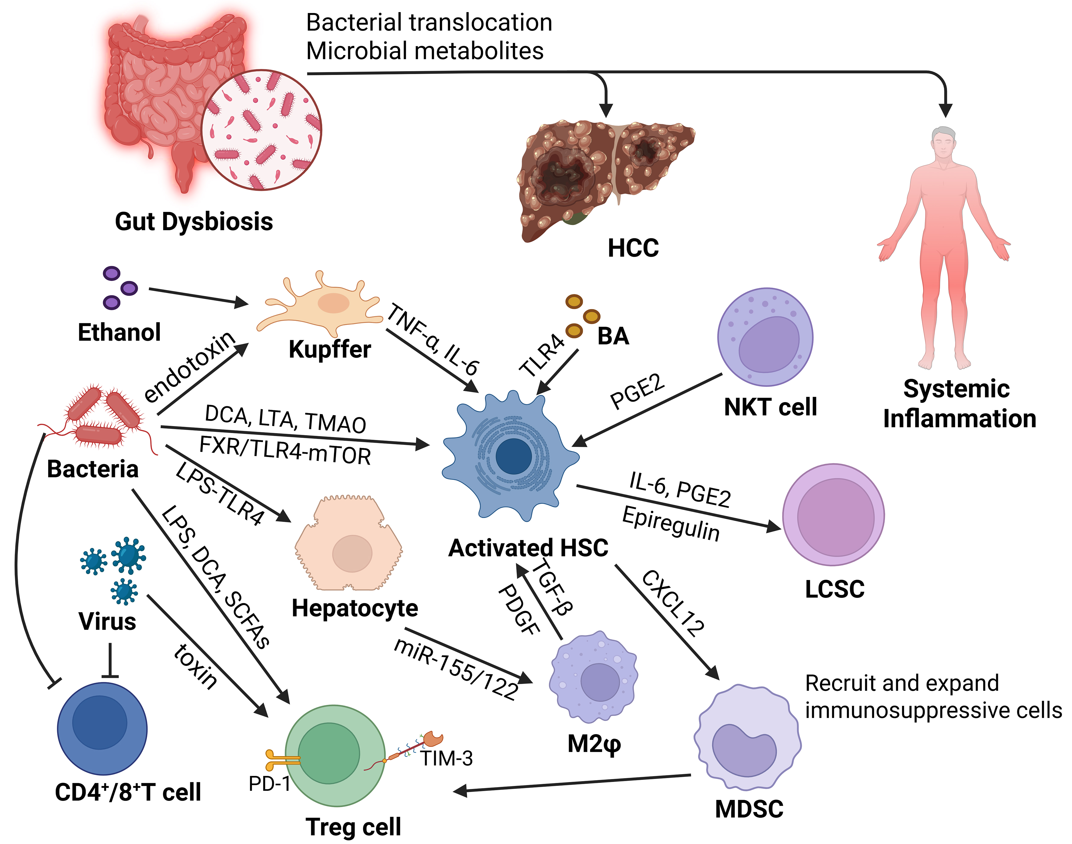
Figure 2. Intestinal microbiota dysbiosis and the immune microenvironment of HCC. Gut-derived LPS, DCA, TMAO, ethanol-related products, bacterial toxins, and fungal products can enter the liver through the gut-liver axis, activate Kupffer cells and HSCs, and shape an inflammatory and immunosuppressive microenvironment. Activated HSCs may promote the self-renewal of LCSCs through the FXR/TLR4-mTOR axis. HSC-derived CXCL12 can recruit CXCR4-expressing MDSCs. PGE2 is displayed separately in the NKT-cell/HSC interaction. Together with M2 macrophage polarization, these signals contribute to CD4+/CD8+ T-cell exhaustion and Treg expansion, forming an immunosuppressive microenvironment that promotes HCC development. Created in BioRender. Zhang, K. (2026) https://BioRender.com/bflwt7r. BA: Bile acid; CD4/CD8: cluster of differentiation 4/8; CXCL12: C-X-C motif chemokine ligand 12; CXCR4: C-X-C chemokine receptor type 4; DCA: deoxycholic acid; FXR: farnesoid X receptor; HCC: hepatocellular carcinoma; HSC: hepatic stellate cell; IL-6: interleukin-6; LCSC: liver cancer stem cell; LPS: lipopolysaccharide; LTA: lipoteichoic acid; MDSC: myeloid-derived suppressor cell; mTOR: mechanistic target of rapamycin; NF-κB: nuclear factor kappa-light-chain-enhancer of activated B cells; NKT: natural killer T; PD-1: programmed cell death protein 1; PGE2: prostaglandin E2; SCFA: short-chain fatty acid; TMAO: trimethylamine N-oxide; TNF-α: tumor necrosis factor-α; TLR4: Toll-like receptor 4; Treg: regulatory T cell; TIM-3: T-cell immunoglobulin and mucin domain-containing protein 3; M2φ:macrophage.