


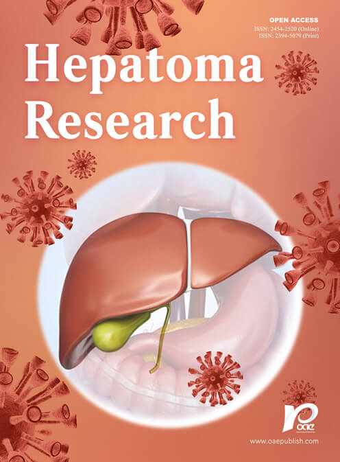
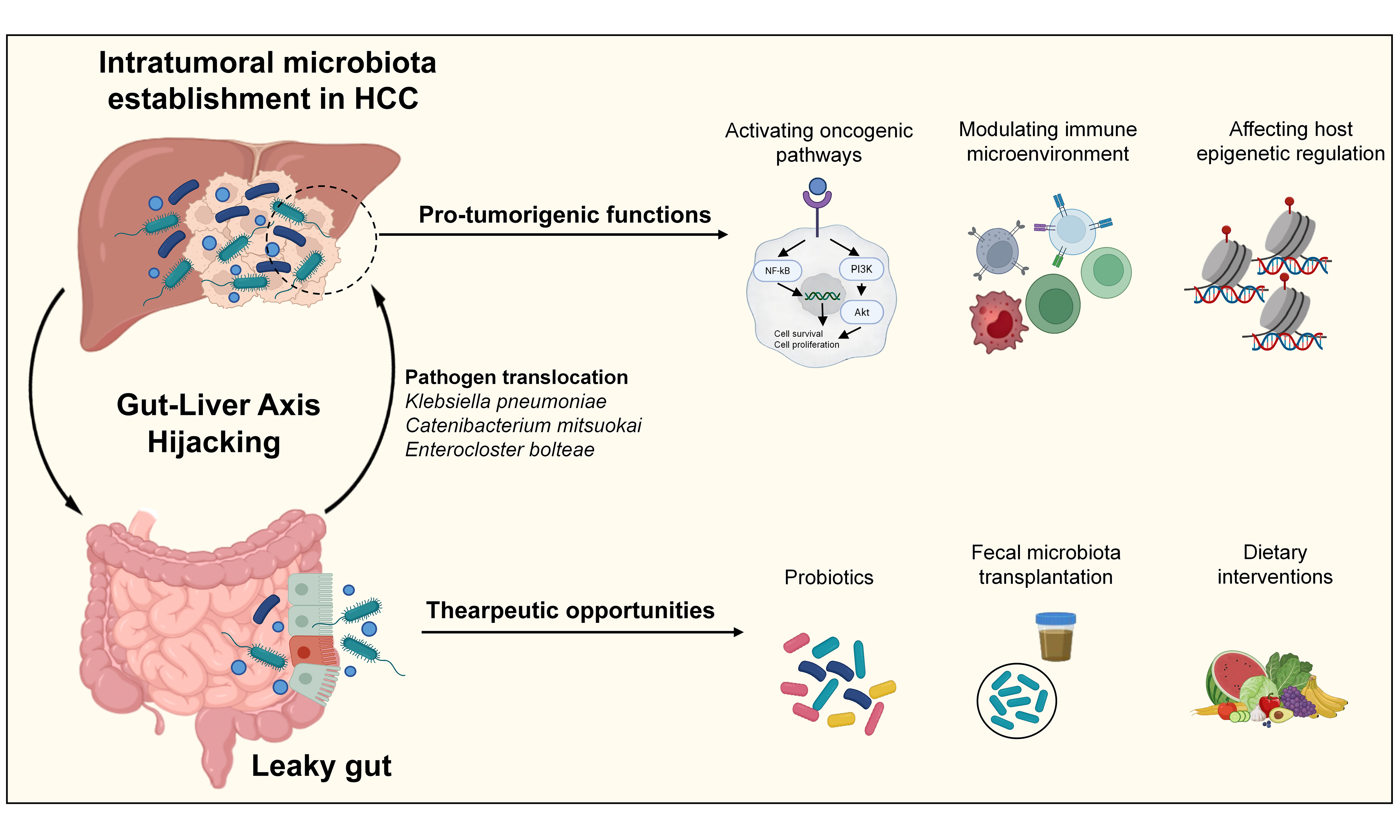
From gut-liver axis hijacking to intratumoral microbiota establishment: mechanisms and therapeutic opportunities in hepatocellular carcinoma
 0
0 Abstract
Hepatocellular carcinoma (HCC), the predominant form of liver cancer, remains a leading cause of global cancer mortality. Recent evidence indicates that intratumoral bacteria reside within HCC tumors, largely originating from the gut. Disruption of the intestinal barrier, coupled with impaired hepatic immune clearance, enables gut-derived microbes to translocate to the liver via the portal vein. Within the tumor microenvironment, these bacteria remain metabolically active and interact directly with host cellular receptors, thereby activating oncogenic signaling cascades, modulating local immune responses, and inducing epigenetic alterations. Notably, distinct bacterial signatures correlate consistently with tumor stage, prognosis, and therapeutic response. This review aims to provide a comprehensive update on the role of intratumoral bacteria in HCC, elucidate their mechanistic involvement in tumor development, and explore the potential of microbiome-targeted strategies for advancing diagnostic and therapeutic modalities.
Keywords
INTRODUCTION
Liver cancer remains a significant threat to global health, with persistently high incidence and mortality rates. Hepatocellular carcinoma (HCC), which constitutes approximately 90% of liver cancer cases, ranks as the fifth most commonly diagnosed malignancy and the third leading cause of cancer-related mortality worldwide[1,2]. While chronic infections with hepatitis B virus (HBV) and hepatitis C virus (HCV) remain well-established primary risk factors for HCC, metabolic dysfunction-associated steatotic liver disease (MASLD) and diabetes mellitus have emerged as the most rapidly increasing etiological contributors to HCC incidence[1,3,4]. Most patients are diagnosed with intermediate or advanced stages, when treatment options are limited and mainly include ablation therapy, transarterial chemoembolization (TACE), targeted therapy, and immunotherapy[5]. The overall prognosis for advanced HCC patients remains poor, with a 5-year survival rate of less than 10%[6,7]. Clinical management is further complicated by challenges such as high rates of tumor recurrence and metastasis, pronounced intratumoral heterogeneity, and both primary and acquired resistance to immunotherapeutic interventions[8]. Consequently, there is an urgent imperative to elucidate novel pathogenic mechanisms underlying HCC progression and to identify innovative prognostic biomarkers and effective therapeutic targets.
The tumor microenvironment (TME) plays a critical role in the progression and therapeutic response of HCC. It consists of tumor cells, cancer-associated fibroblasts, immune cells, extracellular matrix (ECM), and various signaling molecules[9,10]. In 2020, Nejman et al. first demonstrated the presence of bacteria within tumors across multiple cancer types, identifying the tumor-resident microbiota (TRM) as a previously unrecognized component of the TME[11]. Subsequent studies using high-throughput sequencing, fluorescence in situ hybridization (FISH), and tissue culture techniques confirmed that HCC tissues harbor abundant and metabolically active intratumoral bacteria[12-15]. These TRM establish long-term, self-sustaining populations within the TME and exhibit distinct spatial heterogeneity. In contrast, transient microbial populations are characterized by low and variable abundance, inconsistent presence across patients, and lack of stable spatial distribution even within different regions of the same tumor. These transient microbes differ fundamentally from true TRM in terms of their biological properties, functional roles, and clinical significance. It is hypothesized that TRM colonize liver tumors primarily via a disrupted gut-liver axis, which encompasses intestinal barrier dysfunction, compromised hepatic immune clearance, and microbial translocation through the portal vein, biliary tract, or hematogenous pathways.
Large-scale cohort studies have demonstrated that HCC tissues possess a unique intratumoral bacterial profile closely associated with clinicopathological features such as tumor grade, stage, and patient survival[12,14,16,17]. Importantly, recent functional studies have begun to establish causal relationships between specific intratumoral microbial populations and the initiation and progression of HCC[13,15,16]. This review aims to provide a systematic overview of the latest advances in this rapidly evolving field, including the routes of microbial translocation to liver tumors, the compositional landscape of the intratumoral microbiota, its pro-tumorigenic mechanisms in HCC, and emerging therapeutics that target the gut-liver axis and intratumoral microbiota.
GUT-LIVER AXIS HIJACKING: THE GATEWAY FOR INTRATUMORAL MICROBIOTA ESTABLISHMENT
The gut-liver axis refers to a bidirectional communication network established between the intestine and the liver via the biliary tract, portal vein, and systemic circulation. This axis plays a critical role in metabolic regulation, immune surveillance, and modulation of systemic inflammatory responses[18-20]. The formation of intratumoral microbiota in HCC occurs through a sequential mechanism involving the pathological disruption of gut-liver axis defenses, compromised hepatic immune surveillance, translocation of intestinal microbes, and their subsequent active colonization within the TME.
Initiation of intestinal barrier disruption
The disruption of the gut-liver axis is initiated by impairment of the intestinal epithelial mucosal barrier. Metabolic disorders, alcohol, viral infection, and high-fat diets downregulate tight junction proteins (Occludin, Claudin, ZO-1) and thin the mucus layer, producing a “leaky gut” with increased permeability[20-24]. This compromised barrier facilitates the translocation of gut microbial products, notably lipopolysaccharide (LPS), into the portal circulation[21]. Concurrently, the gut vascular barrier (GVB), a semipermeable endothelial layer, is also compromised, permitting whole bacteria to circumvent portal filtration and gain direct access to the systemic bloodstream[25,26].
Compromised hepatic immune clearance
In the healthy liver, resident Kupffer cells and innate lymphoid cells constitute a highly efficient surveillance system that rapidly eliminates gut-derived microbes arriving via the portal circulation. However, this clearance mechanism becomes progressively impaired during chronic liver disease and hepatocarcinogenesis. In a tumor-permissive state, chronic liver inflammation induces M2-type polarization and immunosuppression of Kupffer cells, compromising their ability to clear translocated microbes[27,28].
Three pathways of microbial translocation into the liver
The translocation of gut microbiota and their metabolites into the liver constitutes a fundamental mechanism underlying the disruption of the gut-liver axis. This phenomenon predominantly transpires via three principal pathways. First, through the portal vein, bacteria and their metabolites [such as LPS and short-chain fatty acids (SCFAs)] pass through the damaged intestinal barrier directly into the portal circulation and reach the liver. Notably, LPS-positive bacterial vesicles have been detected in portal blood, and fecal microbiota transplantation (FMT) from patients with HCC has been shown to introduce viable bacteria into the liver via this route[15,29]. Second, hematogenous dissemination occurs when damage to the GVB permits bacteria to enter the systemic circulation, where they utilize erythrocytes as vehicles to evade immune detection and facilitate transport to hepatic tissues[25,26]. Third, the retrograde biliary pathway allows duodenal bacteria to ascend through a dysfunctional sphincter of Oddi, a process particularly implicated in the pathogenesis of intrahepatic cholangiocarcinoma and reflected in the microbial resemblance observed between HCC tumors and the upper gastrointestinal tract[30-32].
From microbial translocation to intratumoral colonization
Upon reaching the liver, translocated microbes actively colonize the TME. Early intrahepatic colonization of translocated microbiota is driven by the TME. Chemokines, ECM components, and metabolic precursors secreted by HCC cells selectively recruit and retain distinct microbial populations[33]. Li et al. defined the intrahepatic metagenomic characteristics of the microbiota in patients with HCC and found that Rhodobacter, Megasphaera, and Lactobacillus were significantly enriched in tumor tissues compared with adjacent non-tumor tissues[33]. Furthermore, Ren et al. mechanistically demonstrated that Enterocloster bolteae directly binds to surface receptors on HCC cells, facilitating robust bacterial adhesion to tumor cells and stable colonization within the TME[34].
Following initial colonization, the emerging intratumoral microbiota must circumvent the residual immune surveillance that remains active within the compromised hepatic environment. Translocated microbiota and their metabolites can suppress antitumor immunity and induce an immunotolerant TME, thereby protecting colonizing intratumoral microbes from clearance[35]. Yoshimoto et al. demonstrated that deoxycholic acid (DCA), a metabolite derived from gut microbiota, induces a senescence-associated secretory phenotype (SASP) in hepatic cells, thereby establishing an immune-privileged niche conducive to the stable colonization of intratumoral microbiota[36]. Loo et al. reported that gut microbiota promote intratumoral microbiota colonization through prostaglandin E2 (PGE2)-dependent suppression of antitumor immunity[35].
The metabolic symbiosis established between tumor cells and the intratumoral microbiota, to a certain extent, sustains the persistence of microbes within the TME. Hepatoma cells continuously supply essential nutrients, such as amino acids and bile acids, which are subsequently metabolized by the resident microbial community. In turn, bacterial metabolites, particularly SCFAs, play a critical role in modulating the metabolic-immune interactions that promote the progression of HCC while concurrently maintaining the stability of the microbial niche within the TME[37,38]. Che et al. confirmed that bile acid metabolism reshapes the metabolic homeostasis of HCC and facilitates long-term survival of the intratumoral microbiota[39]. Similarly, Pan et al. reported that SCFAs regulate HCC progression through metabolic-immune crosstalk, establishing a metabolic symbiotic state that supports both tumor growth and the sustained persistence of the intratumoral microbiota[37,39].
Through a sequential process involving selective recruitment, immune evasion, and metabolic mutualism, gut-derived microorganisms collectively transition from transient invaders to stable, TRM within the context of hepatocarcinogenesis. These microbes not only directly promote the progression of HCC but also systemically modulate the hepatic immune microenvironment and affect responses to immune checkpoint inhibitors (ICIs) through the secretion of metabolites and activation of pattern recognition receptors[40].
THE COMPOSITION AND CHARACTERISTICS OF INTRATUMORAL MICROBIOTA IN HCC
The intratumoral microbiota of HCC exhibits distinct taxonomic composition that differs substantially from those observed in normal liver tissue and adjacent non-tumorous regions, both in terms of overall microbial abundance and the enrichment of specific microbial taxa [Table 1].
Characteristics of intratumoral microbiota in HCC
| Samples | Methods | Enriched taxa | Reduced taxa | Ref. |
| 46 HCC, 28 paired paracancerous and 33 normal tissues | 16S rRNA sequencing | Stenotrophomonas maltophilia Enterococcus Phyllobacterium Lactobacillus | Acinetobacter | [13] |
| 28 normal, 64 paired HCC tumor and peritumoral tissues | 16S rRNA sequencing Fresh tissue culture | Streptococcaceae Lactococcus Staphylococcus | Robiota Chlamydiae | [12] |
| 91 HCC and 65 paratumor tissues | 16S rRNA sequencing | Gammaproteobacteria Bacilli | Deinococcus thermus Acidobacteriota | [41] |
| 12 CHB liver biopsies; 29 paired HCC tumor and adjacent normal tissues | Metagenomic sequencing | Escherichia coli Shigella dysenteriae Babesia bigemina Pannonibacter phragmitetus | Phylum level: Firmicutes Actinobacteria Klebsiella variicola | [42] |
| 99 HCC patients with paired samples | MiSeq sequencing 16S rRNA sequencing | Fusobacterium Neisseria | Pseudomonas | [14] |
| 46 HCC, 42 paracancerous and 11 normal tissues | 16S rRNA sequencing | Streptococcus parasanguinis | Akkermansia | [43] |
| 3 paired HCC tumor and normal tissues | 2bRAD-M sequencing | Mycoplasma sp. HU2014 Escherichia coli | Pseudomonas | [38] |
| 19 paired HCC tumor tissues and adjacent normal tissues | 16S rRNA sequencing | Rhodobacter Megasphaera | Pseudochrobactrum | [33] |
| 19 HCC and 15 normal liver tissues | 16S rRNA sequencing | NA | Bacteroides Prevotella Parabacteroides | [44] |
| 172 paired HCC tumor and adjacent normal tissues | 16S rRNA sequencing | Intestinimonas Brachybacterium Rothia | NA | [17] |
| 16 HCC, 7 non-tumor adjacent tissues | 16S rRNA sequencing | Hoylesella Agathobacter Sphingobium Cardiobacterium Photobacterium Serratia | Campylobacteria Bacteroida | [31] |
| 90 HCC patients | 16S rRNA sequencing | Proteobacteria | Akkermansia | [45] |
| 8 HCC with paired peritumoral and normal liver tissues | 16S rRNA sequencing | Rhodococcus sp. B513 | Akkermansia Lactobacillus Muribaculaceae Dubosiella | [46] |
| 54 HCC tumor tissues | Culturomics | Klebsiella pneumoniae Escherichia coli Enterococcus faecalis Enterococcus casseliflavus Staphylococcus warneri Actinomyces oris Enterobacter bugandensis Enterobacter cloacae Odoribacter splanchnicus Staphylococcus epidermidis Staphylococcus hominis Klebsiella variicola Phocaeicola vulgatus Streptococcus mitis oralis | NA | [15] |
| 58 paired multifocal HCC tumors and adjacent nontumor tissues | 16S rRNA sequencing | Enterococcus Streptococcus | Stenotrophomonas Corynebacterium Burkholderia Clostridium | [16] |
| 48 HCC patients and 17 patients with hepatic hemangioma | 2bRAD-M sequencing | Ralstonia Klebsiella Acinetobacter Stenotrophomonas Bacillus | Firmicutes Actinobacteria | [47] |
Multiple independent profiling studies have converged on a core signature: at the phylum level, the HCC microenvironment is dominated by Proteobacteria, Firmicutes, Actinobacteriota, and Bacteroidetes, with Proteobacteria consistently exhibiting the highest relative abundance in tumor tissues[12,14,41,48]. Notably, the microbiota composition in HCC displays heterogeneity contingent upon distinct etiological factors. For instance, Liu et al. examined HBV-associated HCC and delineated a unique intratumoral microbiota and immune microenvironment signature characteristic of this subtype[49]. Specific genera, such as Cutibacterium, were significantly enriched in HBV-related HCC, with their abundance closely correlating with the phenotypic profiles of tumor-infiltrating immune cells. Furthermore, Dang et al. established a correlation between intratumoral bacterial populations and HBV viral load, identifying taxa such as Bacteroides that are associated with HBV levels; these bacteria may serve as prognostic biomarkers for stratifying clinical outcomes in HCC patients[50]. In the context of MASLD-related HCC, Liu et al. reported a specific enrichment of Lactobacillus johnsonii[51]. Subsequent research by Zhang et al. demonstrated that Catenibacterium mitsuokai (C. mitsuokai), which selectively accumulates in HCC, may promote the progression of metabolic dysfunction-associated steatohepatitis (MASH)-related HCC[52]. Regarding alcohol-associated liver disease (ALD)-related HCC, Magdy Wasfy et al. employed culturomics to characterize microbial profiles in patients with alcohol-associated HCC, revealing a significant enrichment of Thomasclavelia ramosa within this cohort[53].
In addition to bacteria, the fungal component of the intratumoral microbiota has recently gained attention. Shen et al. reported a significant enrichment of Malassezia spp. within HCC tissues relative to adjacent non-tumorous tissues, with their abundance showing a strong association with stromal and immune characteristics of the TME[54]. Nonetheless, several challenges remain, notably the low microbial biomass present within tumors, which heightens the risk of contamination from reagents, surgical procedures, and environmental sources. To accurately differentiate authentic tumor-resident microbes from contaminants, four standardized methodological approaches have been proposed: (1) implementation of sterile pre-analytical procedures alongside the use of blank controls, paraffin controls, and paired non-tumor liver samples to monitor reagent-derived contaminants; (2) application of FISH, bacterial immunohistochemistry (IHC), and electron microscopy to spatially verify microbial localization within tumor lesions; (3) utilization of computational filtering techniques to exclude contaminants originating from blank controls while retaining taxa enriched in tumor samples; (4) cultivation of tumor tissue to isolate viable bacteria, thereby excluding DNA from non-viable contaminant sources.
FUNCTIONAL MECHANISMS OF INTRATUMORAL MICROBIOTA IN PROMOTING LIVER CANCER
Following stable colonization within the TME of HCC, the intratumoral microbiota acts as an oncogenic promoter independent of tumor origin. It facilitates HCC initiation, progression, invasion, and metastasis via three principal mechanisms: modulation of host oncogenic signaling pathways, remodeling of the tumor immune microenvironment (TIME), and alteration of host epigenetic regulation.
Direct binding and secreted metabolites modulate host oncogenic pathways
Tumorigenesis is closely associated with the widespread activation of oncogenic signaling pathways, which facilitate the transformation of normal cells into malignant phenotypes and support tumor progression[55]. Accumulating evidence indicates that the intratumoral microbiota and its metabolites, as key components of the TME, specifically interact with host cells and promote the activation of critical pro-oncogenic signaling pathways, thereby playing pivotal roles in the initiation and progression of HCC[56,57] [Figure 1]. Mechanistically, this pro-tumorigenic interplay operates through two primary modes: direct binding of microbial surface proteins to host receptors, and secretion of bioactive metabolites that activate intracellular signaling. Wang et al., using metagenomic analysis and bacterial culture of HCC tissues, found increased abundance of Klebsiella pneumoniae (K. pneumoniae) in both HCC patients and mice transplanted with microbiota from these patients[15]. The K. pneumoniae surface protein PBP1B was shown to bind directly to Toll-like receptor 4 (TLR4) on HCC cells, thereby promoting tumor cell proliferation and activating downstream oncogenic pathways[15] [Figure 1A]. Similarly, Ren et al. reported that Enterocloster bolteae adheres to HCC cells via the surface adhesion protein PbpT, which impairs the tumor-suppressive activity of desmoglein-1 (DSG1), thus accelerating HCC progression[34]. Extending the mechanistic spectrum, Zhang et al. observed elevated levels of Catenibacterium in both tumor tissues and fecal samples of HCC patients compared with healthy individuals[52]. C. mitsuokai can disrupt the intestinal barrier and migrate to the liver as live bacteria. Its surface proteins Gtr1/RagA interact with γ-catenin receptors on HCC cells, facilitating its colonization within HCC tissues. Furthermore, C. mitsuokai secretes the metabolite quinolinic acid, which promotes HCC progression by binding to TIE2 and activating the downstream phosphatidylinositol 3-kinase/protein kinase B (PI3K/AKT) pathway[52] [Figure 1B]. Collectively, these findings highlight that the intratumoral microbiota contributes to hepatocarcinogenesis through two complementary mechanisms: direct receptor binding and paracrine metabolite signaling.
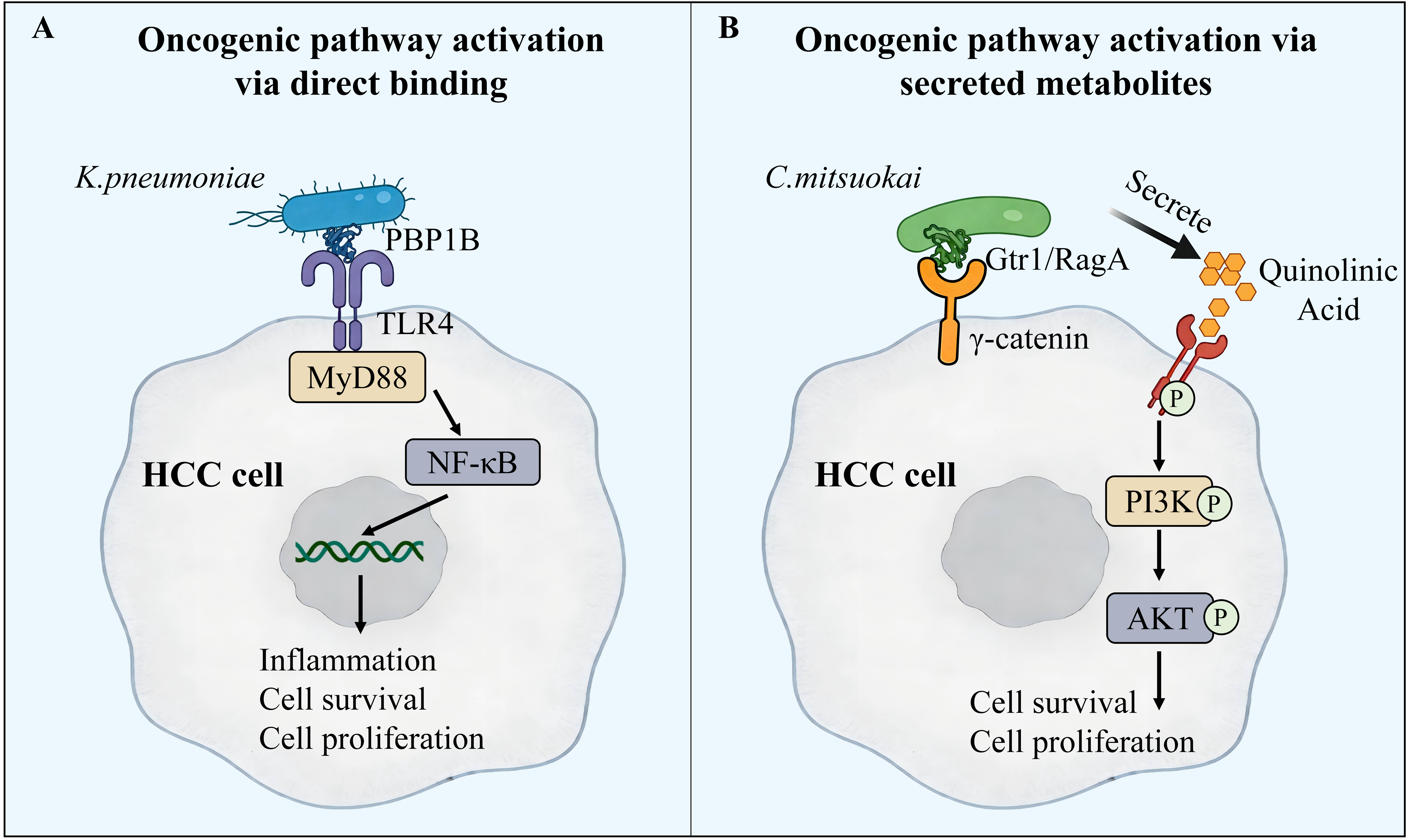
Figure 1. Mechanisms by which two tumor-resident microbes promote HCC progression by modulating host oncogenic pathways. (A) Direct binding-mediated pathway activation. K. pneumoniae binds to HCC cells via PBP1B and activates the TLR4/NF-κB signaling pathway, leading to inflammation, cell survival, proliferation, and ultimately tumorigenesis; (B) Secreted metabolite-mediated pathway activation. C. mitsuokai surface proteins Gtr1/RagA bind to the γ-catenin receptor on HCC cells, leading to tumor colonization. It secretes quinolinic acid to activate the PI3K/AKT signaling pathway by binding to TIE2, influencing cell survival and proliferation. HCC: Hepatocellular carcinoma; PBP1B: penicillin-binding protein 1B; TLR4: toll-like receptor 4; MyD88: myeloid differentiation primary response protein 88; NF-κB: nuclear factor kappa-B; PI3K: phosphatidylinositol 3-kinase; AKT: protein kinase B.
Dual immunomodulatory roles of the intratumoral microbiota
Accumulating evidence indicates that the intratumoral microbiota, which inhabits both tumor and immune cells, plays an active role in modulating the TIME through mechanisms involving activation, inhibition, and bystander effects[58-62] [Figure 2]. In the context of immune suppression, Stenotrophomonas maltophilia is markedly enriched in cirrhosis-associated HCC and accelerates tumor progression by triggering NOD-, LRR- and pyrin domain-containing protein 3 (NLRP3) inflammasome assembly[13]. Ma et al. demonstrated that dysbiosis of the gut-liver axis microbiota disrupts bile acid metabolism, leading to the suppression of natural killer T (NKT) cell activity within the TIME, thereby promoting HCC progression[63]. Reduced intratumoral microbiota diversity is closely associated with an immunosuppressive phenotype[42]. FMT from HCC patients into germ-free mice induces colonic inflammation and increases hepatic infiltration of Th1, Th2, and Th17 cells, creating a proinflammatory microenvironment that promotes tumor progression[15]. Additionally, bacteria enriched in metastatic HCC, including Enterococcus faecalis and Streptococcus anginosus, have been shown to directly recruit and activate myeloid-derived suppressor cells (MDSCs), thereby enhancing their immunosuppressive functions while concurrently impairing CD8+ T cell proliferation and cytotoxicity. This interaction contributes to an immune imbalance characterized by reduced antitumor immunity[16] [Figure 2A].

Figure 2. Intratumoral microbiota and gut microbiotaderived factors exert opposing effects on the TIME. (A) Immunosuppression: Intratumoral bacteria such as K. pneumoniae, S. anginosus, and E. faecalis promote an immunosuppressive microenvironment by modulating immune cell functions, including inhibiting the activity of NKT cells, increasing the infiltration of Th1, Th2, and Th17 cells, recruiting MDSCs, and impairing the proliferation of CD8+ T cells, leading to HCC progression; (B) Immunoactivation: Intestinal microbiota that can influence the TIME, such as A. muciniphila, activate anti-tumor immunity by stimulating CD8+ T cells and reducing Treg infiltration, leading to HCC regression. HCC: Hepatocellular carcinoma; TIME: tumor immune microenvironment; NKT: natural killer T cell; Th: T helper cell; MDSC: myeloid-derived suppressor cell; Treg: regulatory T cell.
Conversely, certain intratumoral microbiota constituents can suppress tumor progression by activating antitumor immune responses [Figure 2B]. Certain gut-derived bacteria also exert an intratumoral immunoactivation effect. For instance, Akkermansia muciniphila enhances the efficacy of programmed death 1 (PD-1) inhibitors in metabolic dysfunction-associated fatty liver disease (MAFLD)-associated HCC by promoting the activation of CD8+ T cells in the TIME and reducing regulatory T cell infiltration[64]. Similarly, Brevibacillus parabrevis contributes to the augmentation of antitumor immunity through the inhibition of natural killer (NK) cell ferroptosis[65]. Metabolites derived from the intratumoral microbiota also play a significant role in immune modulation; SCFAs produced by Lactobacillus species influence immune cell function[66]. Additionally, the enzyme fpPRPS, produced by Faecalibacterium prausnitzii, depletes ATP within tumor cells, resulting in the degradation of Rab11a and disruption of programmed cell death ligand 1 (PD-L1) membrane trafficking, thereby enhancing CD8+ T cell-mediated antitumor responses[67]. Furthermore, rapamycin-induced xenophagy promotes the major histocompatibility complex (MHC) class II-restricted presentation of neoantigens derived from Bifidobacterium longum, eliciting potent CD4+ T cell activation and tumor-specific cytotoxicity[68]. Collectively, these studies demonstrate that the intratumoral microbiota can activate diverse antitumor immune pathways through direct cellular interactions, metabolite secretion, and neoantigen presentation, thereby presenting multiple avenues for therapeutic intervention.
Modulation of host epigenetics by the intratumoral microbiota
Epigenetic modifications, including DNA methylation and histone alterations, regulate a wide array of cellular functions such as proliferation, apoptosis, invasion, and senescence, with their dysregulation recognized as a hallmark of cancer[69]. Emerging evidence indicates that the intratumoral microbiota and its metabolites can modulate the expression of relevant genes by influencing host epigenetic modifications, such as DNA methylation and histone modifications, thereby affecting HCC progression [Figure 3]. A seminal investigation by Li et al. provided the first direct evidence linking the composition of the intratumoral microbiota to host DNA methylation patterns in HBV-related HCC, demonstrating that specific microbial taxa correlate with aberrant methylation of genes implicated in hepatocarcinogenesis[42]. Expanding upon these findings, Chen et al. conducted integrative analyses of intratumoral microbial characteristics and DNA methylation-driven gene expression in HCC, revealing that the abundance of particular intratumoral bacteria, such as Pseudomonas and Stenotrophomonas, correlates positively with the expression of DNA methyltransferases (DNMT1, DNMT3A) and is linked to hypermethylation of tumor suppressor gene promoters[70].
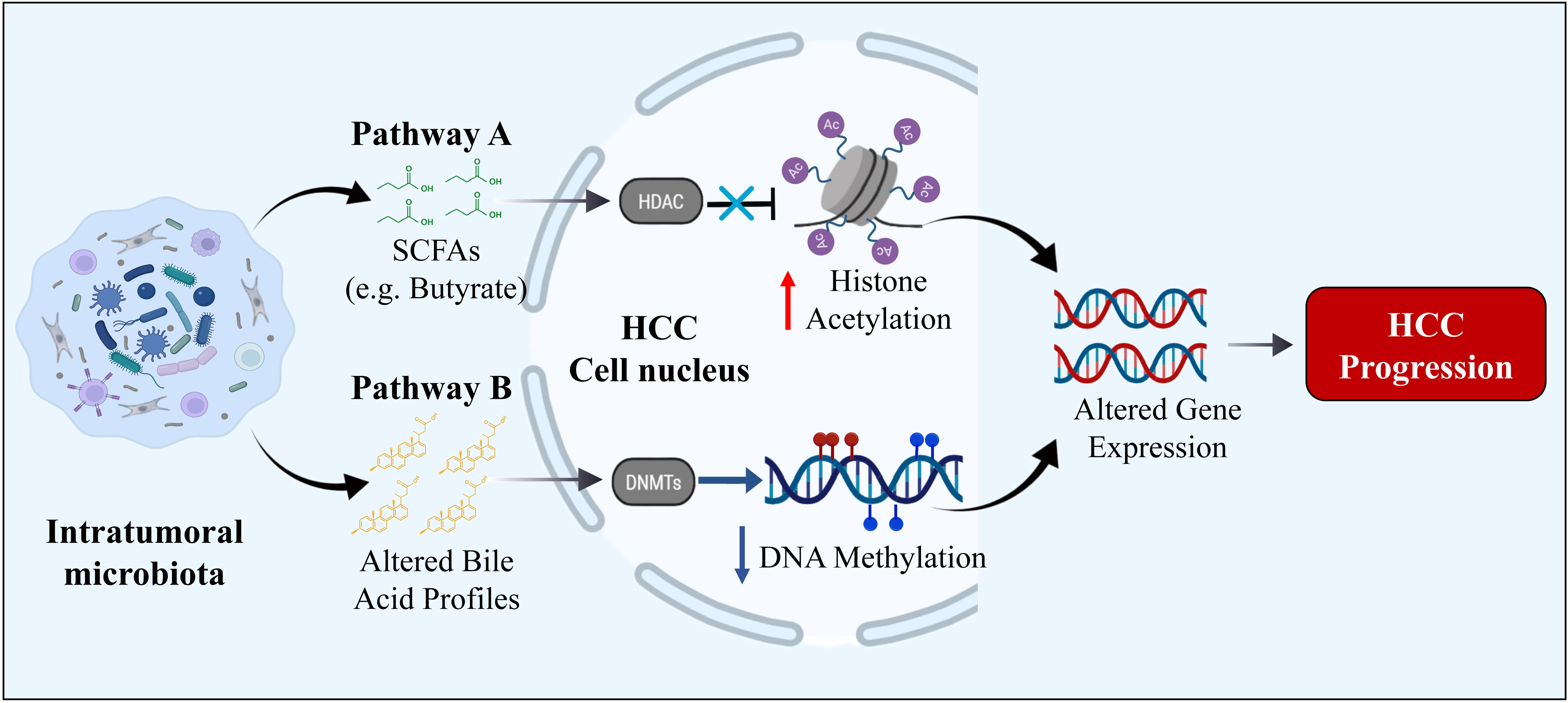
Figure 3. The proposed mechanisms of intratumoral microbiota promoting HCC by affecting host epigenetic regulation. Intratumoral microbiota affects host epigenetic regulation mainly through two pathways. Pathway A: Intratumoral microbiota secretes SCFAs (e.g., butyrate), which inhibit HDAC and increase histone acetylation, leading to altered gene expression in the HCC cell nucleus, resulting in tumorigenesis. Pathway B: Intratumoral microbial activity alters bile acid metabolism, which modulates DNMT function and leads to hypermethylation or hypomethylation of DNA, further altering gene expression, resulting in tumorigenesis. HCC: Hepatocellular carcinoma; SCFAs: short-chain fatty acids; HDAC: histone deacetylase; DNMT: DNA methyltransferase.
In addition to DNA methylation, metabolites derived from intratumoral microbiota, particularly short-chain fatty acids such as butyrate, can function as histone deacetylase inhibitors, modulating histone acetylation in HCC cells and consequently affecting the transcription of oncogenes and tumor suppressor genes[66]. Importantly, the epigenetic modifications induced by microbiota appear to differ according to the etiology of HCC: in HBV-associated HCC, microbial factors may interact with viral elements to epigenetically silence antiviral and tumor suppressor genes, whereas in non-alcoholic fatty liver disease (NAFLD)-associated HCC, microbial metabolites predominantly affect genes involved in metabolic pathways[42,66]. Although these observations provide valuable correlative evidence, there remains a lack of direct mechanistic validation elucidating how specific components of the intratumoral microbiota modulate host epigenetic landscapes. Future investigations should focus on elucidating the causal molecular mechanisms, ideally within the context of HCC etiology-specific microenvironments.
CLINICAL CORRELATION BETWEEN INTRATUMORAL MICROBIOTA AND HCC
The relationship between dysbiosis of the intestinal microbiota, intratumoral microbiota, and HCC has been substantiated across multiple clinical cohorts. Ren et al. conducted a multi-stage cohort study involving 486 participants encompassing chronic hepatitis B, liver cirrhosis, and HCC cases[71]. This study was the first to demonstrate a significant enrichment of genera such as Gemmiger and Parabacteroides in the fecal samples of early-stage HCC patients, concomitant with a reduction in butyrate-producing genera. Utilizing these findings, the authors developed a diagnostic model for early HCC detection, which achieved an area under the curve (AUC) of 80.64%, thereby providing clinical evidence supporting the utility of intestinal microbiota as a non-invasive biomarker for HCC. Building upon this work, Huang et al. integrated gut metagenomic data with host liver transcriptomic profiles from 32 Chinese patients with HBV-related HCC. Their analysis revealed specific enrichment of Bacteroides, Lachnospiracea incertae sedis, and Clostridium XIVa, with the abundance of these genera correlating with unfavorable clinical outcomes and the establishment of an immunosuppressive TME. Notably, six key microbial markers associated with the TIME or bile acid metabolism demonstrated potential prognostic value, achieving an AUC of 81%[48]. Regarding intratumoral microbiota, Huang et al. performed 16S rRNA sequencing on paired tumor and adjacent non-tumor tissues from 32 HCC patients. Their systematic analysis confirmed that the relative abundance of Proteobacteria, Gammaproteobacteria, Firmicutes, Actinobacteriota, and Saccharimonadia was significantly associated with the clinicopathological features of HCC[48].
The composition and burden of the intratumoral microbiota are closely linked to the clinicopathological characteristics and prognosis of patients with liver cancer, underscoring its potential role as both a biomarker and a therapeutic target. In terms of tumor progression, Jo et al. demonstrated that the intratumoral microbiota composition differs significantly between early-stage and advanced-stage HCC, with Sphingomonadaceae and Skermanella more abundant in advanced tumors[72]. Regarding prognosis, Sun et al. constructed a genus-level scoring system based on the relative abundance of intratumoral microbiota, revealing that patients in the high-score group had significantly shorter overall survival[41]. Jiang et al. reported that intratumoral microbiota α-diversity is inversely associated with overall survival, with greater diversity linked to poorer outcomes, possibly reflecting more complex immune-modulatory effects[17]. The intratumoral microbiota also influences the TIME in a species-specific manner. For instance, enrichment of Lachnoclostridium is positively correlated with the formation of tertiary lymphoid structures, which serve as markers of antitumor immune activation[73]. Moreover, accumulating evidence suggests that the intratumoral microbiota can enhance the efficacy of current targeted therapies and immunotherapies for HCC. In the context of immunotherapy, Liu et al. demonstrated that intratumoral Lactobacillus johnsonii increases the sensitivity of HCC to PD-1 inhibitor treatment by promoting the expansion of CD8+ T cells within the TME[51]. Wu et al. further showed that Akkermansia muciniphila improves therapeutic responses to PD-1 blockade in MASH-related HCC[64]. Compared to immunotherapy, research on the regulatory effects of intratumoral microbes on targeted agents for HCC remains nascent. Nonetheless, existing data indicate that intratumoral Enterococcus faecium enhances the synergistic activity of sorafenib against advanced HCC by inducing ferroptosis in tumor cells through increased infiltration of IFN-γ-producing CD8+ T cells in the TME[74]. Most current findings have been validated primarily in preclinical models, and prospective interventional clinical trials are needed to confirm whether intratumoral bacteria can directly influence the clinical efficacy of targeted therapies or ICIs in HCC patients. Collectively, these multidimensional clinical associations emphasize the intratumoral microbiota as a functionally significant and potentially targetable component within the liver cancer ecosystem.
THERAPEUTIC OPPORTUNITIES: TARGETING THE GUT-LIVER AND INTRATUMORAL MICROBIOTA
Insights into gut-liver axis hijacking and the tumor-promoting functions of the intratumoral microbiota have opened new avenues for HCC prevention and treatment. Contemporary therapeutic approaches can be broadly classified into two main strategies: preventing microbial translocation by preserving gut-liver axis integrity, and precisely eliminating established intratumoral microbes.
Modulating the gut-liver axis to prevent microbial hijacking
Interventions aimed at inhibiting the translocation of microbes from the gastrointestinal tract to the liver constitute a fundamental preventive approach. The principal goals of such strategies include the restoration of intestinal barrier integrity, the reestablishment of gut microbial equilibrium, and the reduction of metabolites that promote carcinogenesis.
Probiotics
Probiotics demonstrate anti-HCC effects through mechanisms including competitive exclusion of pathogenic microbes, reinforcement of intestinal barrier function, and modulation of the immune response[75-77]. MASLD, the predominant non-viral driver of HCC, establishes a unique immune microenvironment characterized by CD4+ T-cell depletion and expansion of lipid-associated macrophages, which collectively attenuate the efficacy of ICIs[78,79]. Within this framework, metabolites derived from the gut microbiota may mitigate metabolic susceptibilities inherent to MASLD-related HCC. For example, Lactobacillus acidophilus produces valeric acid, which disrupts tumor energy metabolism in NAFLD-associated HCC[80]. Similarly, acetate generated by Bifidobacterium pseudolongum inhibits MASLD-HCC progression by suppressing inflammatory and oncogenic signaling pathways[81]. Consequently, Lactobacillus acidophilus and Bifidobacterium pseudolongum represent potential prophylactic agents against metabolic dysfunction-associated fatty liver disease related hepatocellular carcinoma (MAFLD-HCC). Additionally, Klebsiella oxytoca competitively inhibits the oncogenic Klebsiella pneumoniae, thereby maintaining intestinal barrier integrity and preventing hepatic bacterial translocation[15]. Lactobacillus rhamnosus GG induces a cGAS/STING-dependent type I interferon response, and its combination with Bifidobacterium enhances anti-PD-1 efficacy in HCC models[82,83]. Collectively, these findings underscore the dual role of probiotics as both protectors of the gut-liver axis and adjuvants that potentiate immunotherapeutic interventions in HCC.
FMT
FMT serves to reestablish intestinal homeostasis through the functional remodeling of the gut microbiota. The administration of encapsulated FMT has been shown to improve clinical outcomes in patients with cirrhosis[84]. Additionally, FMT derived from female donors has been demonstrated to restore intestinal permeability and mitigate hepatic inflammation in experimental models of alcoholic liver disease[85]. Although direct evidence in HCC is limited, the ongoing FLORA trial (NCT05690048) is evaluating FMT to overcome resistance to atezolizumab/bevacizumab in advanced HCC[86].
Dietary and pharmacological interventions
Dietary and pharmacological strategies aimed at directly strengthening the intestinal barrier represent a promising prophylactic approach. Pharmacological agents targeting tight junction proteins, such as Occludin and Claudin, have been shown to reduce LPS translocation[21]. Nutritional interventions, including high-fiber and low-fat diets, promote the proliferation of butyrate-producing bacteria while concurrently suppressing the generation of pro-carcinogenic metabolites[87]. Furthermore, natural compounds such as polyphenols, flavonoids, and saponins contribute to the preservation of gut-liver axis integrity and exhibit protective effects against HCC[24,88]. Beyond the use of probiotics and FMT, prebiotics - specifically indigestible carbohydrates like inulin-type fructans and galactooligosaccharides - selectively enhance the growth of intestinal Bifidobacterium and Lactobacillus species. These prebiotics facilitate the production of SCFAs, which are critical for maintaining intestinal barrier function and modulating immune cell activity, thereby representing a novel avenue for therapeutic intervention targeting the gut-liver axis[89].
Precise therapy targeting intratumoral microbiota
Following the establishment of the intratumoral microbiota, direct antimicrobial strategies become necessary. These include conventional antibiotics, engineered antimicrobial polymers, and highly specific phage therapy.
Antibiotics
Selective elimination of pro-tumorigenic bacteria can suppress HCC. Broad-spectrum antibiotics partially reverse Enterococcus faecalis and Streptococcus anginosus-driven tumor progression[16]. Antibiotics also enhance immunotherapy: a phase II trial of nivolumab, tadalafil, and oral vancomycin reshaped the gut microbiota and TIME in advanced HCC[90]. Mechanistically, antibiotic treatment mitigates dysbiosis-induced expansion of MDSCs and facilitates the restoration of T cell functionality[91]. Additionally, vancomycin administration reduces serum levels of secondary bile acids, which contributes to the attenuation of HCC progression in murine models[92]. Future efforts should focus on precision antibiotic regimens tailored to individual microbial profiles.
Antibacterial polymer therapy
Antibacterial polymer therapy involves the use of engineered macromolecules, such as antimicrobial polymers and peptides, which are designed to selectively eliminate pathogenic bacteria while minimizing adverse off-target effects. For instance, tellurium-containing polycarbonate carriers have been shown to overcome chemoresistance in colorectal cancer by eradicating intratumoral microbiota[93]. Similarly, gemcitabine micelles crosslinked with colistin have demonstrated efficacy in eliminating bacteria responsible for drug resistance[94]. In the context of liver cancer, nanoparticle-encapsulated antimicrobial peptides have successfully cleared intratumoral microbiota and facilitated remodeling of the TME[95]. Additionally, peptides derived from bacteriophages have exhibited activity against HCC cells as well as multidrug-resistant Acinetobacter baumannii[96]. Collectively, these findings underscore the potential for developing liver-targeted antimicrobial polymer systems as therapeutic strategies for HCC.
Phage therapy
Phage therapy utilizes bacteriophages to achieve highly specific bacterial lysis, exhibiting a minimal likelihood of resistance development[97,98]. In preclinical studies, phage VA7 demonstrated targeted lysis of Bacteroides fragilis, thereby restoring chemosensitivity in colorectal cancer models[99]. Although the application of phage therapy in HCC remains largely underexplored, its specificity and potential synergistic interactions with existing treatments merit further comprehensive investigation.
DISCUSSION
This review synthesizes the current understanding of the intratumoral microbiota in HCC, outlining its development from disruptions in the gut-liver axis to stable colonization within the TME. A key controversy remains regarding whether the intratumoral microbiota actively contribute to HCC oncogenesis or merely represent passive colonizers of established tumors. The review presents three lines of evidence supporting a driver role for specific gut-derived bacteria, such as Klebsiella pneumoniae and C. mitsuokai: (1) FMT and monocolonization animal models induce liver tumor formation; (2) bacterial adhesins and metabolites activate oncogenic, epigenetic, and immunosuppressive pathways; (3) tumor-specific microbial signatures are detectable prior to clinical diagnosis of HCC. Nonetheless, heterogeneity is evident, as certain taxa with antitumor properties, such as Akkermansia muciniphila, do not function as oncogenic drivers, and the majority of low-abundance intratumoral microbes appear to be neutral passengers. To definitively distinguish driver from passenger microbes across different etiologies of HCC - including HBV-related, MASLD-related, and alcohol-related cases - future research employing strain- and etiology-specific functional studies is warranted.
We have summarized how intestinal barrier damage, impaired hepatic immune clearance, and microbial translocation via portal vein, hematogenous, and biliary routes collectively enable gut-derived microbes to seed liver tumors. Once established within the TME, the intratumoral microbiota contributes to HCC progression via three interrelated mechanisms: direct activation of oncogenic pathways such as TLR4 and PI3K/AKT via surface proteins or secreted metabolites, remodeling of the immune microenvironment toward immunosuppression or immune activation depending on the bacterial species, and modulation of host DNA methylation and histone acetylation. The distinct taxonomic profiles of the intratumoral microbiota, with consistent enrichment of Proteobacteria and specific genera like Klebsiella and Stenotrophomonas, and their correlation with clinicopathological features, support the potential of the intratumoral microbiota as a source of prognostic biomarkers and therapeutic targets.
The field has advanced from descriptive characterization to functional and causal investigations. Experimental approaches such as FMT, bacterial monocolonization, and live-bacteria tracking have yielded direct evidence implicating specific microbial taxa, including Klebsiella pneumoniae and C. mitsuokai, in the promotion of HCC progression[15,52]. Innovative therapeutic strategies, including probiotics that competitively exclude pathogenic bacteria, precision antibiotics, antibacterial polymers, and phage therapy, have shown promise in preclinical models. Nonetheless, these interventions currently lack personalization tailored to the heterogeneous clinical profiles of HCC patients during clinical translation. Safety considerations remain paramount, particularly given that the majority of HCC patients present with cirrhosis and portal hypertension, conditions that compromise gut barrier integrity and facilitate bacterial translocation[100]. The administration of live probiotics in this vulnerable population carries risks of opportunistic infections and bacteremia, especially when combined with ICIs. Although FMT holds therapeutic potential, it necessitates rigorous donor screening and standardized procedural protocols to mitigate the risk of transmitting drug-resistant and virulent bacterial strains. Antimicrobial resistance (AMR) constitutes a significant obstacle; while broad-spectrum antibiotics may eradicate target pathogens, they concurrently promote the emergence of resistant clones - a risk heightened in cirrhotic patients with prior infections or antibiotic exposure. Furthermore, antimicrobial polymers and bacteriophages offer targeted antimicrobial activity but face practical limitations, including poor in vivo stability, inadequate tumor accumulation, and rapid development of phage resistance. Collectively, these challenges underscore that promising preclinical findings do not inherently translate into clinical efficacy. Comprehensive assessments of safety profiles, resistance development, and real-world applicability are imperative. Consequently, rigorously designed early-phase clinical trials that stratify HCC patients based on liver disease etiology and severity of hepatic dysfunction are urgently required to validate microbiota-targeted therapeutic strategies.
Despite these encouraging associations, translating intratumoral microbial signatures into reliable clinical biomarkers for HCC remains fraught with challenges. A principal impediment to clinical application is the lack of standardized protocols encompassing pre-analytical, analytical, and post-analytical stages. Variability in sample collection methods - ranging from surgical resection to biopsy and fine-needle aspiration - introduces substantial heterogeneity, as microbial communities differ markedly between tumor cores, peripheral margins, and even among multifocal lesions within the same liver. This spatial heterogeneity, coupled with the inherently low microbial biomass of liver tissue, renders microbiota profiling highly susceptible to contamination from environmental sources, reagents, and adjacent non-tumorous tissues. As a result, the reproducibility of microbial biomarkers is significantly compromised across different institutions and studies. To advance the field, future research must prioritize establishing and validating standardized protocols that delineate tissue sampling strategies (e.g., number and anatomical location of biopsies), DNA extraction methodologies, selection of 16S rRNA gene hypervariable regions or metagenomic sequencing parameters, and rigorous contamination control measures employing appropriate negative controls.
Future research should elucidate the molecular pathways through which specific intratumoral microbiota members contribute to oncogenic signaling, immune evasion, and metabolic reprogramming in HCC. Integrating multi-omics approaches with spatial profiling will be critical for mapping microbial localization and activity within the TME. From a translational standpoint, several key priorities arise. It is imperative to develop liver-targeted delivery systems capable of transporting antimicrobial agents directly to tumor sites. Additionally, the engineering of tumor-colonizing probiotics or bacteriophages to selectively target oncogenic bacterial populations represents a promising therapeutic avenue. Given the pronounced heterogeneity of HCC in terms of both etiology and microbial composition, personalized microbiome profiling may facilitate the design of customized treatment regimens. Collectively, these approaches aim to concurrently target malignant cells and the pathogenic bacteria that support tumor progression, thereby potentially overcoming drug resistance and enhancing clinical outcomes.
CONCLUSION
The intratumoral microbiota in HCC is established through disruptions in the gut-liver axis and serves as an active constituent of the TME. This microbiota facilitates cancer progression by directly activating oncogenic signaling pathways, modulating the immune microenvironment, and inducing host epigenetic modifications. Specific microbial profiles correlate with clinical features, highlighting their potential as biomarkers. Therapeutic strategies that restore the gut-liver barrier or precisely eliminate oncogenic bacteria, such as probiotics, phage therapy, and antimicrobial polymers, offer promising new directions. Given the high heterogeneity of HCC, personalized microbiome-based interventions may ultimately improve treatment responses and patient outcomes.
DECLARATIONS
Acknowledgments
Some scientific illustrations used in Figures 1-3 and the graphical abstract were adapted from materials available on SciDraw (https://scidraw.io/), which are distributed under CC-BY 4.0.
Authors’ contributions
Conceptualized and designed: Chen W
Drafted and revised the manuscript: Chen W, Li X
Wrote the paper: Xu S, Li W
Availability of data and materials
Not applicable.
AI and AI-assisted tools statement
During the preparation of this manuscript, the AI tool Copilot (version 1.8.3, released 2025-05-06) was used solely for paraphrase and grammar check. The tool did not influence the study design, data collection, analysis, interpretation, or the scientific content of the work. All authors take full responsibility for the accuracy, integrity, and final content of the manuscript.
Financial support and sponsorship
This study was supported by the National Natural Science Foundation of China (82504174, 82372596) and the Guangdong Basic and Applied Basic Research Foundation (2026A1515010163).
Conflicts of interest
All authors declare that there are no conflicts of interest.
Ethical approval and consent to participate
Not applicable.
Consent for publication
Not applicable.
Copyright
© The Author(s) 2026.
REFERENCES
1. Llovet JM, Kelley RK, Villanueva A, et al. Hepatocellular carcinoma. Nat Rev Dis Primers. 2021;7:6.
2. Mauro E, de Castro T, Zeitlhoefler M, et al. Hepatocellular carcinoma: epidemiology, diagnosis and treatment. JHEP Rep. 2025;7:101571.
3. Ohtani N, Kamiya T, Kawada N. Recent updates on the role of the gut-liver axis in the pathogenesis of NAFLD/NASH, HCC, and beyond. Hepatol Commun. 2023;7:e0241.
5. Petrowsky H, Fritsch R, Guckenberger M, De Oliveira ML, Dutkowski P, Clavien PA. Modern therapeutic approaches for the treatment of malignant liver tumours. Nat Rev Gastroenterol Hepatol. 2020;17:755-72.
6. Yilma M, Houhong Xu R, Saxena V, et al. Survival outcomes among patients with hepatocellular carcinoma in a large integrated US health system. JAMA Netw Open. 2024;7:e2435066.
7. Lim RY, Koh B, Ng CH, et al. ; Liver Cancer Research Network. Hepatocellular carcinoma surveillance and survival in a contemporary Asia-Pacific cohort. JAMA Netw Open. 2025;8:e2520294.
8. Singal AG, Kanwal F, Llovet JM. Global trends in hepatocellular carcinoma epidemiology: implications for screening, prevention and therapy. Nat Rev Clin Oncol. 2023;20:864-84.
9. Su X, Yan X, Zhang H. The tumor microenvironment in hepatocellular carcinoma: mechanistic insights and therapeutic potential of traditional Chinese medicine. Mol Cancer. 2025;24:173.
10. Franzè MS, Saffioti F, Mavroeidis VK. Interactions between tumor microenvironment and resistance to transarterial and systemic treatments for HCC. Cancer Drug Resist. 2025;8:33.
11. Nejman D, Livyatan I, Fuks G, et al. The human tumor microbiome is composed of tumor type-specific intracellular bacteria. Science. 2020;368:973-80.
12. Huang JH, Wang J, Chai XQ, et al. The intratumoral bacterial metataxonomic signature of hepatocellular carcinoma. Microbiol Spectr. 2022;10:e0098322.
13. Liu B, Zhou Z, Jin Y, et al. Hepatic stellate cell activation and senescence induced by intrahepatic microbiota disturbances drive progression of liver cirrhosis toward hepatocellular carcinoma. J Immunother Cancer. 2022;10:e003069.
14. He Y, Zhang Q, Yu X, Zhang S, Guo W. Overview of microbial profiles in human hepatocellular carcinoma and adjacent nontumor tissues. J Transl Med. 2023;21:68.
15. Wang X, Fang Y, Liang W, et al. Gut-liver translocation of pathogen Klebsiella pneumoniae promotes hepatocellular carcinoma in mice. Nat Microbiol. 2025;10:169-84.
16. Lu Y, Xu L, Chen W, et al. Intrahepatic microbial heterogeneity in multifocal hepatocellular carcinoma and its association with host genomic and transcriptomic alterations. Cancer Discov. 2025;15:1630-48.
17. Jiang F, Dang Y, Zhang Z, et al. Association of intratumoral microbiome diversity with hepatocellular carcinoma prognosis. mSystems. 2025;10:e0076524.
19. Giraud J, Saleh M. Host-microbiota interactions in liver inflammation and cancer. Cancers. 2021;13:4342.
20. Jiang N, Xiao Y, Yuan K, Wang Z. Effect of intestinal microbiota on liver disease and its related future prospection: From the perspective of intestinal barrier damage and microbial metabolites. J Gastroenterol Hepatol. 2023;38:1056-71.
21. An L, Wirth U, Koch D, et al. The role of gut-derived lipopolysaccharides and the intestinal barrier in fatty liver diseases. J Gastrointest Surg. 2022;26:671-83.
22. Cui Y, Wang Q, Chang R, Zhou X, Xu C. Intestinal barrier function-non-alcoholic fatty liver disease interactions and possible role of gut microbiota. J Agric Food Chem. 2019;67:2754-62.
23. Feng D, Zhang H, Jiang X, et al. Bisphenol A exposure induces gut microbiota dysbiosis and consequent activation of gut-liver axis leading to hepatic steatosis in CD-1 mice. Environ Pollut. 2020;265:114880.
24. Ming Z, Ruishi X, Linyi X, Yonggang Y, Haoming L, Xintian L. The gut-liver axis in fatty liver disease: role played by natural products. Front Pharmacol. 2024;15:1365294.
25. Spadoni I, Zagato E, Bertocchi A, et al. A gut-vascular barrier controls the systemic dissemination of bacteria. Science. 2015;350:830-4.
26. Bertocchi A, Carloni S, Ravenda PS, et al. Gut vascular barrier impairment leads to intestinal bacteria dissemination and colorectal cancer metastasis to liver. Cancer Cell. 2021;39:708-24.e11.
27. Liu N, Wang X, Steer CJ, Song G. MicroRNA-206 promotes the recruitment of CD8+ T cells by driving M1 polarisation of Kupffer cells. Gut. 2022;71:1642-55.
29. Jain H, Kumar A, Almousa S, et al. Characterisation of LPS+ bacterial extracellular vesicles along the gut-hepatic portal vein-liver axis. J Extracell Vesicles. 2024;13:e12474.
30. Elvevi A, Laffusa A, Gallo C, Invernizzi P, Massironi S. Any role for microbiota in cholangiocarcinoma? A comprehensive review. Cells. 2023;12:370.
31. Schulz C, Vilchez-Vargas R, Öcal E, et al. Profiling of the tumor-associated microbiome in patients with hepatocellular carcinoma. Gut Pathog. 2025;17:53.
32. Tsuei J, Chau T, Mills D, Wan YJ. Bile acid dysregulation, gut dysbiosis, and gastrointestinal cancer. Exp Biol Med. 2014;239:1489-504.
33. Li J, Zhai X, Chen C, Zhang R, Huang X, Liu Y. The intrahepatic bacterial metataxonomic signature of patients with hepatocellular carcinoma. Sci Rep. 2024;14:29077.
34. Ren X, Zheng G, Liu Y, et al. Intratumoral bacterium Enterocloster bolteae promotes hepatocellular carcinoma progression by directly binding tumor cells. Cell Host Microbe. 2026;34:406-24.e10.
35. Loo TM, Kamachi F, Watanabe Y, et al. Gut microbiota promotes obesity-associated liver cancer through PGE2-mediated suppression of antitumor immunity. Cancer Discov. 2017;7:522-38.
36. Yoshimoto S, Loo TM, Atarashi K, et al. Obesity-induced gut microbial metabolite promotes liver cancer through senescence secretome. Nature. 2013;499:97-101.
37. Pan D, Bao Y, Lu X, Gu Q, Zhang Y, Zheng Y. Short-chain fatty acids regulate hepatocellular carcinoma progression: a metabolic perspective on tumor immunity (Review). Int J Mol Med. 2025;56:214.
38. Wu L, Feng J, Li J, et al. The gut microbiome-bile acid axis in hepatocarcinogenesis. Biomed Pharmacother. 2021;133:111036.
39. Che Z, Xue W, Zhao X, Hu C, Tian Y. Regulatory role and biomarker potential of gut microbiota metabolites in the progression of metabolic dysfunction-associated steatotic liver disease to hepatocellular carcinoma. Clin Transl Gastroenterol. 2025;16:e00914.
40. Wang J, Jiang R. Targeting gut microbiota to overcome immunotherapy resistance in hepatocellular carcinoma: from mechanisms to clinical practice. Hepatoma Res. 2026;12:25.
41. Sun L, Ke X, Guan A, et al. Intratumoural microbiome can predict the prognosis of hepatocellular carcinoma after surgery. Clin Transl Med. 2023;13:e1331.
42. Li S, Xia H, Wang Z, et al. Intratumoral microbial heterogeneity affected tumor immune microenvironment and determined clinical outcome of HBV-related HCC. Hepatology. 2023;78:1079-91.
43. Yang J, He Q, Lu F, et al. A distinct microbiota signature precedes the clinical diagnosis of hepatocellular carcinoma. Gut Microbes. 2023;15:2201159.
44. Lin H, Ma Z, Li J, et al. Community characteristics and relationship between gut microbiota and intratumoral microbiota in hepatocellular carcinoma. Front Immunol. 2024;15:1500863.
45. Chen Q, Wang Y, Wu D, Zhang H, Xia Q, Qu D. Integrated microbiome-metabolome profiling unveils a predictive signature for early recurrence in hepatocellular carcinoma. Front Microbiol. 2025;16:1653249.
46. Liu Q, Wang J, Yao Y, et al. Intratumor Rhodococcus sp. B513 drives DEN-induced hepatocellular carcinoma progression by modulating gut microbiota and angiogenesis. Curr Res Microb Sci. 2025;9:100428.
47. Li L, Wang M, Jiang Z, Yi C, Yi Y. Intratumoral microbial heterogeneity in HCC reveals a potential therapeutic target. JHEP Rep. 2025;7:101538.
48. Huang H, Ren Z, Gao X, et al. Integrated analysis of microbiome and host transcriptome reveals correlations between gut microbiota and clinical outcomes in HBV-related hepatocellular carcinoma. Genome Med. 2020;12:102.
49. Liu Y, Kim ES, Guo H. Hepatitis B virus-related hepatocellular carcinoma exhibits distinct intratumoral microbiota and immune microenvironment signatures. J Med Virol. 2024;96:e29485.
50. Dang Y, Huang J, Peng X, Wang Y, Wang J. Identification of intratumoral bacteria correlated with hepatitis B virus (HBV) levels: a prognostic indicator for patient outcomes in hepatocellular carcinoma patients. Microbiol Spectr. 2025;13:e0118825.
51. Liu Q, Liu Y, Zhou Y, et al. Intratumoral Lactobacillus johnsonii enhances sensitivity to PD-1 blockade by inducing CD8+ T-cell expansion in hepatocellular carcinoma. Cancer Res. 2026;86:1939-55.
52. Zhang Y, Liu W, Wong CC, et al. Catenibacteriummitsuokai promotes hepatocellular carcinogenesis by binding to hepatocytes and generating quinolinic acid. Cell Metab. 2025;37:1998-2013.e7.
53. Magdy Wasfy R, Abdoulaye A, Borentain P, et al. Thomasclavelia ramosa and alcohol-related hepatocellular carcinoma: a microbial culturomics study. Gut Pathog. 2025;17:27.
54. Shen W, Li Z, Wang L, et al. Tumor-resident Malassezia can promote hepatocellular carcinoma development by downregulating bile acid synthesis and modulating tumor microenvironment. Sci Rep. 2025;15:15020.
55. Zheng J, Wang S, Xia L, et al. Hepatocellular carcinoma: signaling pathways and therapeutic advances. Signal Transduct Target Ther. 2025;10:35.
56. Yang L, Li A, Wang Y, Zhang Y. Intratumoral microbiota: roles in cancer initiation, development and therapeutic efficacy. Signal Transduct Target Ther. 2023;8:35.
57. Zhang H, Fu L, Leiliang X, et al. Beyond the gut: the intratumoral microbiome’s influence on tumorigenesis and treatment response. Cancer Commun. 2024;44:1130-67.
58. Schorr L, Mathies M, Elinav E, Puschhof J. Intracellular bacteria in cancer-prospects and debates. NPJ Biofilms Microbiomes. 2023;9:76.
59. Fu Y, Li J, Cai W, et al. The emerging tumor microbe microenvironment: from delineation to multidisciplinary approach-based interventions. Acta Pharm Sin B. 2024;14:1560-91.
60. Kwon SY, Thi-Thu Ngo H, Son J, Hong Y, Min JJ. Exploiting bacteria for cancer immunotherapy. Nat Rev Clin Oncol. 2024;21:569-89.
61. Baruch EN, Wang J, Wargo JA. Gut microbiota and antitumor immunity: potential mechanisms for clinical effect. Cancer Immunol Res. 2021;9:365-70.
62. Sepich-Poore GD, Zitvogel L, Straussman R, Hasty J, Wargo JA, Knight R. The microbiome and human cancer. Science. 2021;371:eabc4552.
63. Ma C, Han M, Heinrich B, et al. Gut microbiome-mediated bile acid metabolism regulates liver cancer via NKT cells. Science. 2018;360:eaan5931.
64. Wu XQ, Ying F, Chung KPS, et al. Intestinal Akkermansia muciniphila complements the efficacy of PD1 therapy in MAFLD-related hepatocellular carcinoma. Cell Rep Med. 2025;6:101900.
65. Pan B, Zhang X, Ye D, et al. Intratumoral Brevibacillus parabrevis enhances antitumor immunity by inhibiting NK cell ferroptosis in hepatocellular carcinoma. Cell Death Dis. 2025;16:407.
66. Fu Y, Bonifacio-Mundaca J, Desterke C, Casafont Í, Mata-Garrido J. Genomic alterations and microbiota crosstalk in hepatic cancers: the gut-liver axis in tumorigenesis and therapy. Genes. 2025;16:920.
67. Ji S, Liu Y, Xu Y, et al. Faecalibacterium prausnitzii enzyme reprograms PD-L1 trafficking and sensitizes colorectal cancer to immunotherapy in mice. Nat Microbiol. 2026;11:1318-35.
68. Li B, Liu G, Zhu Y, et al. Leveraging intratumoral probiotics for pancreatic cancer immunotherapy via xenophagy. Cell Host Microbe. 2026;34:657-71.e7.
69. Cheng Y, He C, Wang M, et al. Targeting epigenetic regulators for cancer therapy: mechanisms and advances in clinical trials. Signal Transduct Target Ther. 2019;4:62.
70. Chen W, Zhang X, Chi M, Zheng Q. Intratumoral microbe correlated with expression of DNA methylation genes in hepatocellular carcinoma. Dig Dis Sci. 2025;70:3383-93.
71. Ren Z, Li A, Jiang J, et al. Gut microbiome analysis as a tool towards targeted non-invasive biomarkers for early hepatocellular carcinoma. Gut. 2019;68:1014-23.
72. Jo HE, Khom S, Lee S, et al. Stage dependent microbial dynamics in hepatocellular carcinoma and adjacent normal liver tissues. Sci Rep. 2024;14:26092.
73. Zhao R, Li J, Chen B, et al. The enrichment of the gut microbiota Lachnoclostridium is associated with the presence of intratumoral tertiary lymphoid structures in hepatocellular carcinoma. Front Immunol. 2023;14:1289753.
74. Yu H, Lin G, Jiang J, et al. Synergistic activity of Enterococcus Faecium-induced ferroptosis via expansion of IFN-γ+CD8+ T cell population in advanced hepatocellular carcinoma treated with sorafenib. Gut Microbes. 2024;16:2410474.
75. Al-Akayleh F, Agha ASAA, Al-Remawi M, et al. What we know about the actual role of traditional probiotics in health and disease. Probiotics Antimicrob Proteins. 2024;16:1836-56.
76. Bubnov RV, Spivak MY, Lazarenko LM, Bomba A, Boyko NV. Probiotics and immunity: provisional role for personalized diets and disease prevention. EPMA J. 2015;6:14.
77. Wan MLY, El-Nezami H. Targeting gut microbiota in hepatocellular carcinoma: probiotics as a novel therapy. Hepatobiliary Surg Nutr. 2018;7:11-20.
78. Llamoza-torres CJ, Fuentes-pardo M, Ramos-molina B. Metabolic dysfunction-associated steatotic liver disease: a key factor in hepatocellular carcinoma therapy response. Metab Target Organ Damage. 2024;4:40.
79. Kalligeros M, Henry LL, Younossi ZM. Intestinal TM6SF2 and the gut-liver axis in MASLD: new insights. Metab Target Organ Damage. 2025;5:31.
80. Lau HC, Zhang X, Ji F, et al. Lactobacillus acidophilus suppresses non-alcoholic fatty liver disease-associated hepatocellular carcinoma through producing valeric acid. EBioMedicine. 2024;100:104952.
81. Song Q, Zhang X, Liu W, et al. Bifidobacterium pseudolongum-generated acetate suppresses non-alcoholic fatty liver disease-associated hepatocellular carcinoma. J Hepatol. 2023;79:1352-65.
82. Chen P, Yang C, Ren K, et al. Modulation of gut microbiota by probiotics to improve the efficacy of immunotherapy in hepatocellular carcinoma. Front Immunol. 2024;15:1504948.
83. Si W, Liang H, Bugno J, et al. Lactobacillus rhamnosus GG induces cGAS/STING- dependent type I interferon and improves response to immune checkpoint blockade. Gut. 2022;71:521-33.
84. Bajaj JS, Salzman N, Acharya C, et al. Microbial functional change is linked with clinical outcomes after capsular fecal transplant in cirrhosis. JCI Insight. 2019;4:133410.
85. Lamas-Paz A, Mesquita M, Garcia-Lacarte M, et al. Fecal microbiota transplantation from female donors restores gut permeability and reduces liver injury and inflammation in middle-aged male mice exposed to alcohol. Front Nutr. 2024;11:1393014.
86. Rauber C, Roberti MP, Vehreschild MJ, et al. Protocol: faecal microbiota transfer in liver cancer to overcome resistance to atezolizumab/bevacizumab - a multicentre, randomised, placebo-controlled, double-blind phase II trial (the FLORA trial). BMJ Open. 2025;15:e097802.
87. Pan Y, Zhang X. Diet and gut microbiome in fatty liver and its associated liver cancer. J Gastroenterol Hepatol. 2022;37:7-14.
88. Hao M, Wang Z, Wang L, et al. Flavonoids as modulators of gut-liver axis: emerging therapeutic strategies for MAFLD. Front Pharmacol. 2025;16:1657751.
89. Wu XQ, Lee TKW. Influence of the gut microbiota on the response to immunotherapy in hepatocellular carcinoma. Hepatoma Res 2025;11:16.
90. Ma C, Awosika JA, Monge C, et al. Altering the gut microbiome and tumor microenvironment in advanced liver cancer: a phase II study of nivolumab, tadalafil and oral vancomycin in patients with refractory primary hepatocellular carcinoma or liver dominant metastatic cancer from colorectal or pancreatic cancers. Cancer Lett. 2025;632:217975.
91. Schneider KM, Mohs A, Gui W, et al. Imbalanced gut microbiota fuels hepatocellular carcinoma development by shaping the hepatic inflammatory microenvironment. Nat Commun. 2022;13:3964.
92. Singh V, Yeoh BS, Abokor AA, et al. Vancomycin prevents fermentable fiber-induced liver cancer in mice with dysbiotic gut microbiota. Gut Microbes. 2020;11:1077-91.
93. Hu J, Ran S, Huang Z, et al. Antibacterial tellurium-containing polycarbonate drug carriers to eliminate intratumor bacteria for synergetic chemotherapy against colorectal cancer. Acta Biomater. 2024;185:323-35.
94. Qiu Q, Lu D, Liu G, et al. Colistin crosslinked gemcitabine micelles to eliminate tumor drug resistance caused by intratumoral microorganisms. Bioconjug Chem. 2022;33:1944-52.
95. Yao H, Ma S, Huang J, et al. Trojan-horse strategy targeting the gut-liver axis modulates gut microbiome and reshapes microenvironment for orthotopic hepatocellular carcinoma therapy. Adv Sci. 2024;11:e2310002.
96. Rothong P, Leungtongkam U, Khongfak S, et al. Antimicrobial activity and synergistic effect of phage-encoded antimicrobial peptides with colistin and outer membrane permeabilizing agents against Acinetobacter baumannii. PeerJ. 2024;12:e18722.
97. Strathdee SA, Hatfull GF, Mutalik VK, Schooley RT. Phage therapy: from biological mechanisms to future directions. Cell. 2023;186:17-31.
98. Olawade DB, Fapohunda O, Egbon E, et al. Phage therapy: a targeted approach to overcoming antibiotic resistance. Microb Pathog. 2024;197:107088.
99. Ding X, Ting NL, Wong CC, et al. Bacteroides fragilis promotes chemoresistance in colorectal cancer, and its elimination by phage VA7 restores chemosensitivity. Cell Host Microbe. 2025;33:941-56.e10.
Cite This Article
 Open Access
How to Cite
Download Citation
Export Citation File:
Type of Import
Tips on Downloading Citation
Citation Manager File Format
Type of Import
Direct Import: When the Direct Import option is selected (the default state), a dialogue box will give you the option to Save or Open the downloaded citation data. Choosing Open will either launch your citation manager or give you a choice of applications with which to use the metadata. The Save option saves the file locally for later use.
Indirect Import: When the Indirect Import option is selected, the metadata is displayed and may be copied and pasted as needed.
About This Article
Special Topic
Copyright

Data & Comments
Data
0









Comments
Comments must be written in English. Spam, offensive content, impersonation, and private information will not be permitted. If any comment is reported and identified as inappropriate content by OAE staff, the comment will be removed without notice. If you have any queries or need any help, please contact us at [email protected].